(25.1) Gross Anatomy of the Kidneys

0.0(0)
studied byStudied by 0 people
0.0(0)
full-widthCall Kai
learnLearn
examPractice Test
spaced repetitionSpaced Repetition
heart puzzleMatch
flashcardsFlashcards
GameKnowt Play
Card Sorting

1/10

encourage image

There's no tags or description

Looks like no tags are added yet.

Study Analytics
Name
Mastery
Learn
Test
Matching
Spaced

No study sessions yet.

11 Terms

1
New cards

List the Organs of the Urinary System

  1. Kidneys

  2. Urters

  3. Urinary bladder

  4. Urethra

<ol><li><p><strong>Kidneys </strong></p></li><li><p><strong>Urters </strong></p></li><li><p><strong>Urinary bladder </strong></p></li><li><p><strong>Urethra</strong></p></li></ol><p></p>
2
New cards

Describe the function of the Kidneys

Major excretory organ, maintain the body’s internal environment by:

  1. Regulating total water volume and total solute concentration in water

  2. Regulating ion concentration in extracellular fluid (ECF)

  3. Ensuring long-term acid-base balance

  4. Excreting metabolic wastes, toxins, drugs

  5. Producing erythropoietin (regulates RBC) and renin (regulates blood pressure) 

  6. Activating vitamin D 

  7. Carrying out gluconeogenesis, if needed

<p><strong><em>Major excretory organ, maintain the body’s internal environment by: </em></strong></p><ol><li><p>Regulating total<span style="color: rgb(0, 0, 0);"> <strong>water volume</strong> and total <strong>solute concentration</strong> in water</span> </p></li><li><p>Regulating ion concentration in extracellular fluid (<strong>ECF</strong>) </p></li><li><p>Ensuring <strong>long-term acid-base balance</strong> </p></li><li><p>Excreting <strong>metabolic wastes</strong>, <strong>toxins</strong>, <strong>drugs</strong></p></li><li><p>Producing <span style="color: blue;"><strong>erythropoietin</strong></span> (regulates RBC) and <span style="color: blue;"><strong>renin</strong></span> (regulates blood pressure)&nbsp;</p></li><li><p>Activating <span style="color: blue;"><strong>vitamin D&nbsp;</strong></span></p></li><li><p>Carrying out <span style="color: purple;"><strong>gluconeogenesis</strong></span>, if needed</p></li></ol><p></p>
3
New cards

Describe the function of the Ureters

Transport urine from kidneys to urinary bladder

<p>Transport urine from kidneys to urinary bladder</p>
4
New cards

Describe the function of the Urinary Bladder

Temporary storage reservoir for urine 

<p>Temporary storage reservoir for urine&nbsp;</p>
5
New cards

Describe the function of the Urethra

Transport urine out of body 

<p>Transport urine out of body&nbsp;</p>
6
New cards

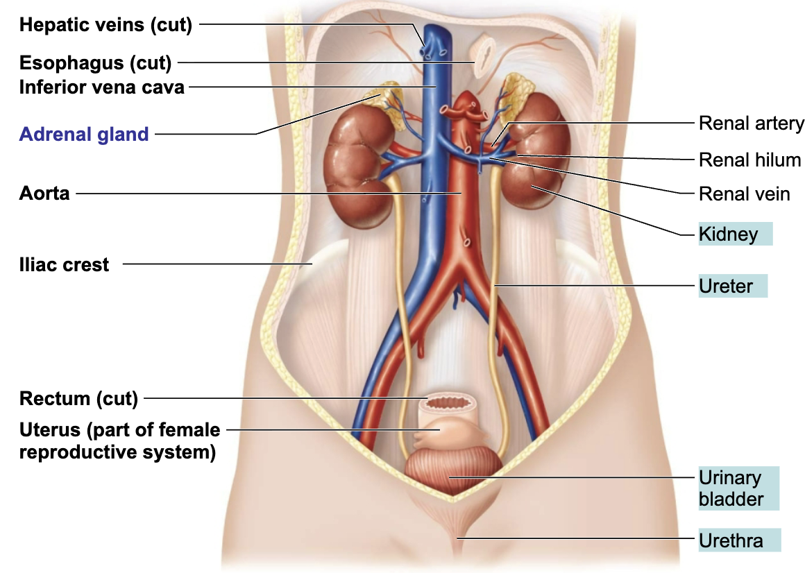
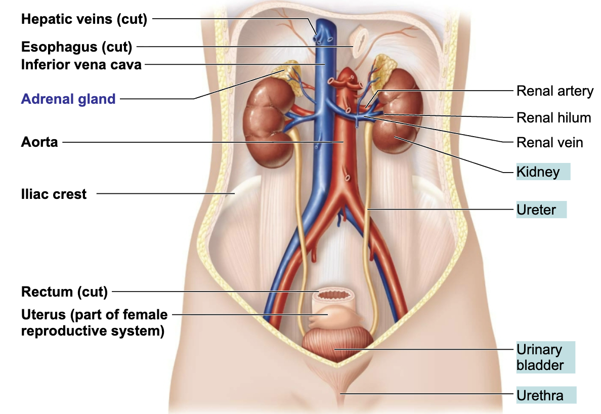
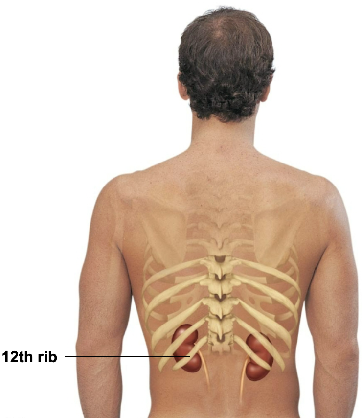
Describe the Location of the Kidneys 

Retroperitoneal position between the dorsal body wall and the parietal peritoneum

<p><span style="color: purple;"><strong><mark data-color="purple" style="background-color: purple; color: inherit;">Retroperitoneal position</mark></strong></span><strong> </strong><span style="color: rgb(0, 0, 0);"><strong><span>→ </span></strong><span>between the dorsal body wall and the parietal peritoneum </span></span></p>
7
New cards

T/F: Kidneys are bean shape and same on both sides

→ FALSE

  • RIGHT kidney is crowded by the liver and lies slightly lower than the left

<p><strong>→ FALSE</strong></p><ul><li><p>RIGHT kidney is crowded by the liver and lies slightly lower than the left</p></li></ul><p></p>
8
New cards

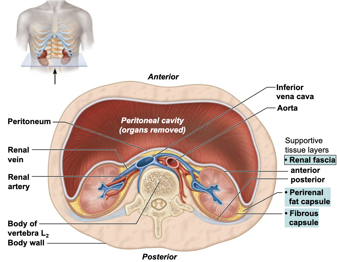
Describe the Location and the Function of the Connective Tissue Layers of the Kidneys

SUPPORTIVE TISSURE LAYERS

  • Renal fascia

    • Outermost layer

    • Anchors the kidney and the adrenal gland to surrounding structures

  • Perirenal fat capsule

    • Middle layer

    • Fatty mass that surrounds the kidney and cushions it against blows

  • Fibrous capsule

    • Innermost layer

    • Transparent capsule that prevents infections in surrounding regions from spreading to the kidney

<p><strong><em>SUPPORTIVE TISSURE LAYERS </em></strong></p><ul><li><p><strong>Renal fascia</strong></p><ul><li><p><span style="color: purple;"><strong><mark data-color="purple" style="background-color: purple; color: inherit;">Outermost layer</mark></strong></span> </p></li><li><p>Anchors the kidney and the adrenal gland to surrounding structures</p></li></ul></li><li><p><strong>Perirenal fat capsule</strong></p><ul><li><p><span style="color: purple;"><strong><mark data-color="purple" style="background-color: purple; color: inherit;">Middle layer </mark></strong></span></p></li><li><p>Fatty mass that surrounds the kidney and cushions it against blows</p></li></ul></li><li><p><strong>Fibrous capsule</strong></p><ul><li><p><span style="color: purple;"><strong><mark data-color="purple" style="background-color: purple; color: inherit;">Innermost layer</mark></strong></span></p></li><li><p>Transparent capsule that prevents infections in surrounding regions from spreading to the kidney</p></li></ul></li></ul><p></p><p></p>
9
New cards

Explain Effect and Cause of Rena ptosis

  • EFFECT

    • Condition in which one or both kidneys drop to lower position

  • CAUSE

    • Can be caused by loss of surrounding fatty tissue capsule that holds kidneys in normal position

    • Seen with emaciation or rapid weight loss

10
New cards

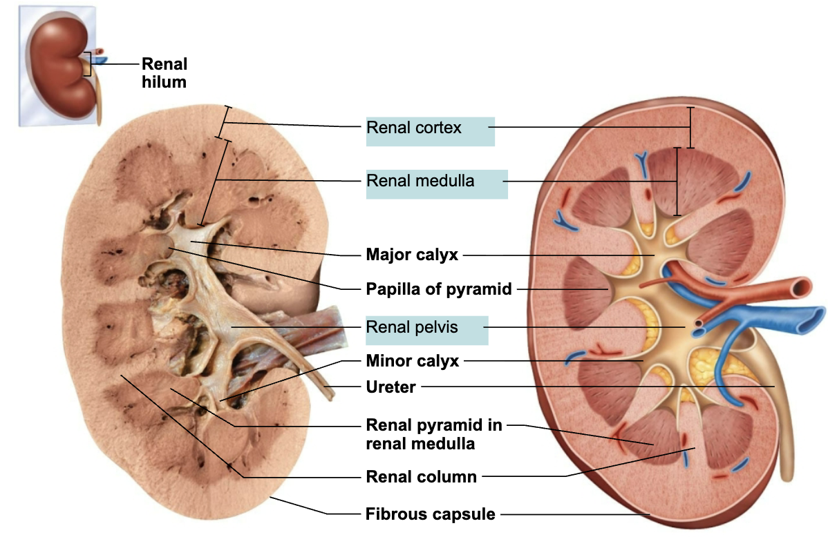
Describe the Cross-Section Anatomy of the Internal Kidney

  1. Renal cortex: granular-appearing superficial region

  2. Renal medulla: deep to cortex, composed of cone-shaped medullary (renal) pyramids

  3. Minor calyces: drain urine into the major calyces

  4. Major calyces: collect urine and drain into the renal pelvis

  5. Renal pelvis: kidney that receives urine from major calyces; continuous with the ureter leaving the renal hilum

<ol><li><p><span style="color: blue;"><strong><span>Renal cortex</span></strong></span>: granular-appearing superficial region</p></li><li><p><span style="color: blue;"><strong><span>Renal medulla</span></strong></span>: deep to cortex, composed of cone-shaped <span style="color: blue;"><strong><span>medullary (renal) pyramids</span></strong></span></p></li><li><p><span style="color: blue;"><strong><span>Minor calyces</span></strong></span><span style="color: rgb(0, 0, 0);"><span>: drain urine into the major calyces</span></span></p></li><li><p><span style="color: blue;"><strong><span>Major calyces</span></strong></span><span style="color: rgb(7, 7, 7);"><span>: collect urine and drain into the renal pelvis</span></span></p></li><li><p><span style="color: blue;"><strong><span>Renal pelvis</span></strong></span><span style="color: rgb(7, 7, 7);"><span>: kidney that receives urine from major calyces; continuous with the ureter leaving the renal hilum</span></span></p></li></ol><p></p>
11
New cards

Know the Pathway of Urine Flow 

knowt flashcard image