Muscle A and P (1)
0.0(0)
Card Sorting
1/101
Earn XP
Description and Tags
Last updated 2:13 AM on 3/21/23
Name | Mastery | Learn | Test | Matching | Spaced | Call with Kai |
|---|
No analytics yet
Send a link to your students to track their progress
102 Terms
1
New cards
Movement
is a fundamental characteristic of all living organisms
2
New cards
Skeletal, Cardiac, Smooth muscle
What are the three types of muscular tissue
3
New cards
Excitability
(responsiveness)—to chemical signals, stretch, and electrical changes across the plasma membrane
4
New cards
Conductivity
local electrical excitation sets off a wave of excitation that travels along the muscle fiber
5
New cards
Contractility
shortens when stimulated
6
New cards
Extensibility
capable of being stretched between contractions
7
New cards
Elasticity
returns to its original rest length after being stretched
\
\
8
New cards
Muscle, Fascicle, Muscle Fiber, Myofibril, Sacromere, Myofilaments
What is the Structural Hierarchy of Skeletal Muscle
9
New cards
Skeletal Muscle
voluntary, striated muscle usually attached to bones
10
New cards
Striations
alternating light and dark transverse bands
\-Results from arrangement of internal contractile proteins
\-Results from arrangement of internal contractile proteins
11
New cards
Voluntary
usually subject to conscious control
12
New cards
Endomysium
connective tissue around muscle cell
13
New cards
Perimysium
connective tissue around muscle fascicle
14
New cards
Epimysium
connective tissue surrounding entire muscle
15
New cards
Sarcolemma
(Muscle fiber)
(Muscle fiber)
plasma membrane of a muscle fiber
16
New cards
Sarcoplasm
(Muscle Fiber)
(Muscle Fiber)
cytoplasm of a muscle fiber
17
New cards
Myofibrils
(Muscle Fiber)
(Muscle Fiber)
long protein cords occupying most of sarcoplasm
18
New cards
Glycogen
carbohydrate stored to provide energy for exercise
19
New cards
Myoglobin
red pigment; provides some oxygen needed for muscle activity
20
New cards
Myoblasts (muscle fiber)
stem cells that fused to form each muscle fiber early in development
21
New cards
Satellite cells
(muscle fiber)
(muscle fiber)
unspecialized myoblasts remaining between the muscle fiber and endomysium
\
\-Play a role in regeneration of damaged skeletal muscle tissue
\
\-Play a role in regeneration of damaged skeletal muscle tissue
22
New cards
Mitochondria
packed into spaces between myofibrils
23
New cards
Multiple nuclei
flattened nuclei pressed against the inside of the sarcolemma
24
New cards
Sarcoplasmic reticulum (SR)
smooth ER that forms a network around each myofibril:
25
New cards
Terminal cisterns
dilated end-sacs of SR which cross the muscle fiber from one side to the other
\- Acts as a calcium reservoir; it releases calcium through channels to activate contraction
\- Acts as a calcium reservoir; it releases calcium through channels to activate contraction
26
New cards
T tubules
tubular infoldings of the sarcolemma which penetrate through the cell and emerge on the other side
27
New cards
Triad
a T tubule and two terminal cisterns associated with it
\
\
28
New cards
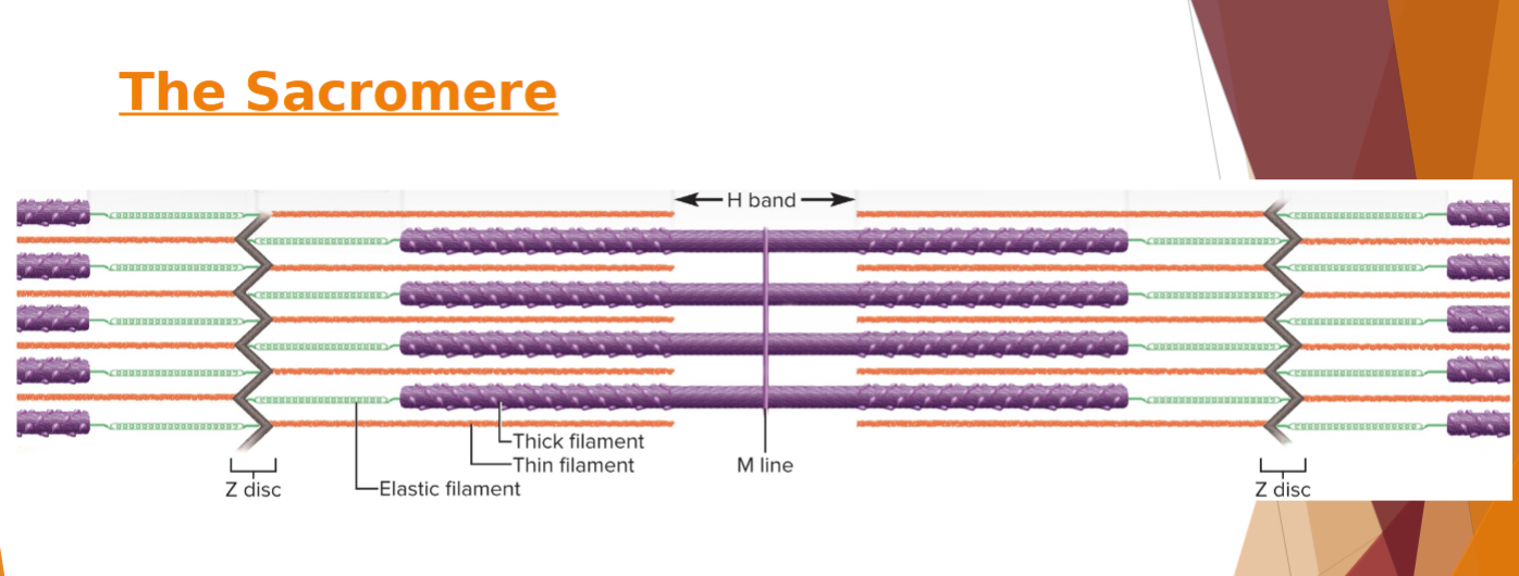
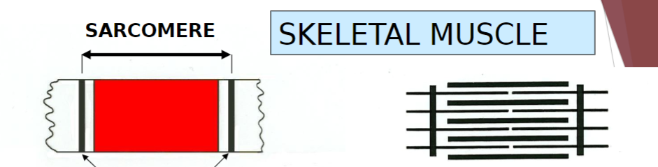
Sarcomere
segment from Z disc to Z disc
29
New cards
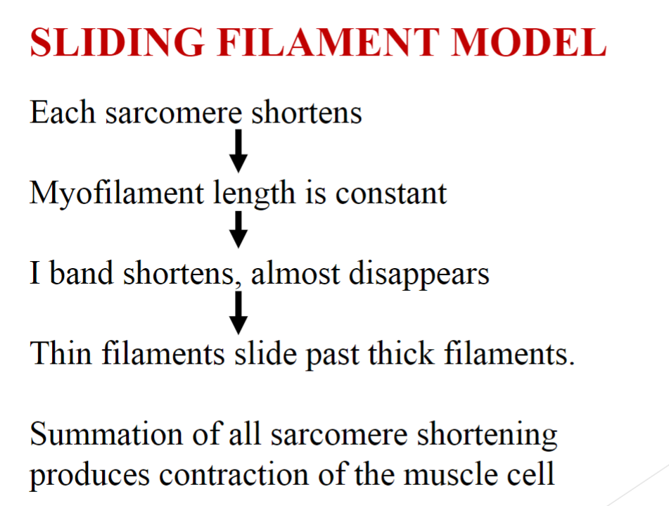
Sarcomere
\-Functional contractile unit of muscle fiber
- Muscle cells shorten because their individual sarcomeres shorten
-Z disc (Z lines) are pulled closer together as thick and thin filaments slide past each other
- Muscle cells shorten because their individual sarcomeres shorten
-Z disc (Z lines) are pulled closer together as thick and thin filaments slide past each other
30
New cards
Sarcomere
Neither thick nor thin filaments change length during shortening
\-Only the amount of overlap changes
\-Only the amount of overlap changes
31
New cards
During shortening of Sarcomere
\-dystrophin and linking proteins also pull on extracellular proteins
Transfers pull to extracellular tissue
\
Transfers pull to extracellular tissue
\
32
New cards
33
New cards
Myofilaments
Thick filaments—made of several hundred myosin molecules
34
New cards
Myofilaments
\-Two chains intertwined
\- Double globular head
\-Heads on one half of the thick filament angle to the left, while heads on other half angle to the right
\- Bare zone
\- Double globular head
\-Heads on one half of the thick filament angle to the left, while heads on other half angle to the right
\- Bare zone
35
New cards
(Thin Filament)- Myofilament
\
Fibrous (F) actin
\
Fibrous (F) actin
\-two intertwined strands
\-String of globular (G) actin subunits each with an active site that can bind to head of myosin molecule
\-String of globular (G) actin subunits each with an active site that can bind to head of myosin molecule
36
New cards
(Thin Filament)-Myofilament
\
Tropomyosin molecules
\
Tropomyosin molecules
\-Each blocking six or seven active sites on G actin subunits
37
New cards
(Thin Filament)- Myofilament
Troponin molecule
Troponin molecule
small, calcium-binding protein on each tropomyosin molecule
38
New cards
(Elastic Filament)- Myofilament
\
Titin
\
Titin
huge, springy protein
\-Run through core of thin filament and anchor it to Z disc and M line
\- Help stabilize and position the thick filament
\- Prevent overstretching and provide recoil
\-Run through core of thin filament and anchor it to Z disc and M line
\- Help stabilize and position the thick filament
\- Prevent overstretching and provide recoil
39
New cards
(Myofilament)
Contractile proteins
Contractile proteins
myosin and actin do the work of contraction
40
New cards
(Myofilament)
Regulatory proteins
Regulatory proteins
tropomyosin and troponin - Act like a switch that determines when fiber can (and cannot) contract
41
New cards
Contraction activated by:
\-release of calcium into sarcoplasm and its binding to troponin
-Troponin changes shape and moves tropomyosin off the active sites on actin
\
-Troponin changes shape and moves tropomyosin off the active sites on actin
\
42
New cards
\
\
\
43
New cards
Contracted
44
New cards
Rest length
45
New cards
Stretched
46
New cards
(Myofilament)
Dystrophin
Dystrophin
clinically important protein
47
New cards
(Myofilament)
Dystrophin
Dystrophin
\- Links actin in outermost myofilaments to membrane proteins that link to endomysium
-Transfers forces of muscle contraction to connective tissue ultimately leading to tendon
\
-Transfers forces of muscle contraction to connective tissue ultimately leading to tendon
\
48
New cards
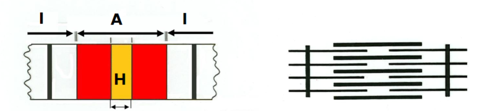
Striations
result from the precise organization of myosin and actin in cardiac and skeletal muscle cells
49
New cards
Z disc
provides anchorage for thin filaments and elastic filaments
50
New cards
YES
Does a Skeletal muscle cannot contract unless stimulated by a nerve
51
New cards
Denervation atrophy
shrinkage of paralyzed muscle when nerve remains disconnected
52
New cards
Somatic motor neurons
Nerve cells whose cell bodies are in the brainstem and spinal cord that serve skeletal muscles
53
New cards
Somatic motor fibers
their axons that lead to the skeletal muscle
54
New cards
YES
Is Each muscle fiber is supplied by only one motor neuron
55
New cards
Motor unit
one nerve fiber and all the muscle fibers innervated by it
56
New cards
Muscle fibers of one motor unit:
\-Provide ability to sustain long-term contraction as motor units take turns contracting
\-Dispersed throughout muscle
\-Effective contraction usually requires contraction of several motor units at once
\
\-Dispersed throughout muscle
\-Effective contraction usually requires contraction of several motor units at once
\
57
New cards
200
How many muscle fibers does the average motor unit contain
58
New cards
Small motor units
fine degree of control
\- Three to six muscle fibers per neuron - Eye and hand muscles
\- Three to six muscle fibers per neuron - Eye and hand muscles
59
New cards
Large motor units
more strength than control
\-Powerful contractions supplied by large motor units with hundreds of fibers - Quadriceps femoris and gastrocnemius have 1,000 muscle fibers per neuron
\-Powerful contractions supplied by large motor units with hundreds of fibers - Quadriceps femoris and gastrocnemius have 1,000 muscle fibers per neuron
60
New cards
Synapse
point where a nerve fiber meets its target cell
61
New cards
Neuromuscular junction (NMJ)
when target cell is a muscle fiber
62
New cards
Axon terminal
swollen end of nerve fiber
-Contains synaptic vesicles with acetylcholine (ACh)
-Contains synaptic vesicles with acetylcholine (ACh)
63
New cards
Synaptic cleft
gap between axon terminal and sarcolemma
64
New cards
Schwann cells
envelope and isolate NMJ
65
New cards
YES
Does the Nerve impulse causes synaptic vesicles to undergo exocytosis releasing Ach (acetylcholine) into synaptic cleft
66
New cards
YES
Does the muscle cells have millions of ACh receptors
67
New cards
Basal lamina
thin layer of collagen and glycoprotein separating Schwann cell and muscle cell from surrounding tissues
68
New cards
YES
Are muscle fibers and neurons are excitability excitable
69
New cards
YES
Does the Their membranes exhibit voltage changes in response to stimulation
70
New cards
Voltage (electrical potential)
a difference in electrical charge from one point to another
71
New cards
Resting membrane potential
\-about −90 mV in skeletal muscle cells -Maintained by sodium–potassium pump
\
\
72
New cards
In an unstimulated (resting) cell
There are more anions (negatively charged particles) on the inside of the membrane than on the outside
\
\-These anions make the inside of the
plasma membrane negatively charged
by comparison to its outer surface
\
\-These anions make the inside of the
plasma membrane negatively charged
by comparison to its outer surface
73
New cards
In an unstimulated (resting) cell
The plasma membrane is electrically polarized (charged) with a negative resting membrane potential (RMP)
74
New cards
In an unstimulated (resting) cell
There are excess sodium ions (Na+) in the extracellular fluid (ECF)
75
New cards
In an unstimulated (resting) cell
There are excess potassium ions (K+) \n in the intracellular fluid (ICF
76
New cards
In a stimulated (active) muscle fiber or nerve cell
1. Na+ ion gates open in the plasma
membrane
2. Na+ flows into cell down its electrochemical
gradient
77
New cards
In a stimulated (active) muscle fiber or nerve cel
3. These cations override the negative
charges in the ICF
4. Depolarization: inside of plasma membrane
becomes positive
78
New cards
In a stimulated (active) muscle fiber or nerve cell
5. Immediately, Na+ gates close and K+ gates
open
6. K+ rushes out of cell partly repelled by
positive sodium charge and partly because
of its concentration gradient
7. Loss of positive potassium ions turns the
membrane negative again (repolarization
79
New cards
A resting membrane potential
s seen in a waiting excitable cell,
80
New cards
action potential
is a quick event seen in a stimulated excitable cell
81
New cards
An action potential
perpetuates itself down the length of a cell’s membrane
82
New cards
Toxins
interfering with synaptic function can paralyze muscles
83
New cards
pesticides
contain cholinesterase inhibitors
84
New cards
pesticides
Bind to acetylcholinesterase and prevent it from degrading ACh
85
New cards
Spastic paralysis
a state of continual contraction of the muscles; possible suffocation
\
\
86
New cards
Tetanus
(lockjaw) is a form of spastic paralysis caused by toxin Clostridium tetani
87
New cards
Tetanus
Does the Glycine in the spinal cord normally stops motor neurons from producing unwanted muscle contractions
88
New cards
YES
Does Tetanus toxin blocks glycine release in the spinal cord and causes overstimulation and spastic paralysis of the muscles
89
New cards
Flaccid paralysis
a state in which the muscles are limp and cannot contract
90
New cards
Curare
competes with ACh for receptor sites, but does not stimulate the muscles
91
New cards
Botulism
type of food poisoning caused by a neuromuscular toxin secreted by the bacterium Clostridium botulinum
92
New cards
Excitation
a process in which nerve action potentials lead to muscle action potentials
93
New cards
Excitation–contraction coupling
events that link the action potentials on the sarcolemma to activation of the myofilaments, thereby preparing them to contract
94
New cards
Contraction
the step in which the muscle fiber develops tension and may shorten
95
New cards
Relaxation
when stimulation ends, a muscle fiber relaxes and returns to its resting length
96
New cards
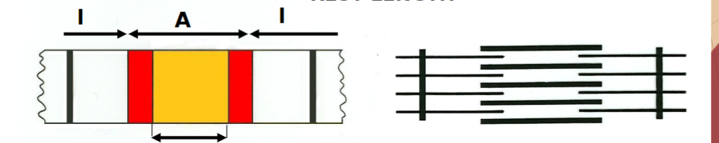
The Length–Tension Relationship and Muscle Tone
the amount of tension generated by a muscle depends on how stretched or shortened it was before it was stimulated
97
New cards
If overly shortened before stimulated
a weak contraction results, as thick filaments just butt against Z discs
98
New cards
If too stretched before stimulated,
a weak contraction results, as minimal overlap between thick and thin filaments results in minimal cross-bridge formation
99
New cards
Optimum resting length
produces greatest force when muscle contracts
100
New cards
Rigor mortis
hardening of muscles and stiffening of
body beginning 3–4 hr after death
1. Deteriorating sarcoplasmic reticulum releases Ca+2
and deteriorating sarcolemma allows Ca+2 to enter
cytosol
2. Ca+2 activates myosin–actin cross-bridging
3. Muscle contracts, but cannot relax
body beginning 3–4 hr after death
1. Deteriorating sarcoplasmic reticulum releases Ca+2
and deteriorating sarcolemma allows Ca+2 to enter
cytosol
2. Ca+2 activates myosin–actin cross-bridging
3. Muscle contracts, but cannot relax