Lec37: Muscle physiology
1/31
There's no tags or description
Looks like no tags are added yet.
Name | Mastery | Learn | Test | Matching | Spaced | Call with Kai |
|---|
No analytics yet
Send a link to your students to track their progress
32 Terms
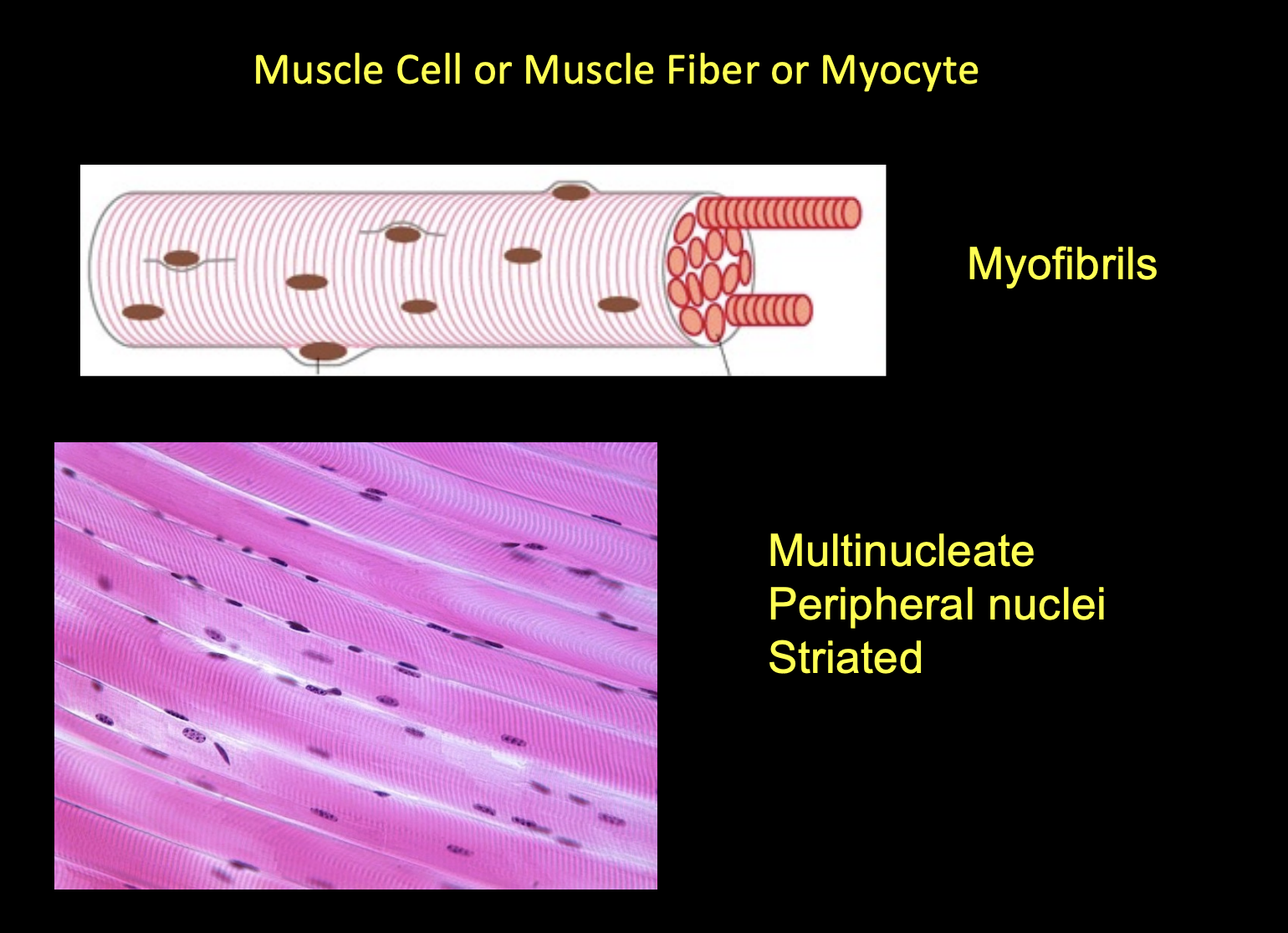
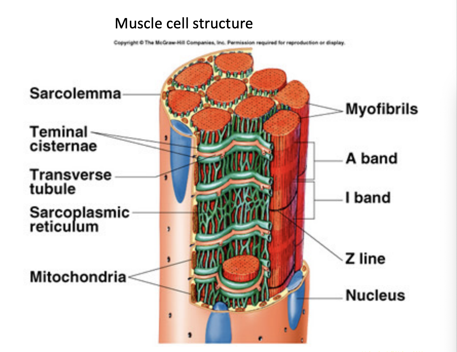
The basic unit of a muscle is the muscle cell, also known as a _______ or _______. These cells are unique because they are __________, meaning they contain many nuclei, which are typically located at the periphery of the cell. They appear _________ under a microscope due to the organized arrangement of internal proteins.
muscle fiber or myocyte
multinucleate
striated (striped)
The outermost connective tissue layer surrounding the entire muscle.
Epimysium

Connective tissue that groups muscle fibers into bundles called fascicles.
Perimysium
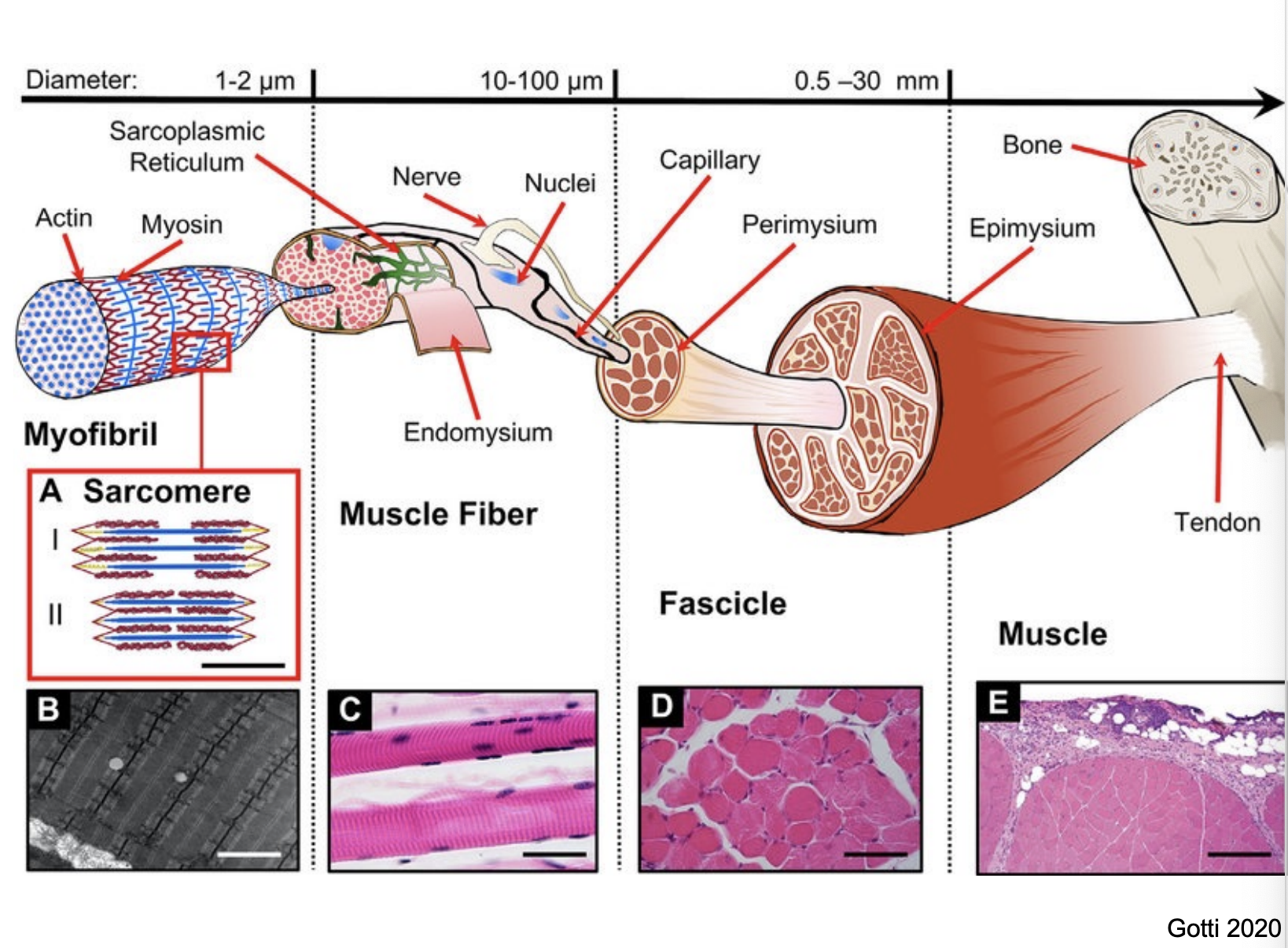
A fine layer of connective tissue that surrounds each individual muscle fiber.
Endomysium

Inside each muscle fiber are _________, which contain the contractile machinery. The fiber also contains a specialized endoplasmic reticulum called the _____________, which stores calcium, and T-tubules, which are deep invaginations of the plasma membrane (sarcolemma) that help transmit electrical signals.
myofibrils
Sarcoplasmic Reticulum (SR)

The _________ is the smallest functional unit of a myofibril, measuring approximately 2.2 µm in length.
sarcomere
Describe the 2 filaments that make up the sacromere.
Filaments: It is composed of two main types of protein filaments:
Thick Filaments (Myosin): Located in the center of the sarcomere (the A-band).
Thin Filaments (Actin): Attached to the Z-discs at the ends of the sarcomere.
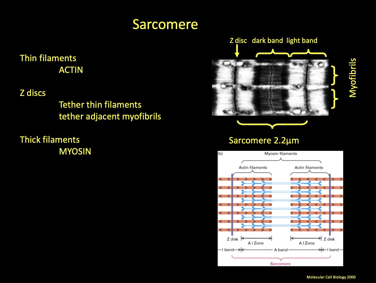
What are the bands and discs that make up the sarcomere?
Z-discs: These define the boundaries of the sarcomere and tether the thin filaments.
I-band: The light region containing only thin filaments.
A-band: The dark region containing the full length of the thick filaments.
Muscle contraction begins with a signal from the __________
nervous system.
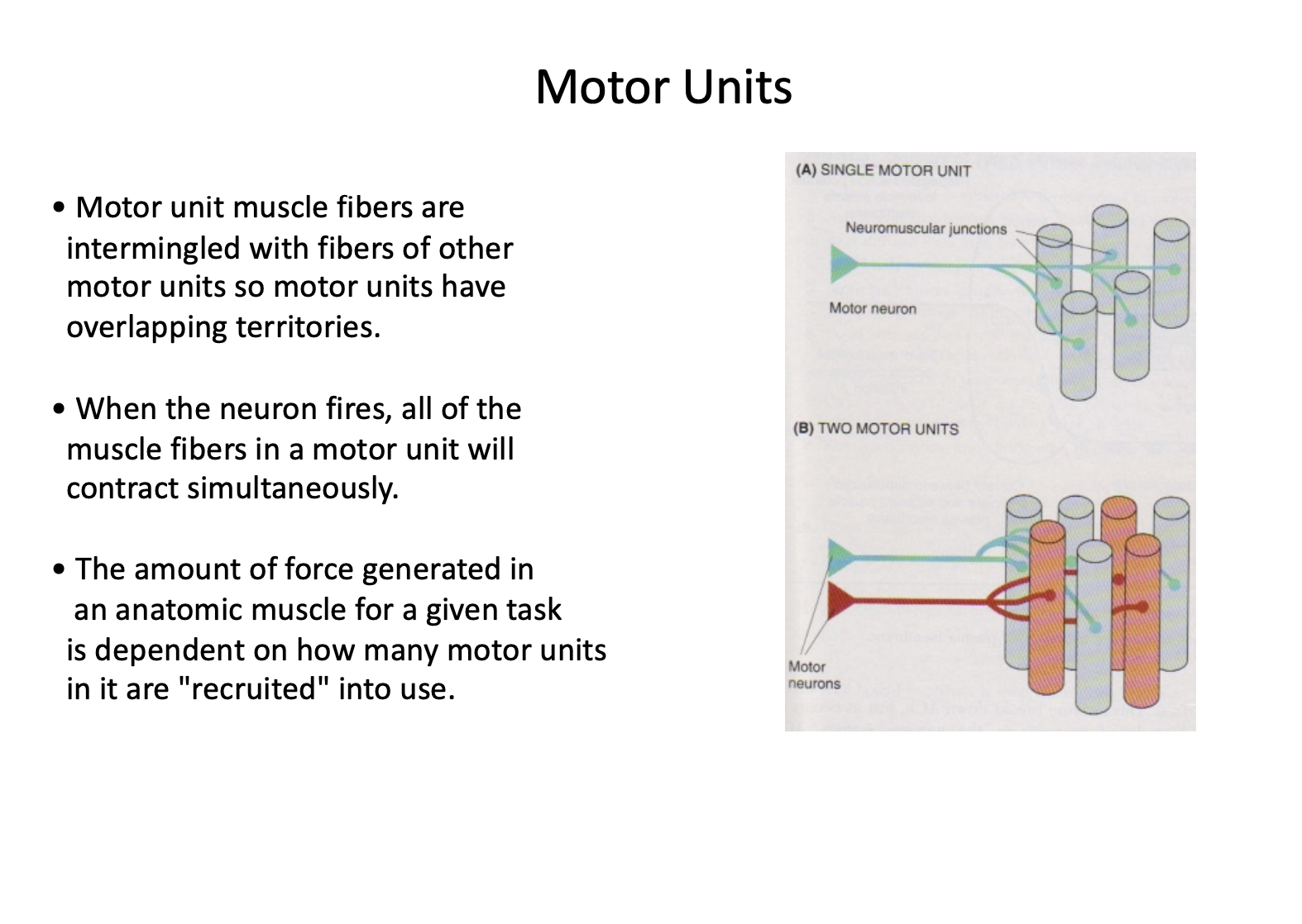
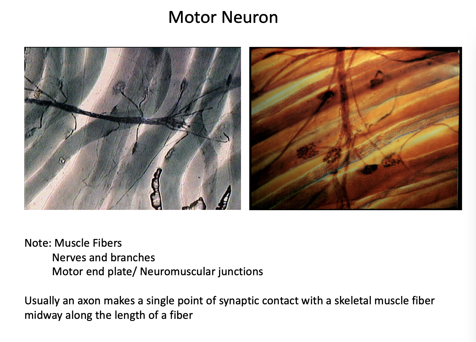
A ________ consists of a single motor neuron and all the individual muscle fibers it innervates.
motor unit
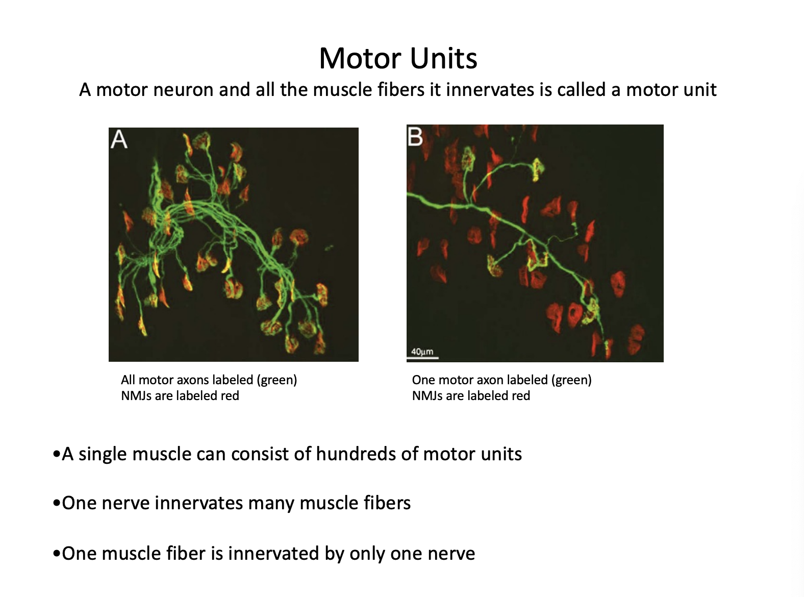
Describe the innervation of the motor unit.
Innervation Rules: While one motor neuron can innervate many muscle fibers, each individual muscle fiber is innervated by only one motor neuron.
T/F: The force generated by a muscle depends on how many motor units are "recruited" or activated.
True
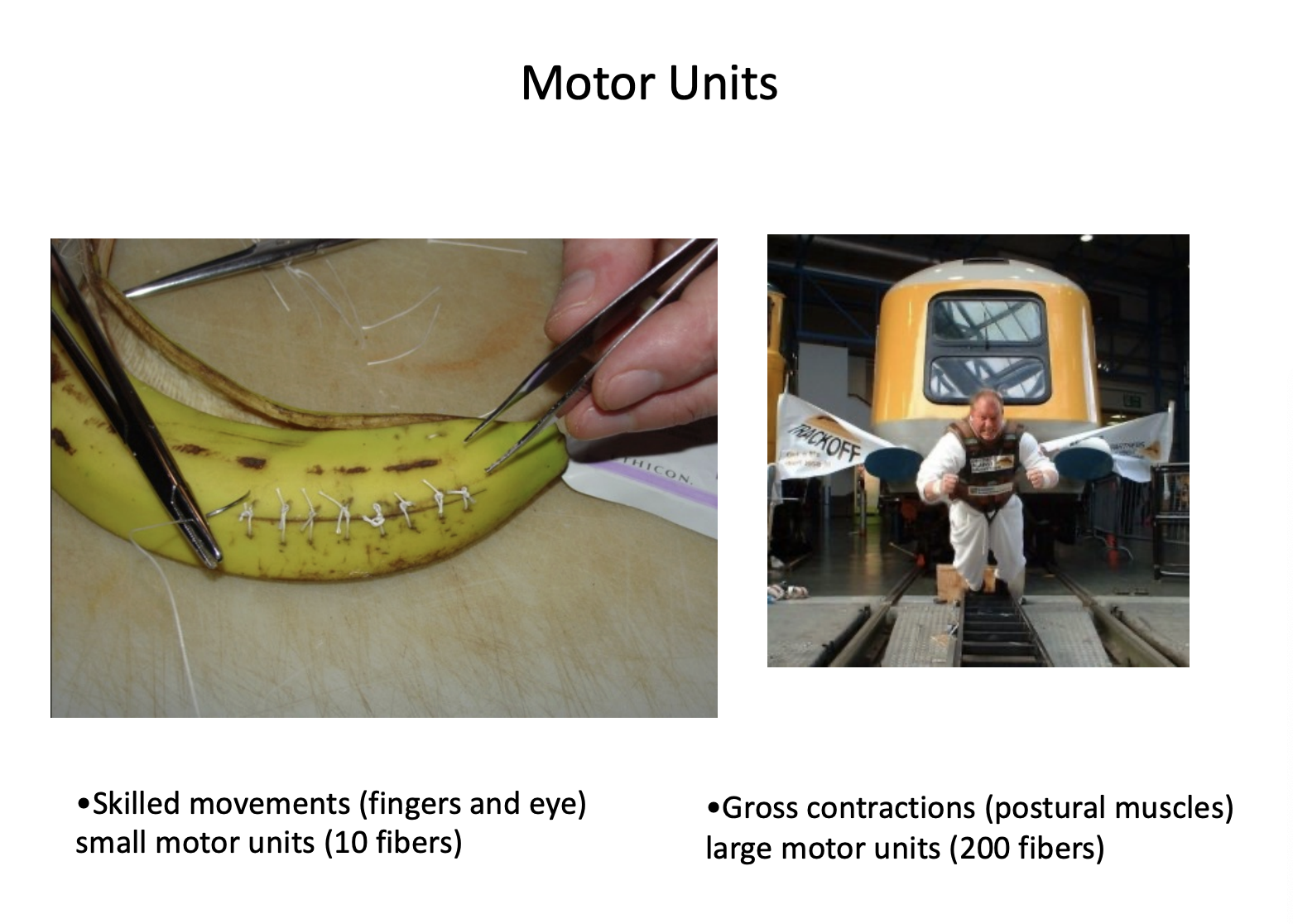
T/F: For skilled movements (like the fingers or eyes), motor units are small (e.g., 10 fibers). For gross contractions and posture (like the back or legs), motor units are large (e.g., 200+ fibers).
True.
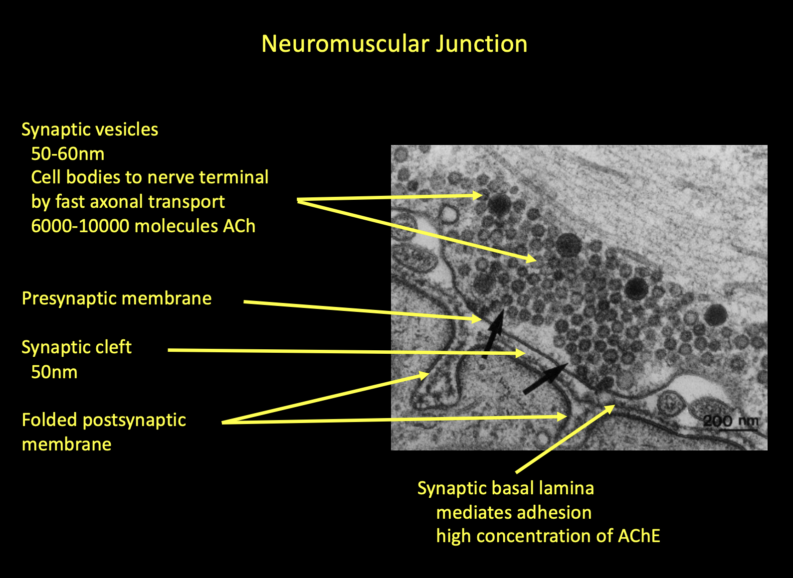
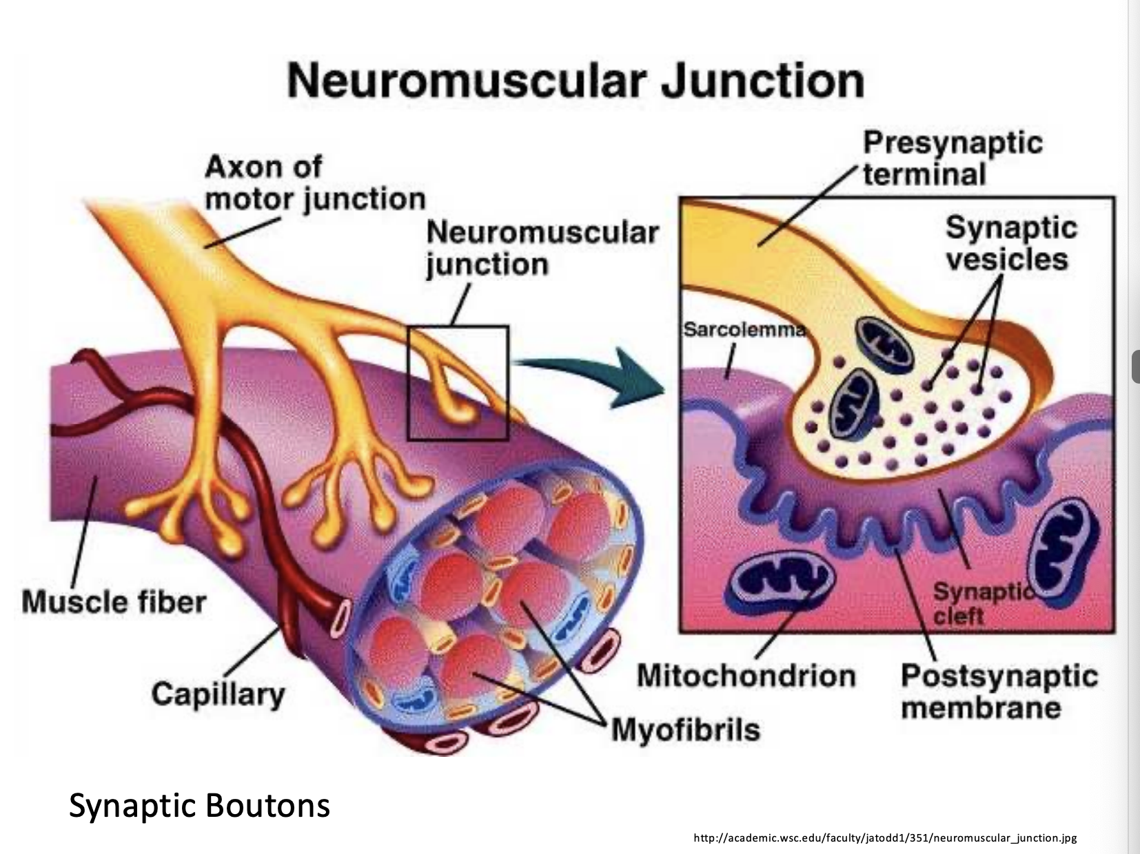
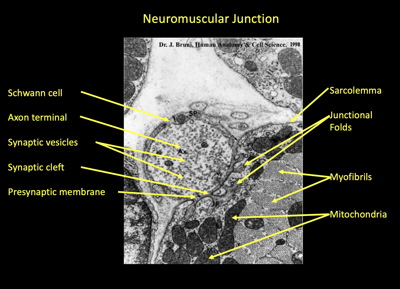
The ___________ is the "synaptic contact" or bridge between the nervous system and the skeletal muscle.
The Neuromuscular Junction (NMJ)
Describe the structure of the NMJ.
It consists of the axon terminal (presynaptic), the synaptic cleft (the gap), and the motor end plate (the folded postsynaptic membrane of the muscle fiber).
The nerve terminal contains vesicles filled with approximately 6,000 to 10,000 molecules of the neurotransmitter ___________
Acetylcholine (ACh).
T/F: The motor neuron is an individual nerve cell. The motor unit is a functional grouping. It is defined as one motor neuron PLUS all the muscle fibers it innervates.
True.
The cell body of the motor neuron is in the ______ root of the spinal cord.
Ventral root of the spinal cord.
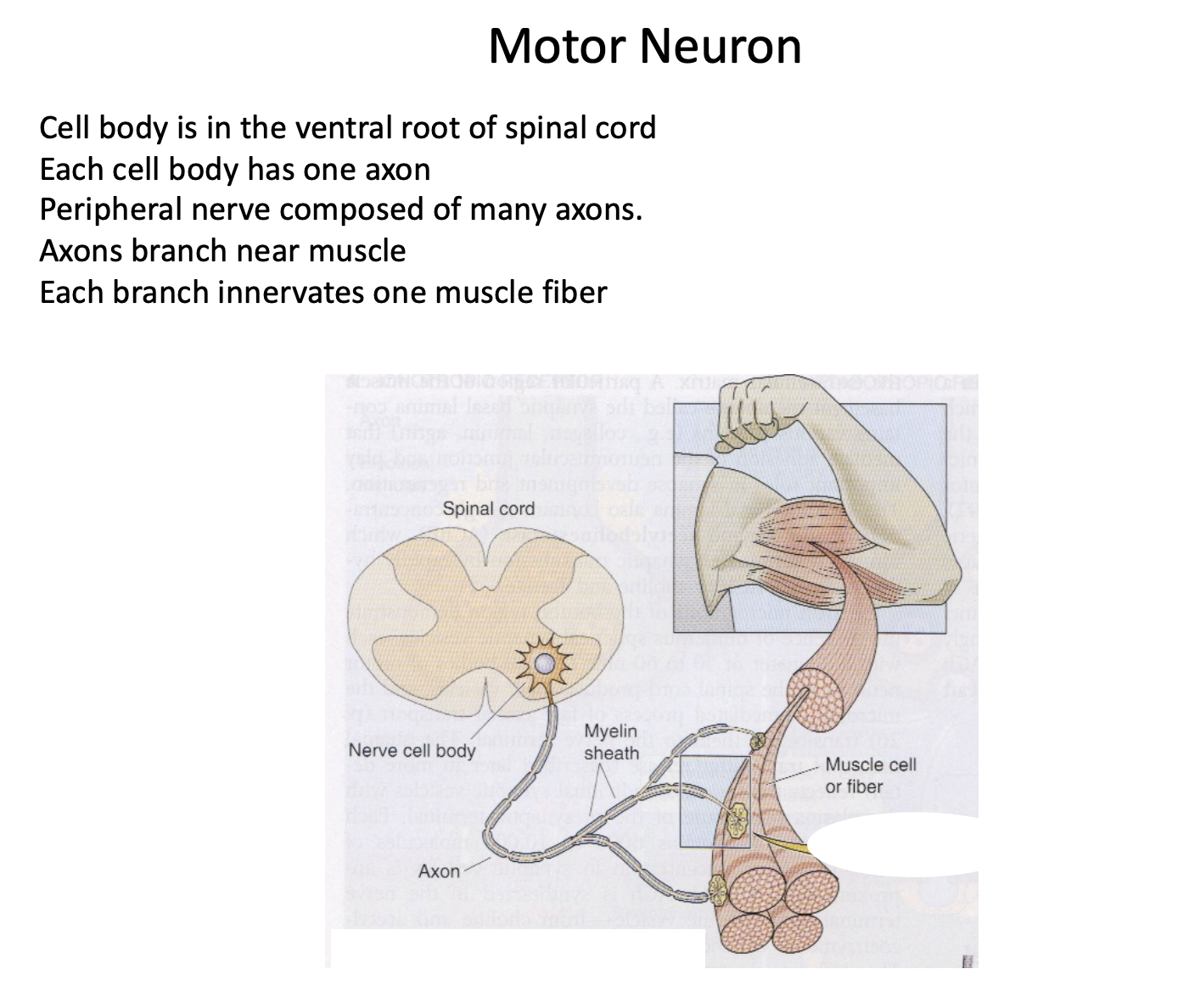
Each cell body has ___ axon
1 axon
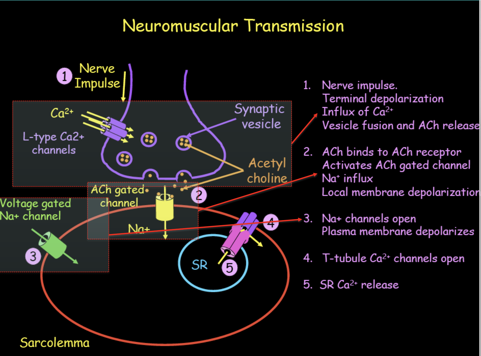
What are the transmission steps at the NMJ?
Transmission Steps:
An electrical nerve impulse reaches the terminal, causing an influx of Calcium (Ca²⁺) through L-type channels.
This triggers vesicles to fuse with the membrane and release ACh into the synaptic cleft.
ACh binds to ACh-gated channels (receptors) on the muscle membrane.
This causes Sodium (Na⁺) influx, leading to a local membrane depolarization.
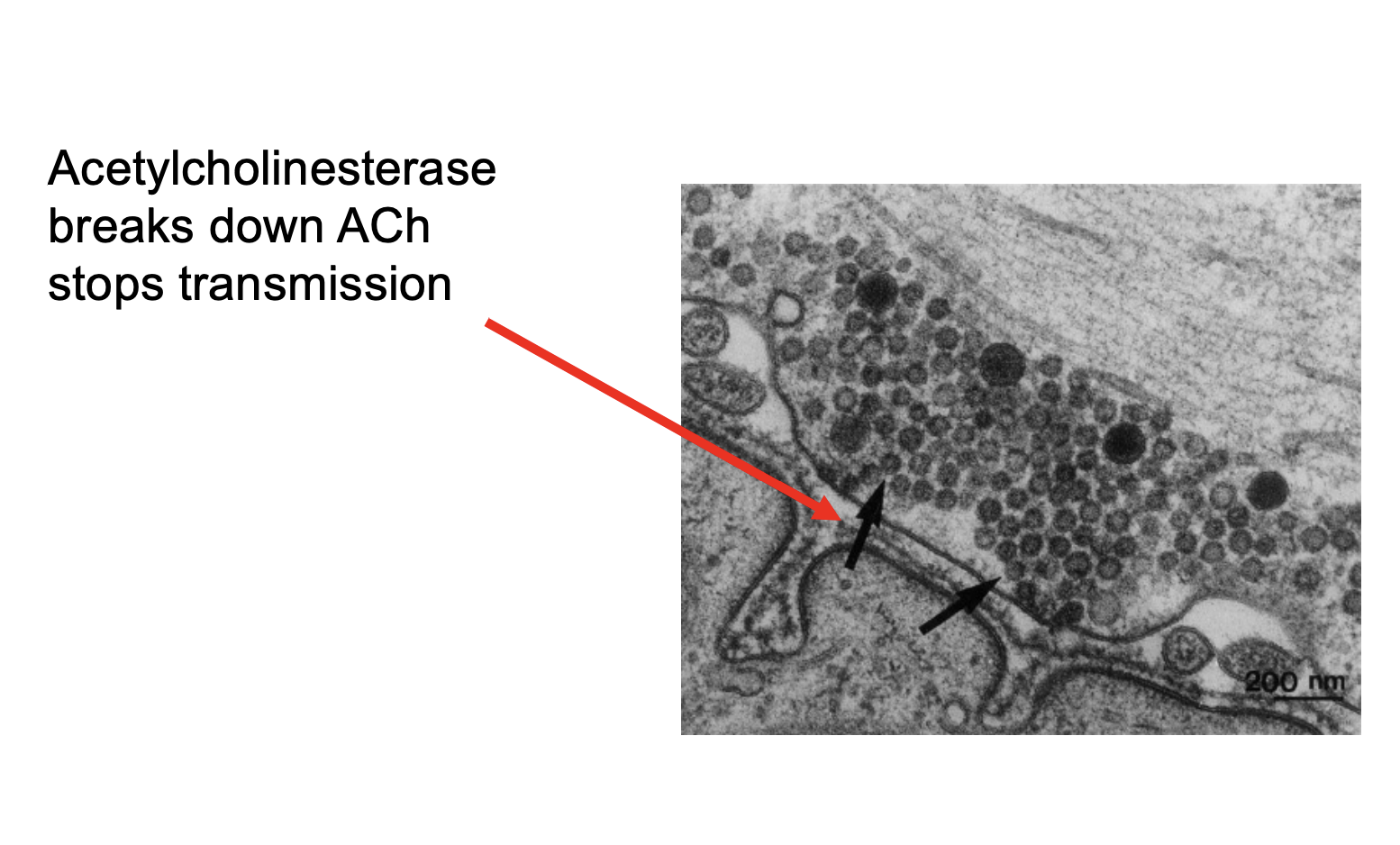
Images of the NMJ
____________ breaks down ACh, stopping transmission
Acetylcholinesterase
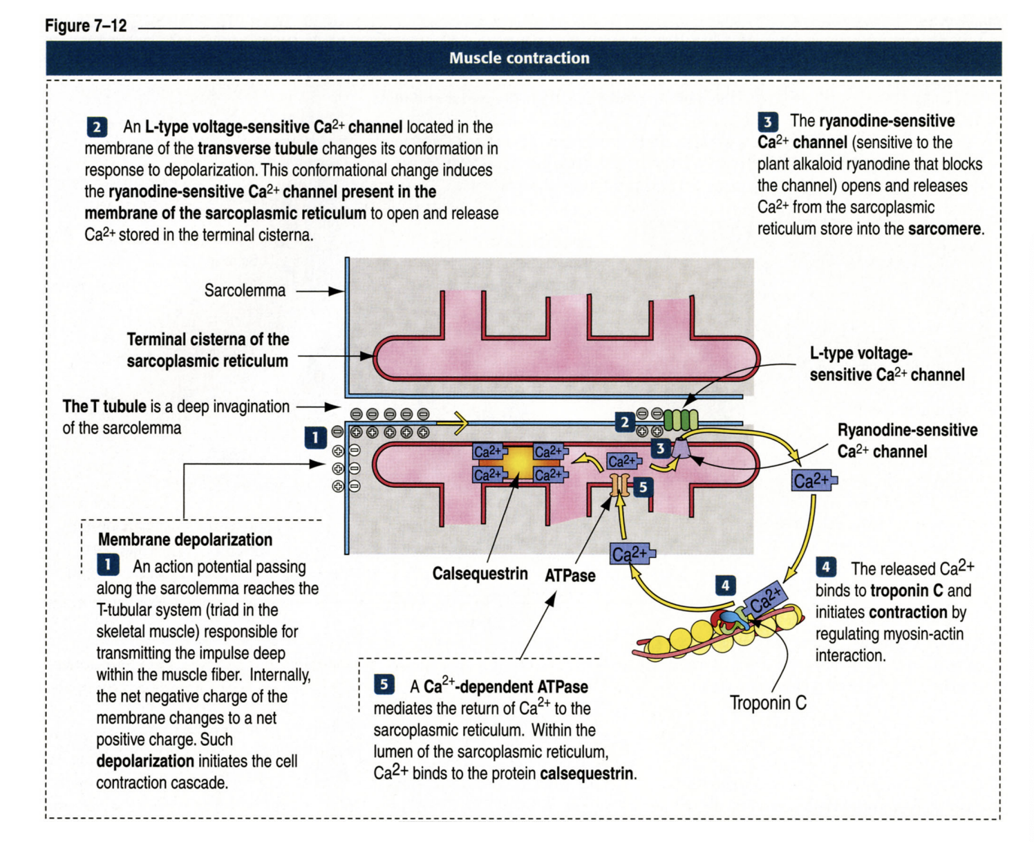
T/F: E-C coupling is the process by which an electrical stimulus (excitation) leads to a mechanical response (contraction).
True.
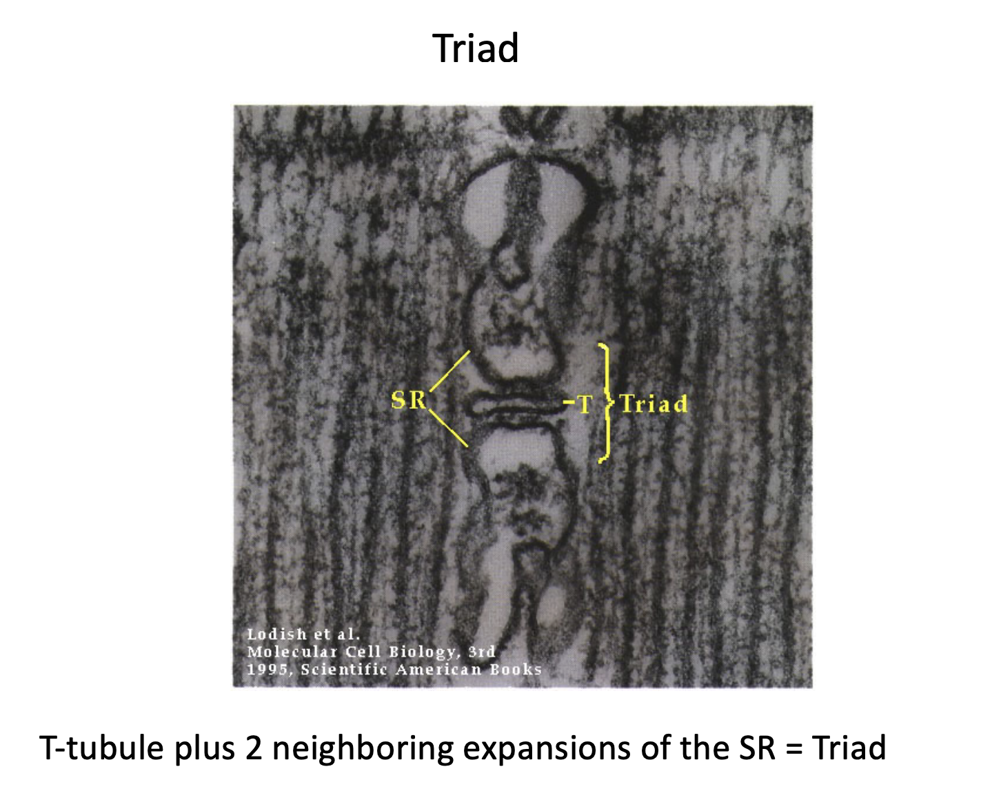
What is the triad?
The Triad: This is a critical structure for signaling, consisting of one T-tubule and two neighboring expansions of the SR called terminal cisternae.
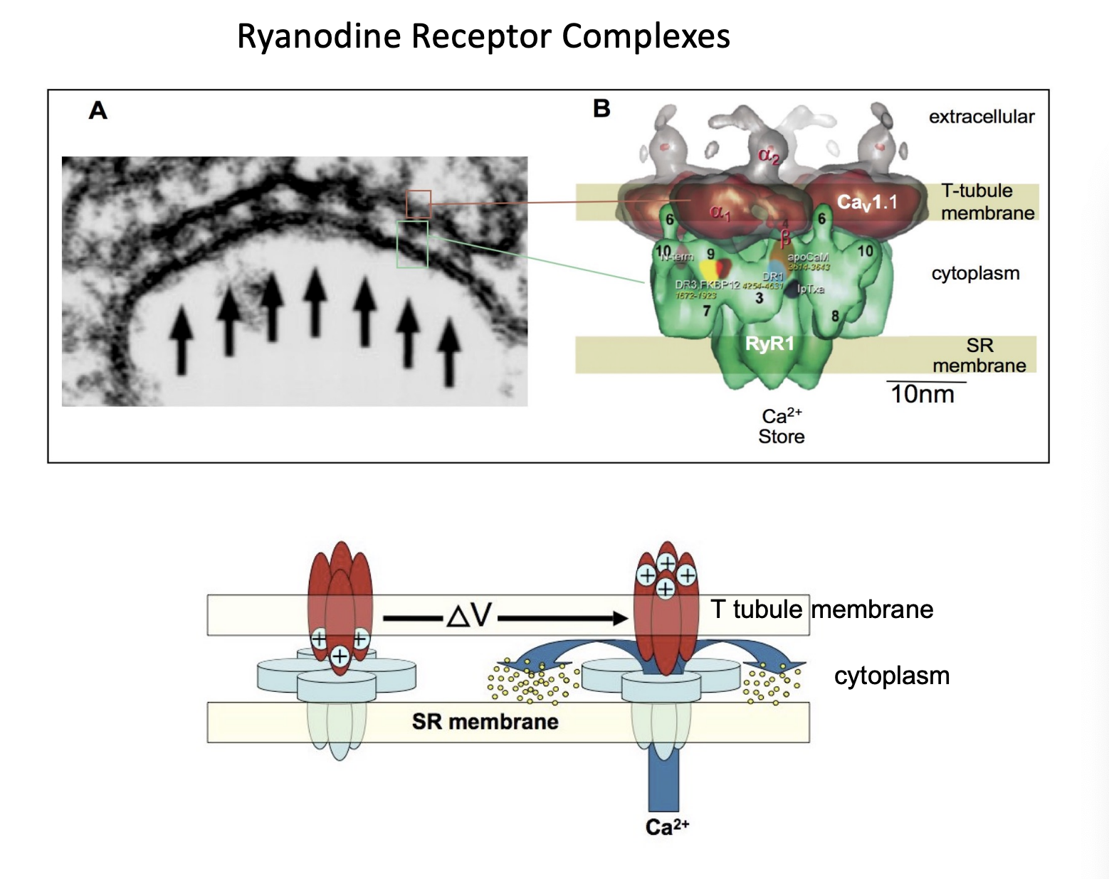
Describe the release of Calcium from the triad.
The Calcium Release Mechanism:
An action potential travels down the T-tubule.
This activates L-type voltage-sensitive Ca²⁺ channels (also known as DHPR) in the T-tubule membrane.
A conformational change in the L-type channel physically triggers the Ryanodine-sensitive Ca²⁺ channels (RyR) on the SR to open.
Calcium is released from the SR stores into the cytosol of the muscle cell.
T/F: Calcium serves as the "on/off" switch for contraction.
True.
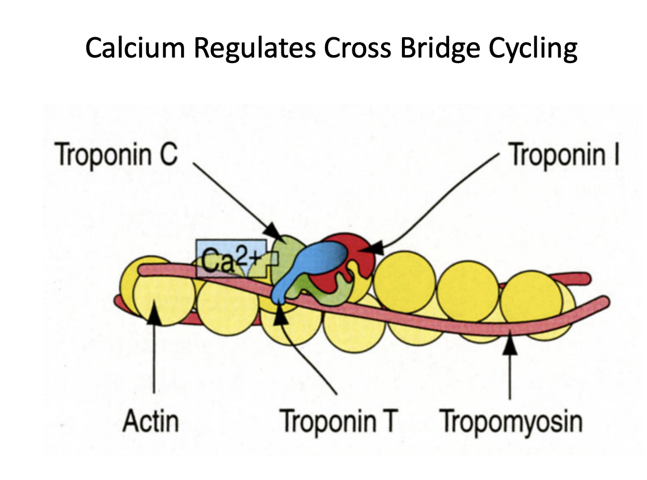
Describe how calcium regulates cross bridge cycling.
Regulation: In a resting state, the interaction between actin and myosin is blocked. When Ca²⁺ is released, it binds to Troponin C. This causes a shift in the troponin-tropomyosin complex, exposing the binding sites on the actin filament.
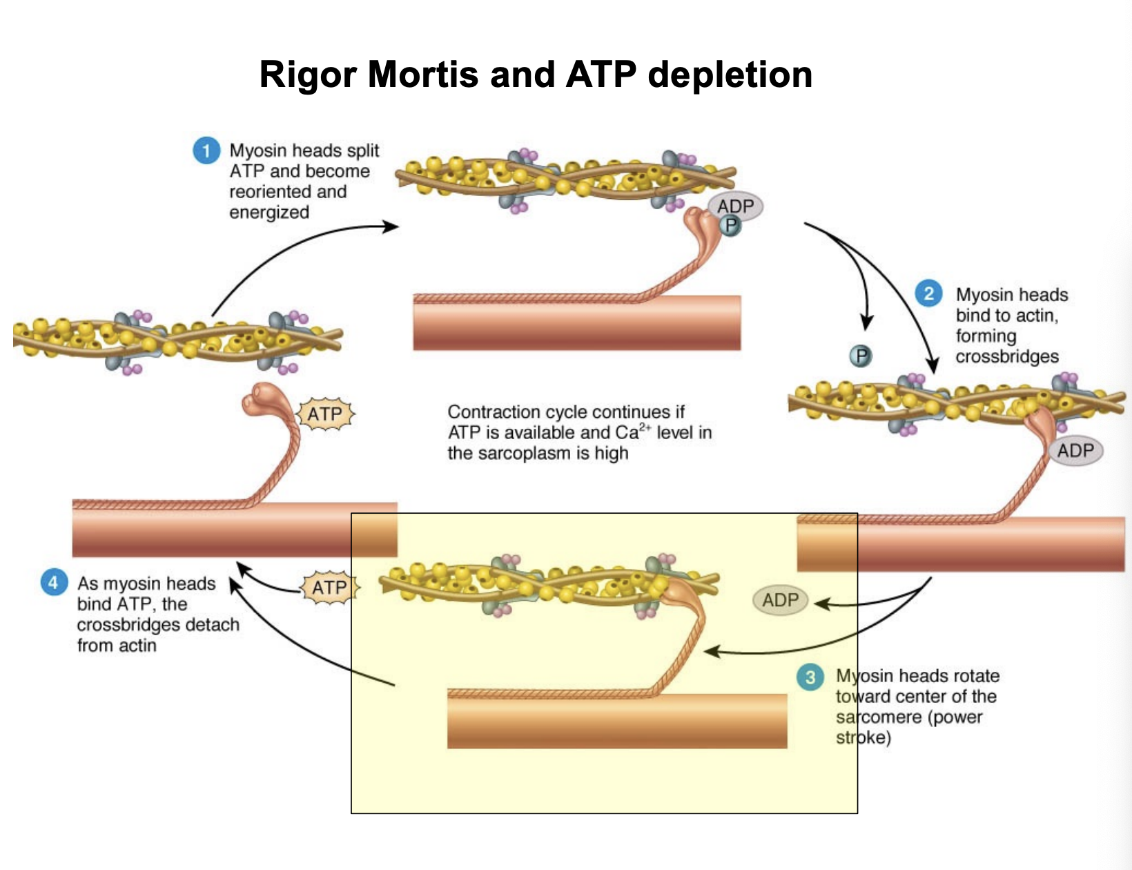
The Cycle: Myosin heads then bind to actin, forming "cross-bridges." They use energy from ATP to pull the actin filaments toward the center of the sarcomere (the power stroke).
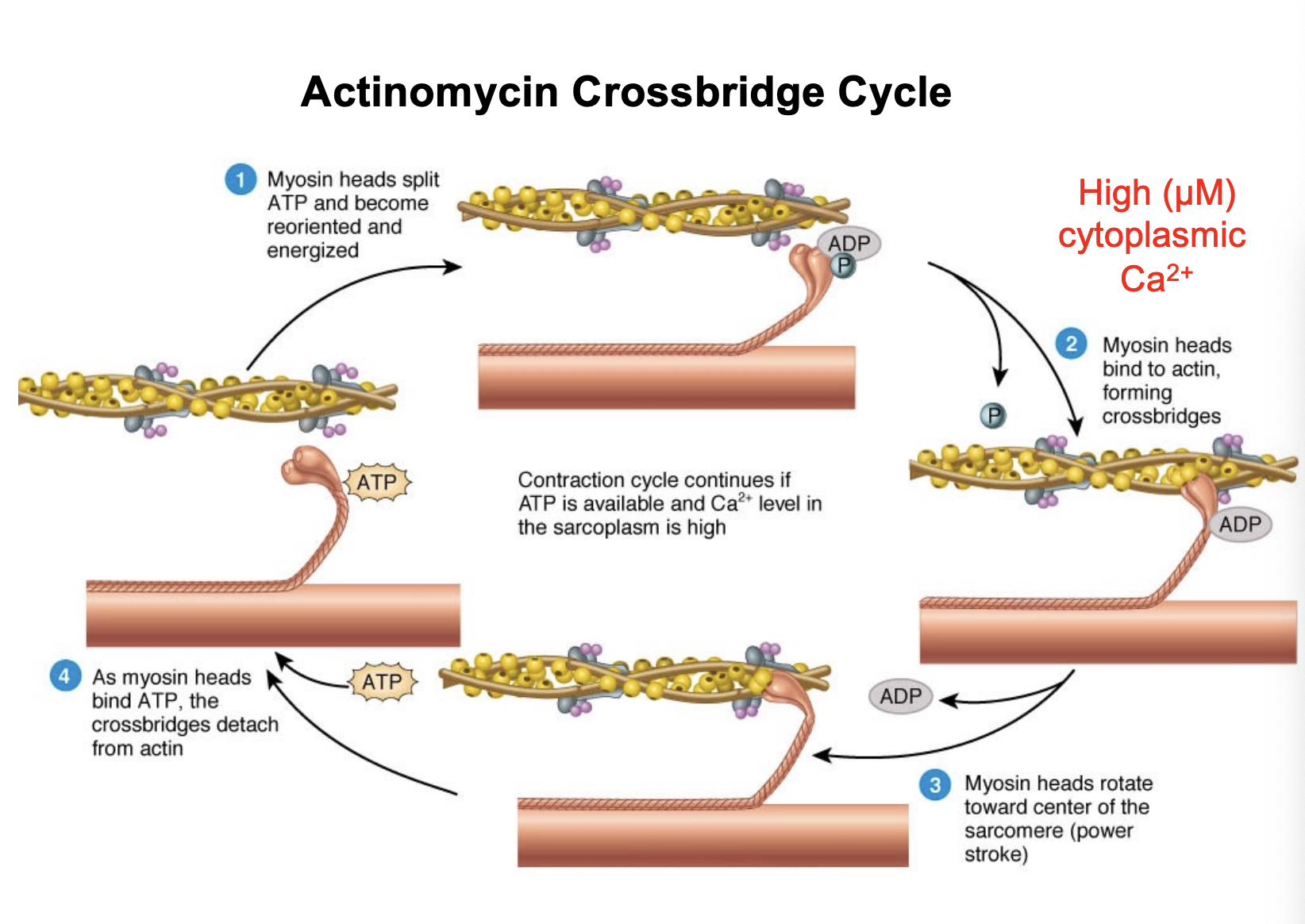
What is rigor mortis?
Rigor Mortis: This occurs when ATP is depleted. Without ATP, the myosin heads cannot detach from the actin filaments, leaving the muscle in a state of permanent contraction or stiffness.
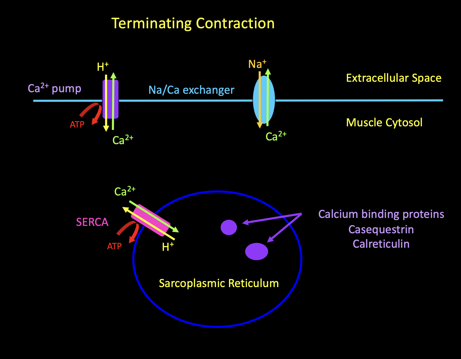
Explain how a contraction is terminated.
To stop a contraction, calcium must be removed from the cytosol.
SERCA Pump: A Ca²⁺-dependent ATPase (SERCA) actively pumps calcium back into the Sarcoplasmic Reticulum.
Calsequestrin: Once inside the SR, calcium binds to the protein calsequestrin for storage.
Acetylcholinesterase (AChE): In the synaptic cleft, this enzyme breaks down ACh to stop the initial electrical signal.
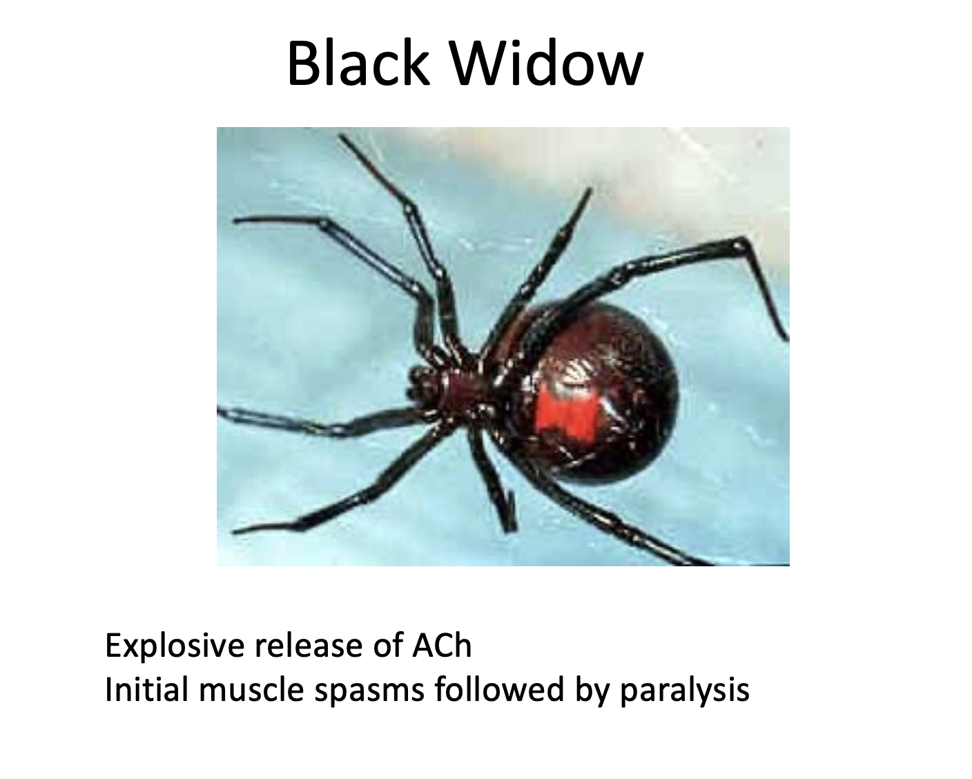
How does black widow venom interfere with the NMJ?
Black Widow Venom: Causes an explosive, uncontrolled release of ACh, leading to initial spasms followed by paralysis.
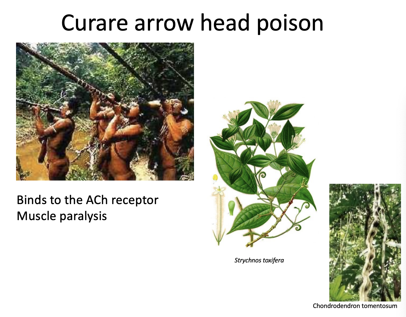
How does curare arrow poison interfere with the NMJ?
Curare: An arrow poison that binds to and blocks ACh receptors, preventing the muscle from receiving the signal to contract.
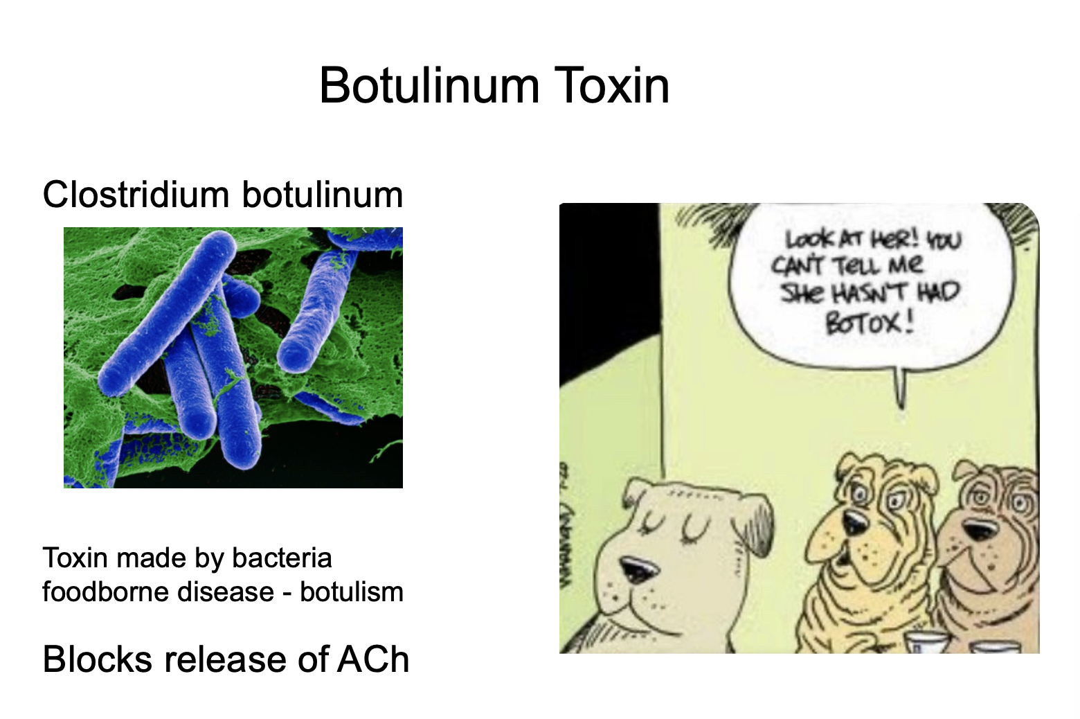
How does Botulinum Toxin interfere with the NMJ?
Botulinum Toxin (Botox): Produced by Clostridium botulinum, it blocks the release of ACh from the nerve terminal, preventing contraction entirely.