BPK 326 - Abdominal Diagrams/Cross Sections
1/83
There's no tags or description
Looks like no tags are added yet.
Name | Mastery | Learn | Test | Matching | Spaced | Call with Kai |
|---|
No analytics yet
Send a link to your students to track their progress
84 Terms
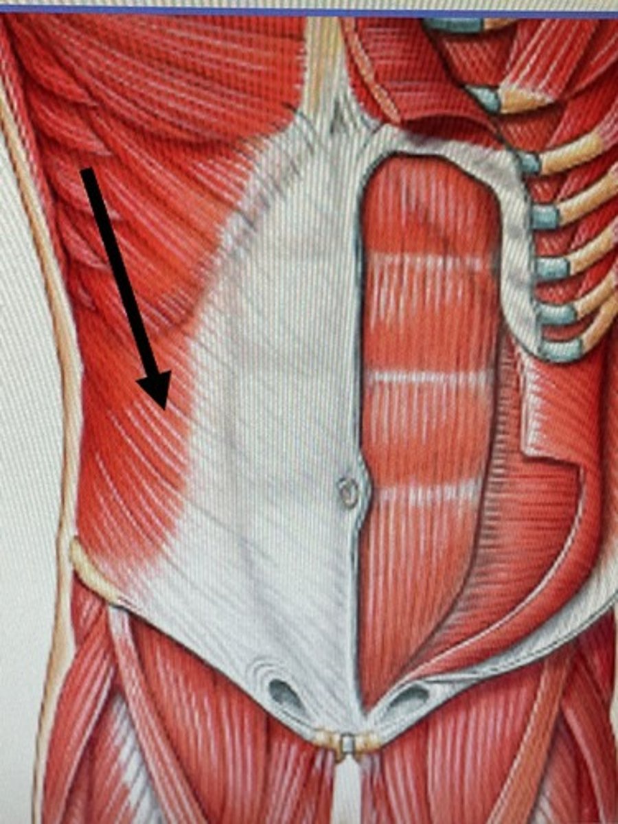
External Oblique Muscles (image)
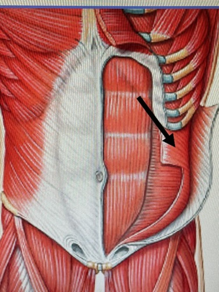
Internal Oblique Muscles (image)
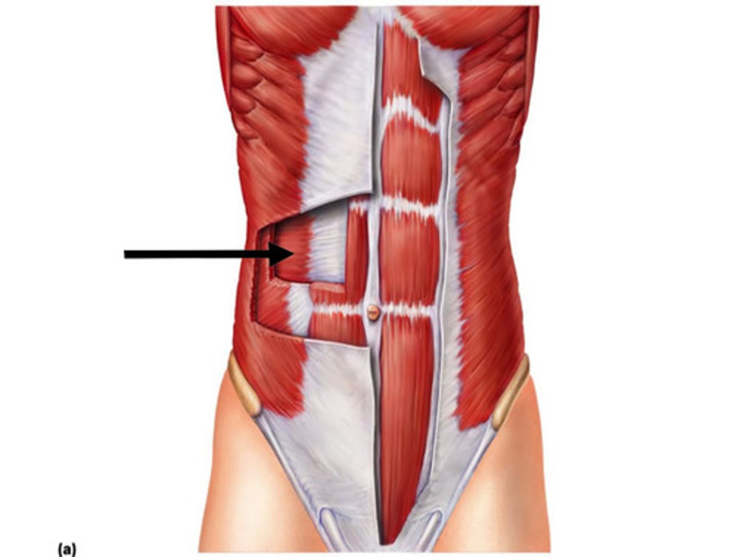
Transverse Abdominis (Image)
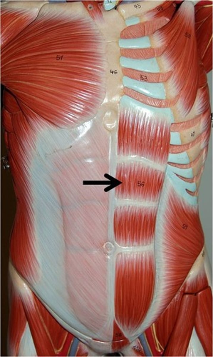
Rectus Abdominis (Image)
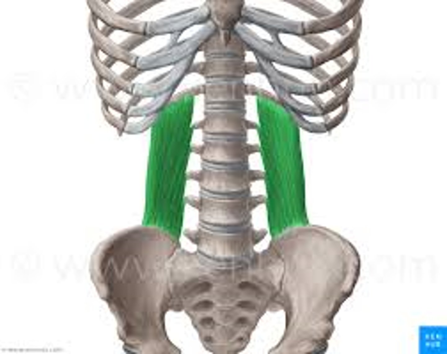
Quadratus Lumborum (image)
INguinal Ligament
ligament extending from pubic bone to anterior superior iliac spine, forming lower border of abdomen

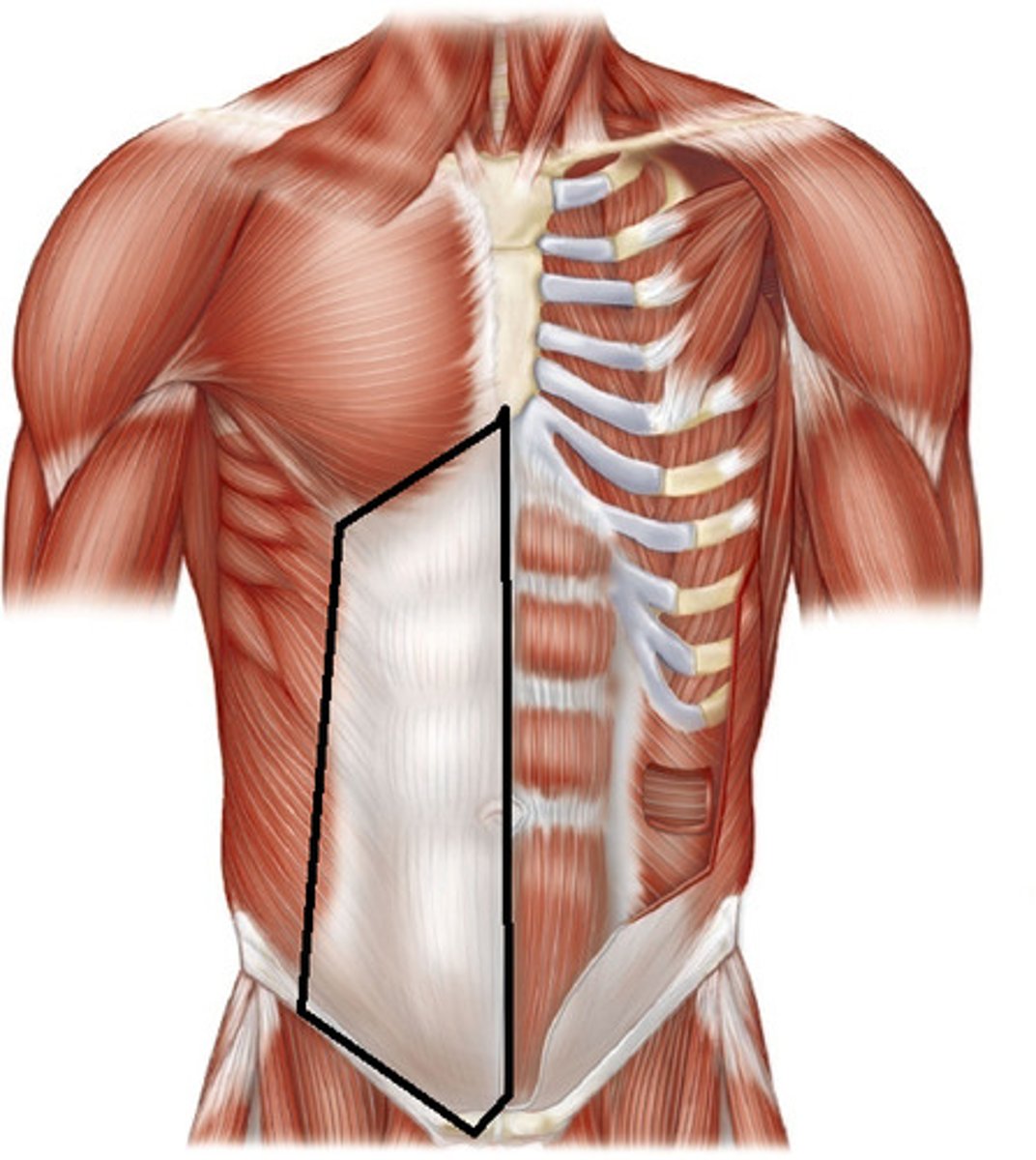
Rectus Sheath
Fibrous sleeve enclosing the rectus abdominis
Mostly T2-L1
Abdominal Muscle Innervation
Cardia
where the stomach meets the esophagus
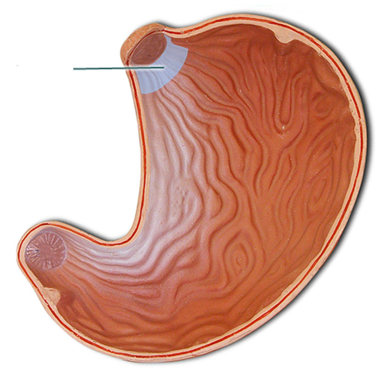
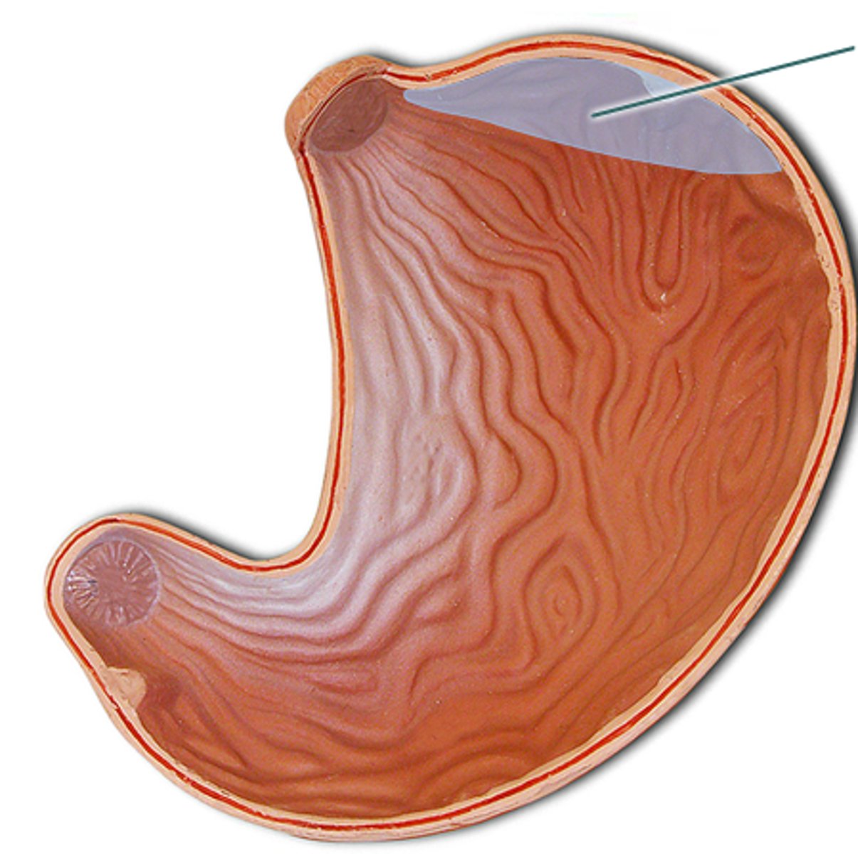
Fundus
Superior round part of organ
Will show up hypodense in CT (full of air)
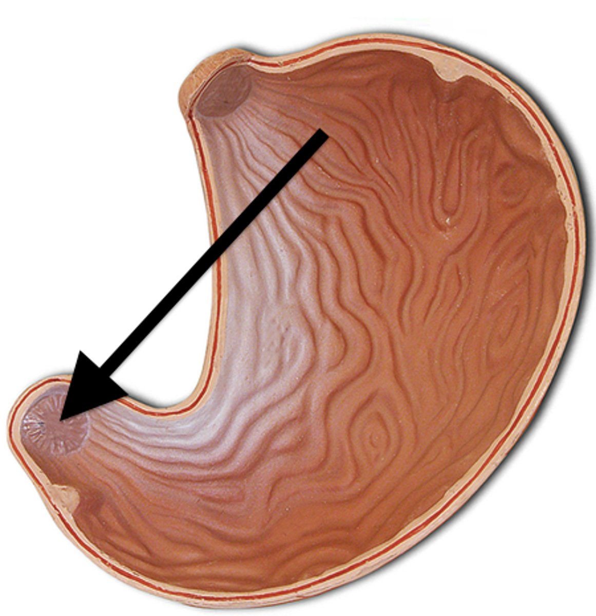
Pyloric Sphincter
seperates the stomach from the duodenum (small intestine)
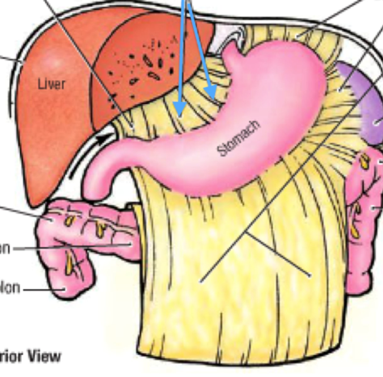
Lesser Omentum
Tethers stomach to liver
Right Hypochondrium Region
1

Right Flank Region
4

Right Inguinal (groin) Region
7

Epigastric Region
2

Umbilical Region
5

Pubic Region
8

Left Hypochondriac Region
3
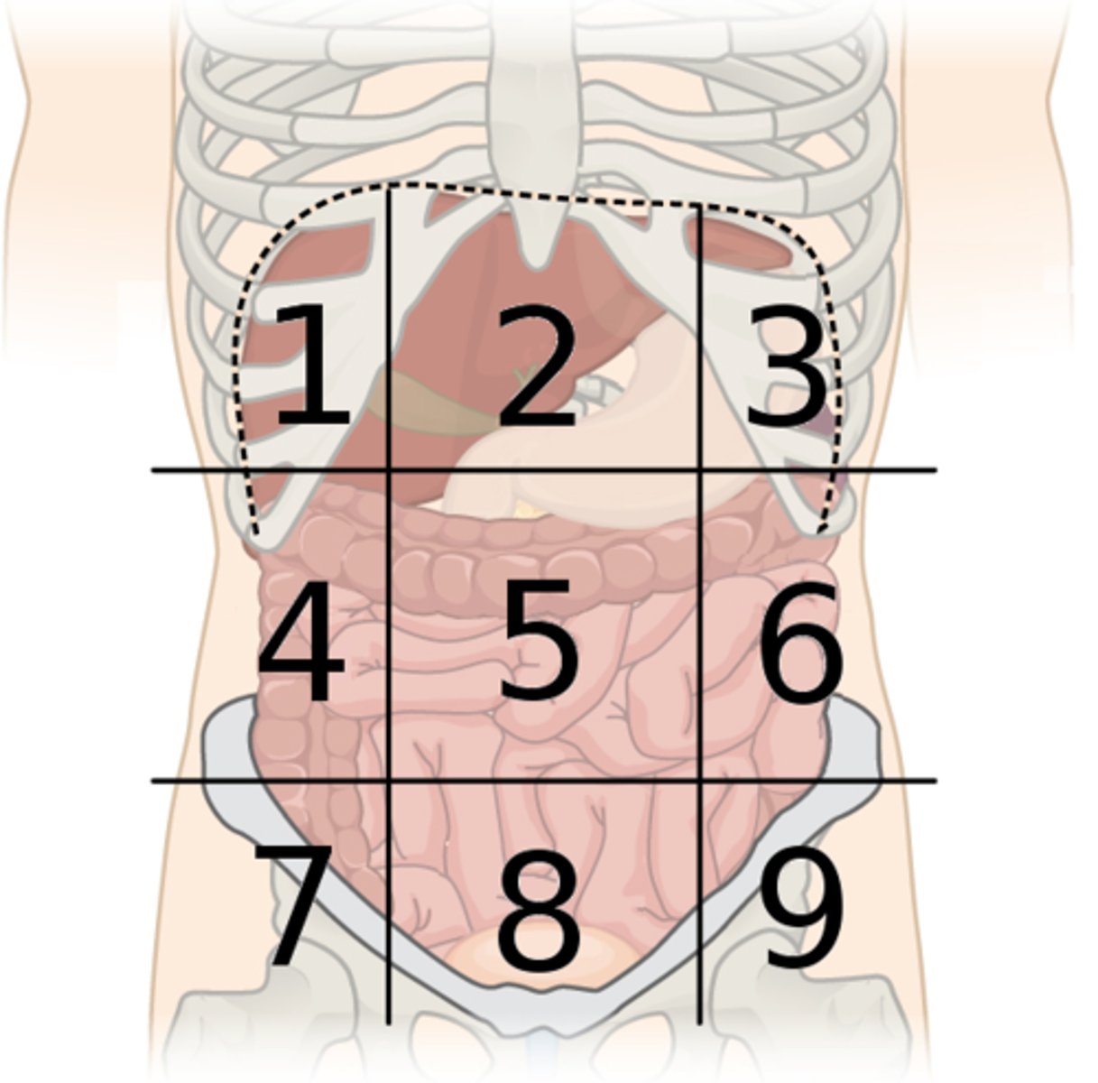
Left Flank Region
6

Left Inguinal Region
9

Transverse (Horizontal) Quadrant Division
Divides the abdomen at the level of the umbilicis (belly button)
Sagittal (Vertical) Quadrant Division
Divides the abdomen at the midline
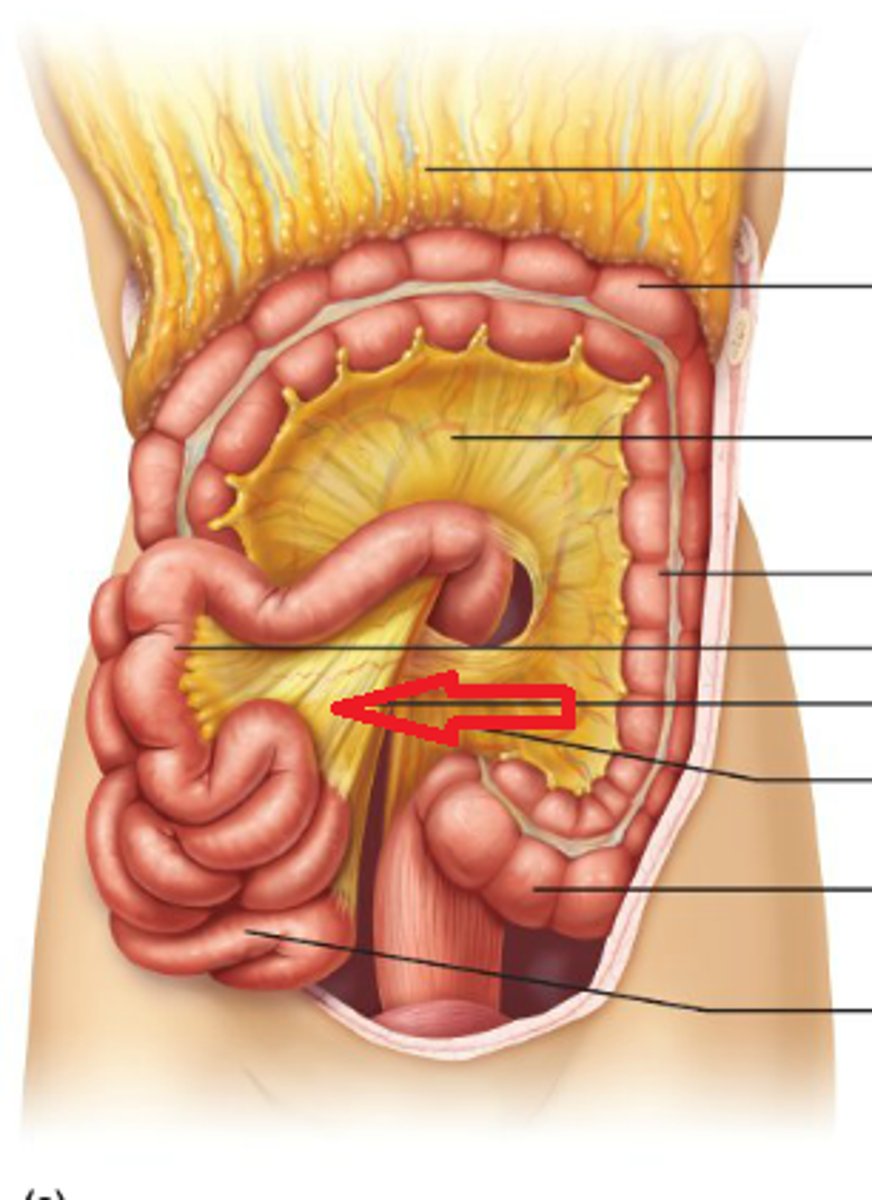
Layer of peritoneum that suspends the jejunum and ileum from the posterior abdominal wall
Mesentery
Descending Aorta (Image)
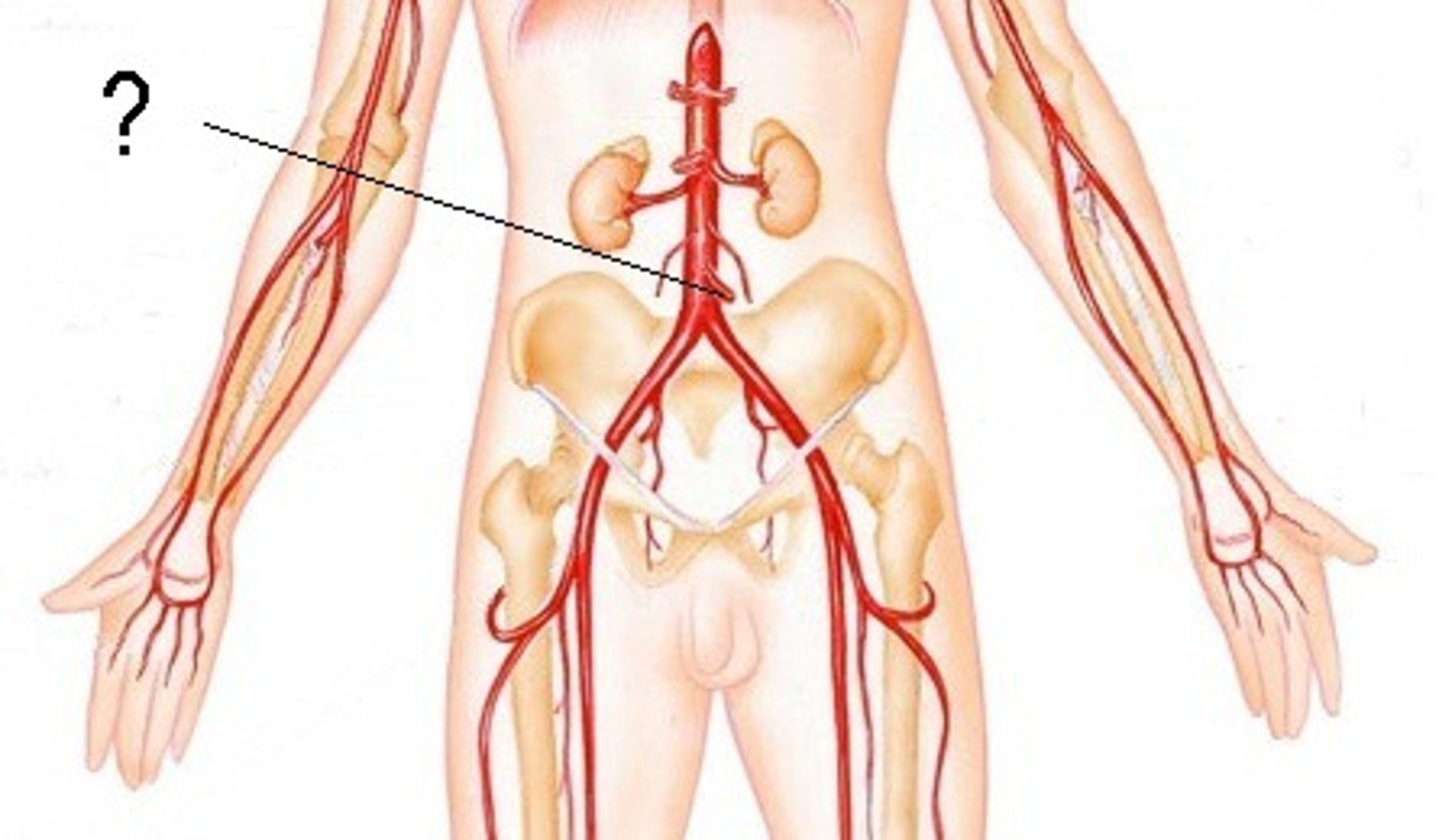
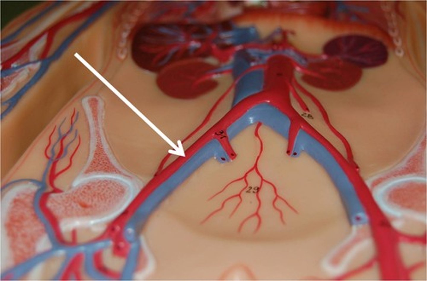
Common Iliac Arteries (Image)
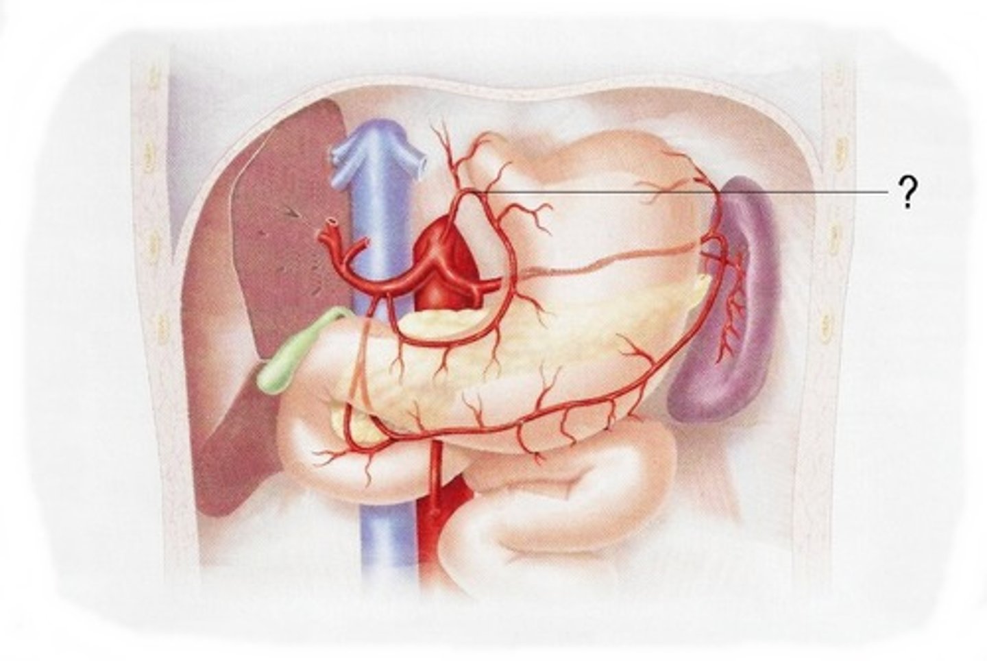
Celiac Trunk
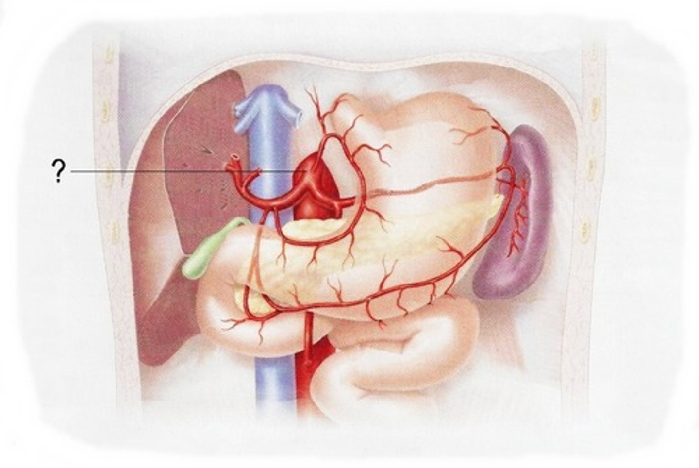
Common Hepatic Artery
Splenic Artery
Left Gastric Artery
Cephalic Trunk Divisions
Common Hepatic Artery
Supplies the liver

Splenic Artery
supplies the spleen

Gastric Artery
supplies the stomach
Superior Mesenteric Artery
supplies the digestive tract from lower duodenum
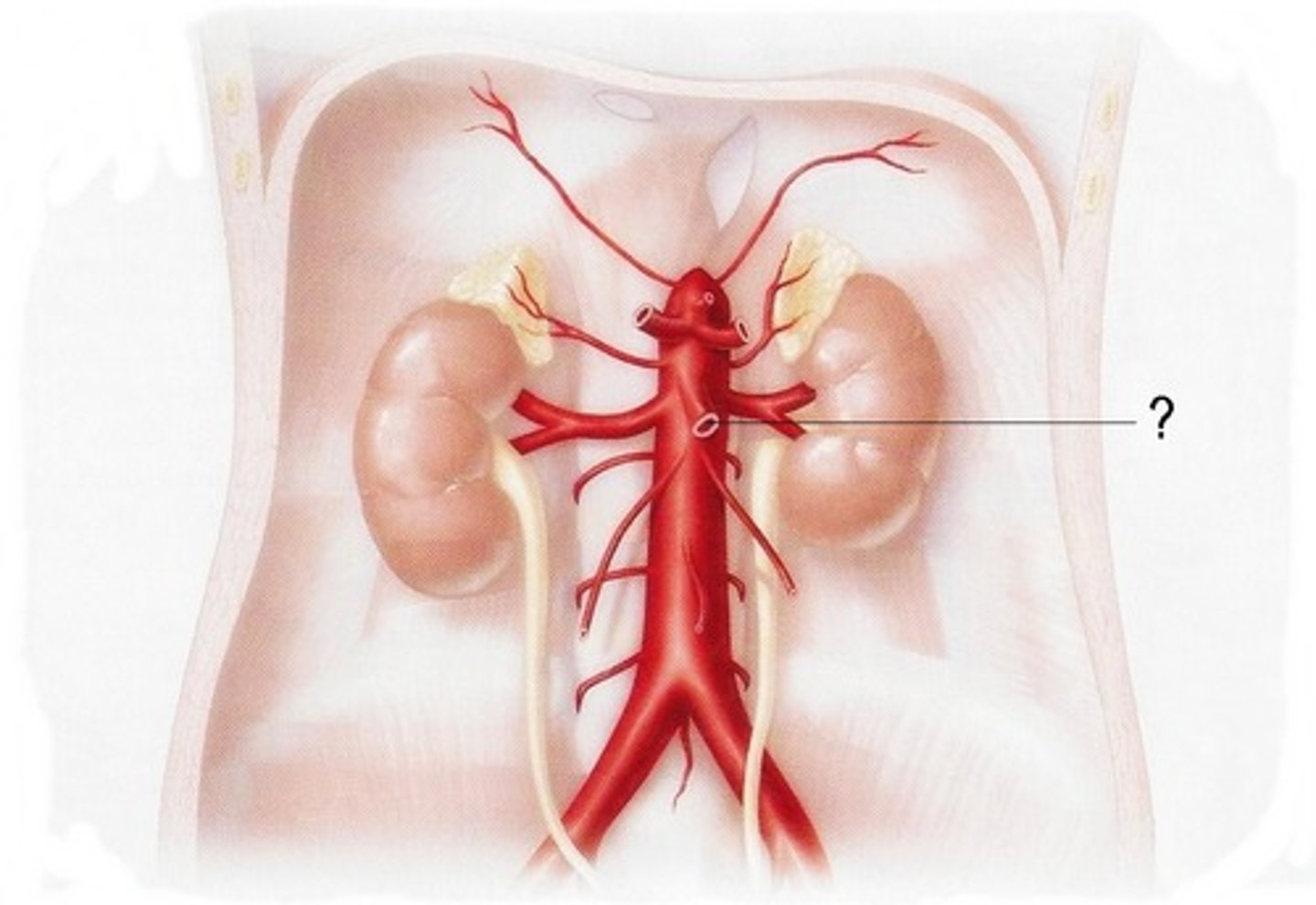
Supplies the descending colon, the sigmoid colon, and part of the rectum
Inferior Mesenteric Artery
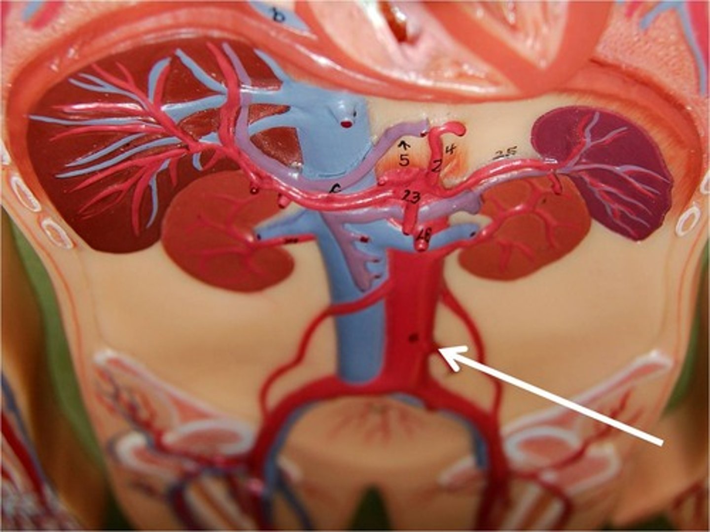
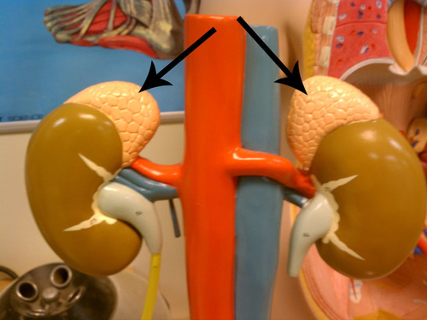
Adrenal Glands (image)
Renal Cortex (image)
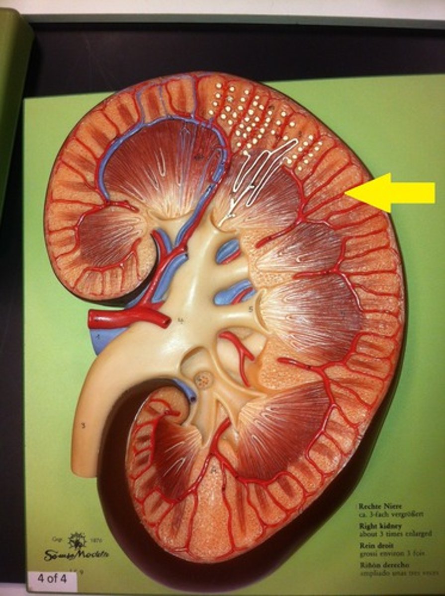
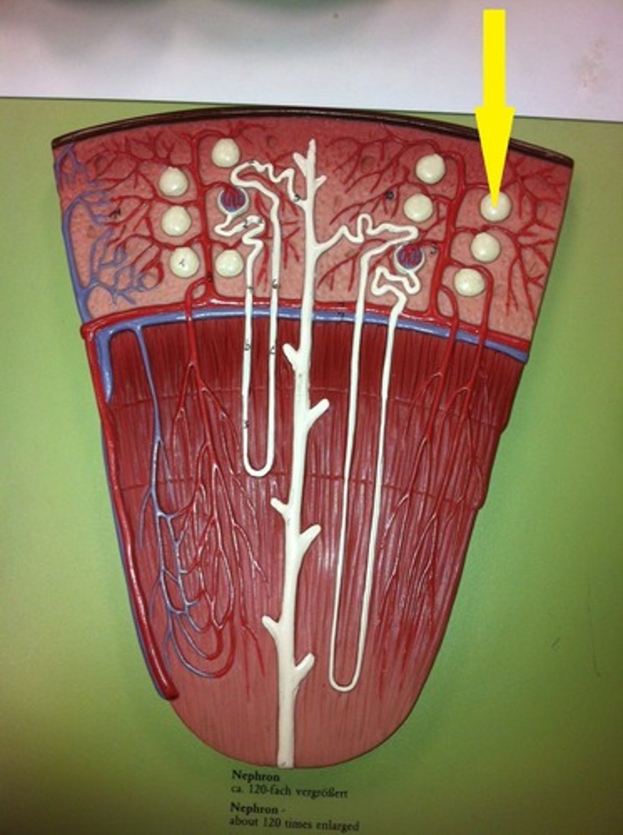
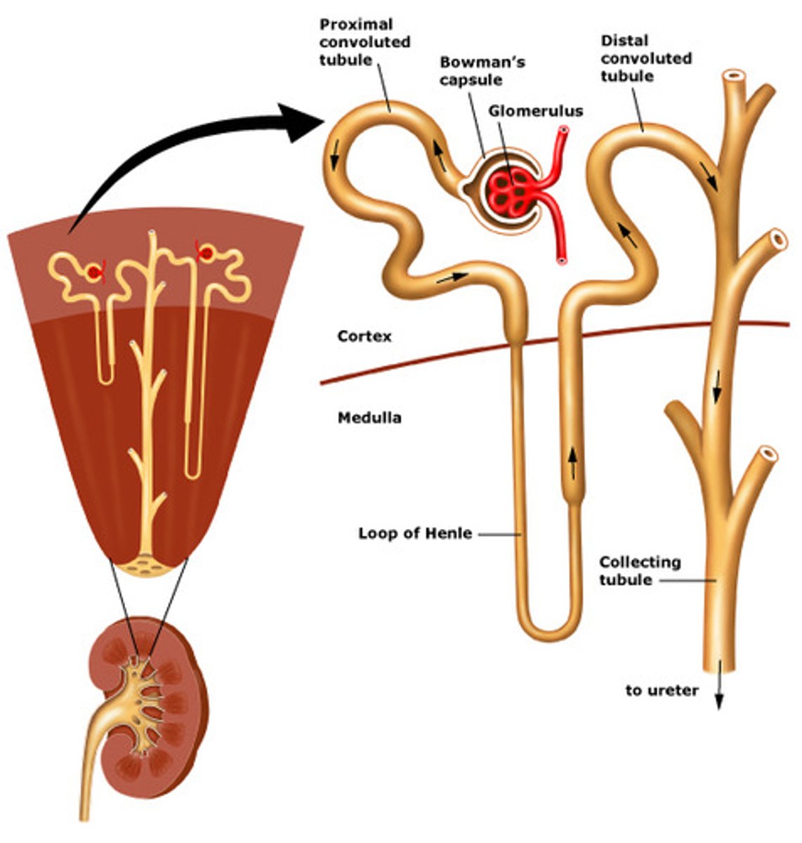
Renal Corpuscles
Aldosterone promotes reabsorption of Na+ from urine by the:
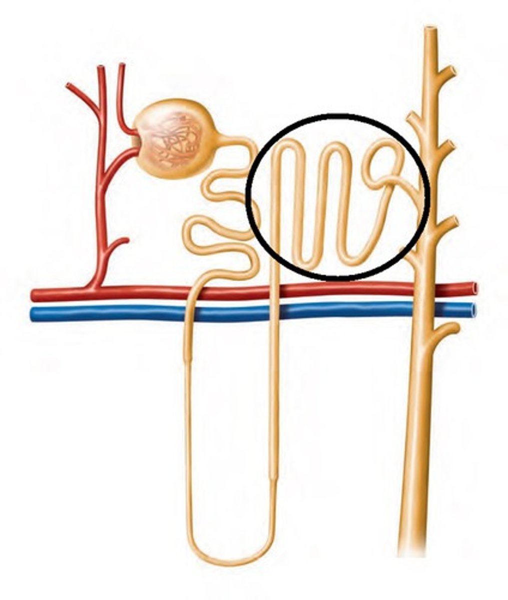
Distal Renal Tubules
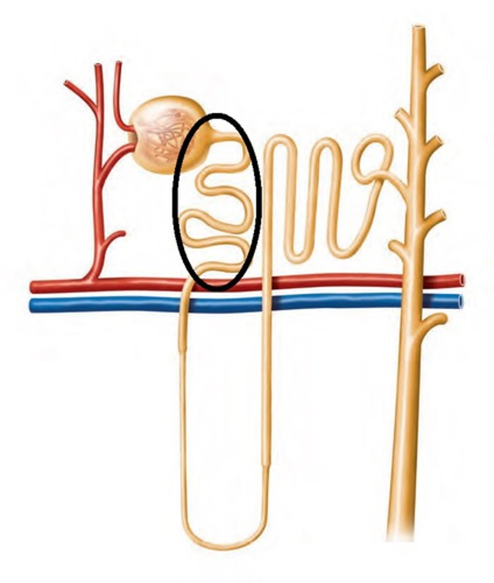
Point of initial reabsorption
Proximal Renal Tubules
Extensions of cortex in between pyramids
Renal Columns
inner portion of the kidney
Contain urinary tubules and Blood vessels
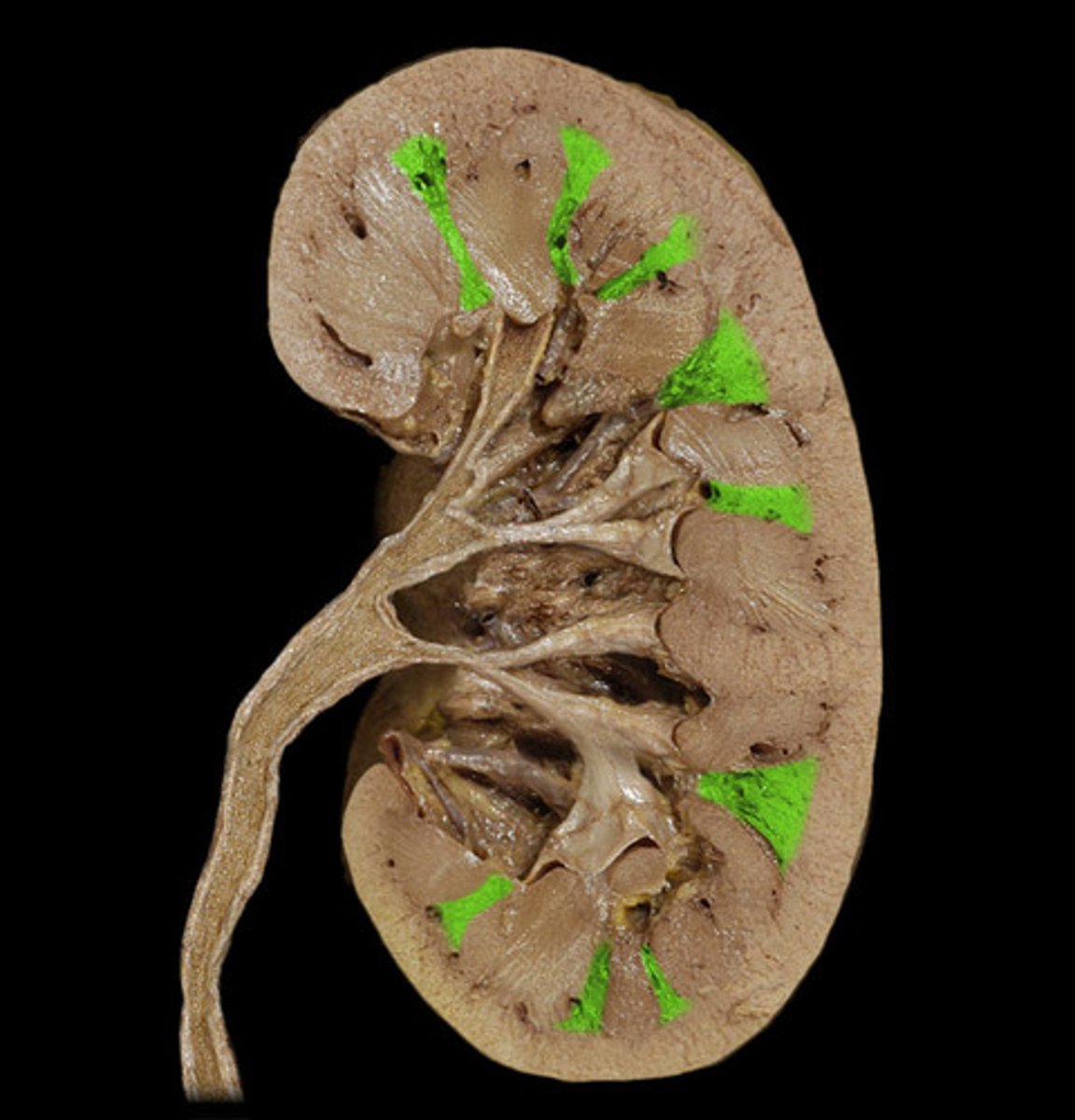
Renal Medulla
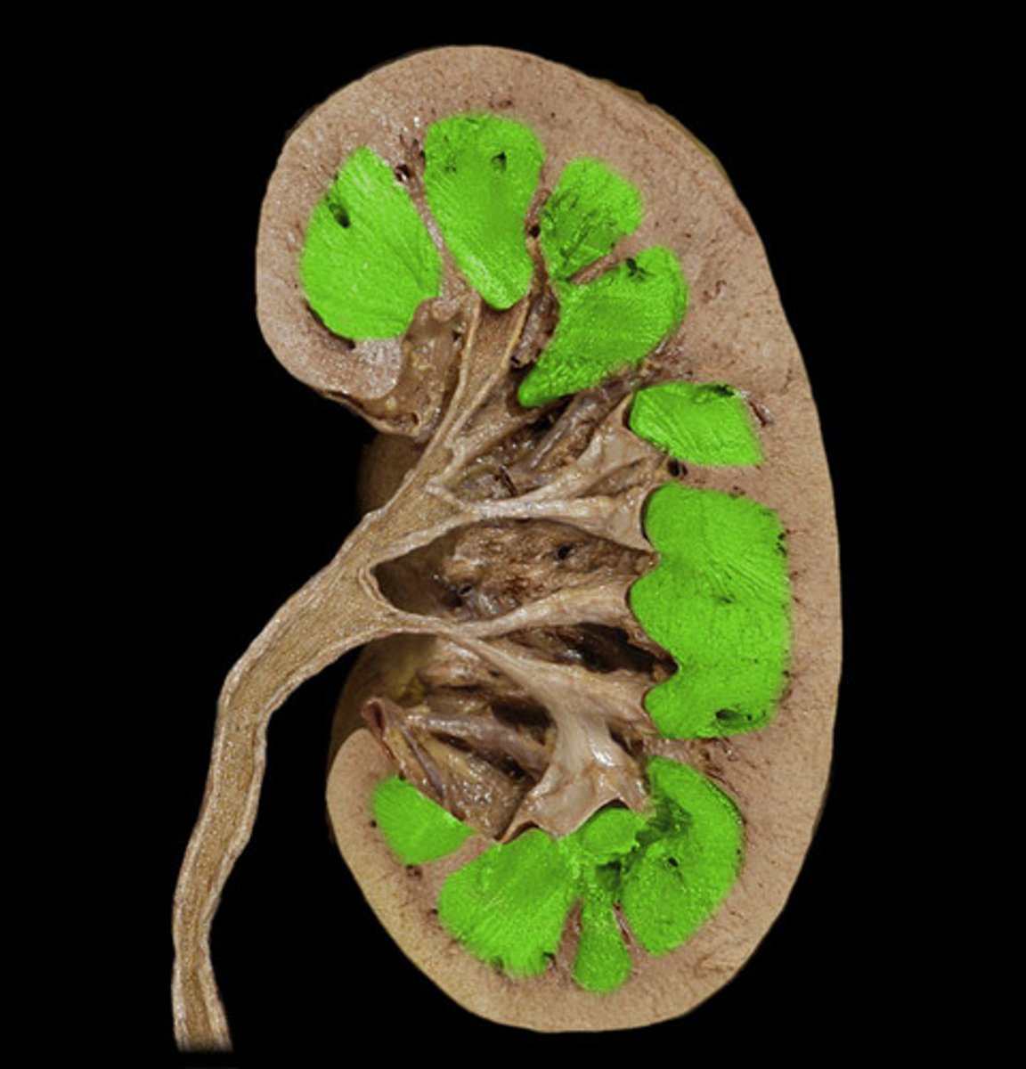
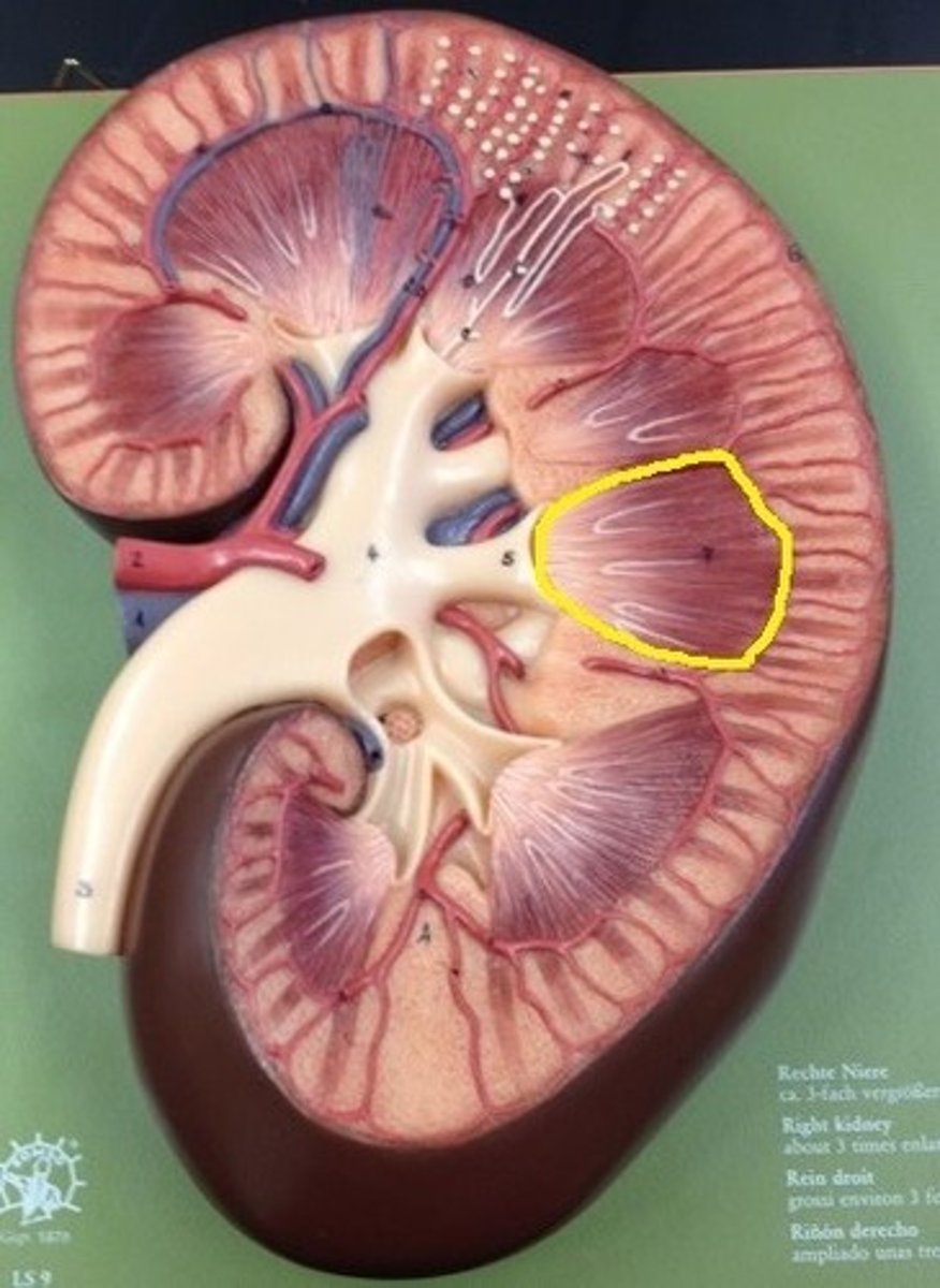
Triangular-shaped areas of tissue in the medulla of the kidney
Renal Pyramids
filters blood to remove waste and filters blood again to return needed nutrients and refine down the actual waste products
Nephron
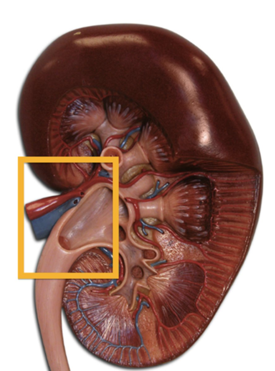
Deepest portion of the kidneys, receives urine
Renal Pelvis
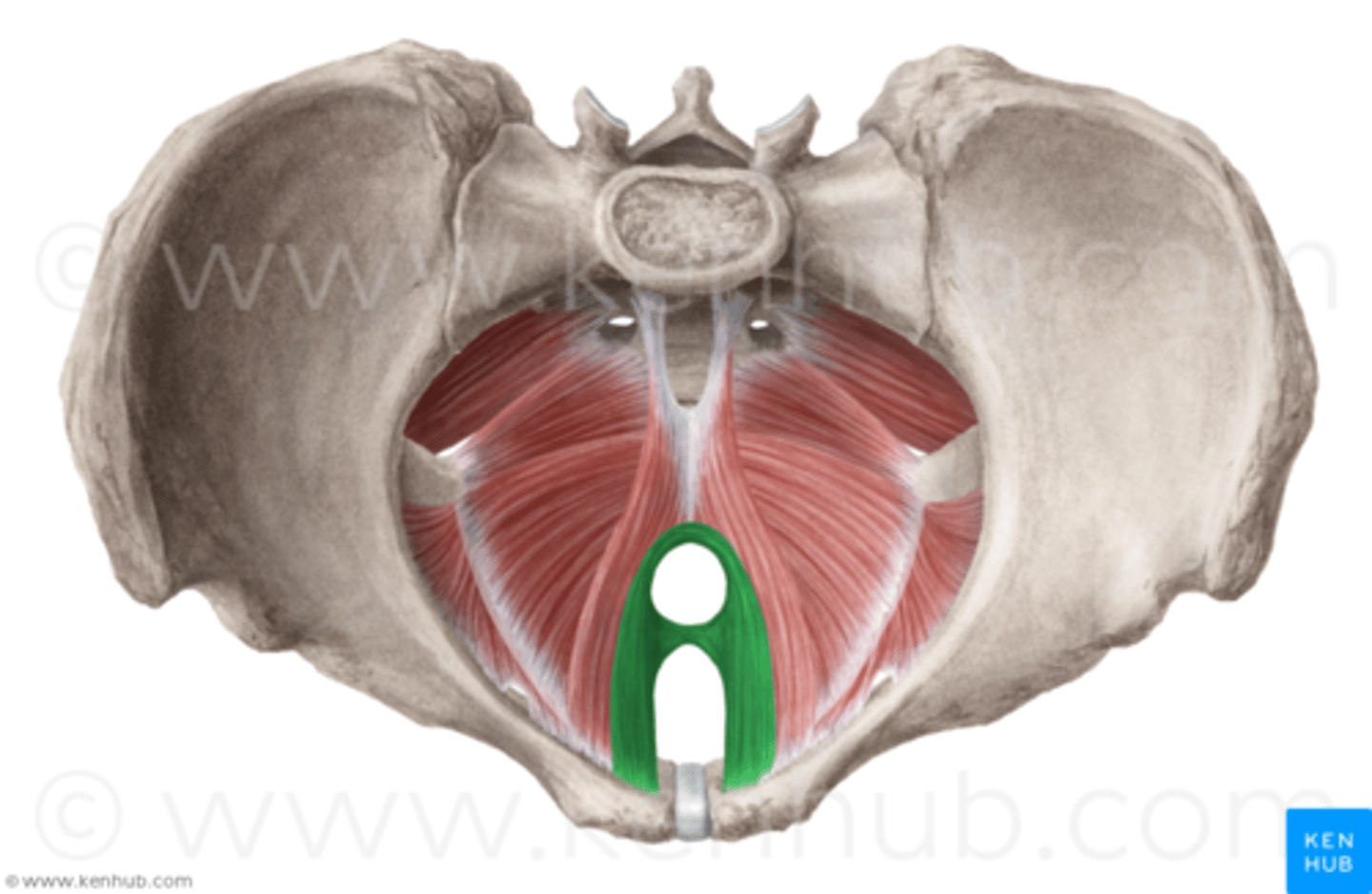
Puborectalis (image)
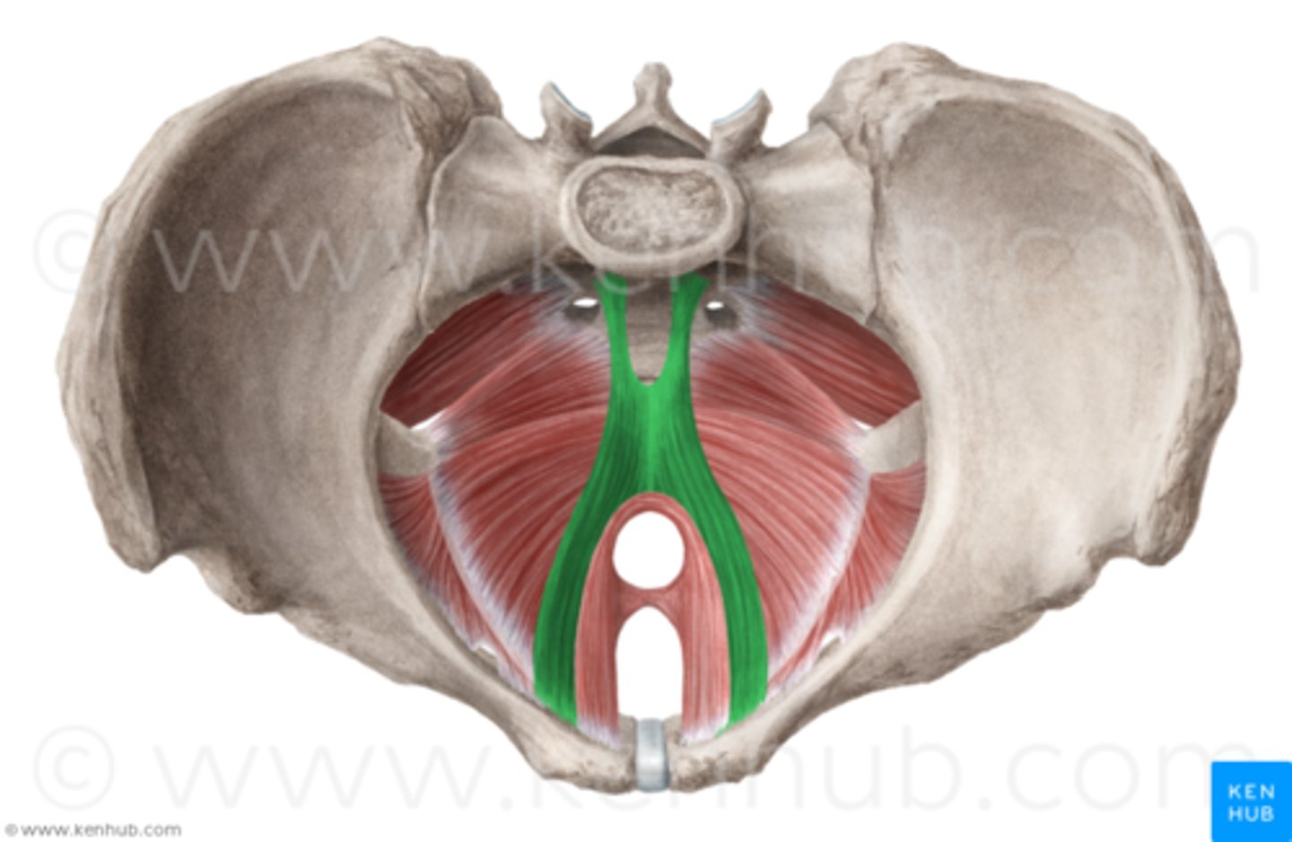
Pubococcygeus (image)
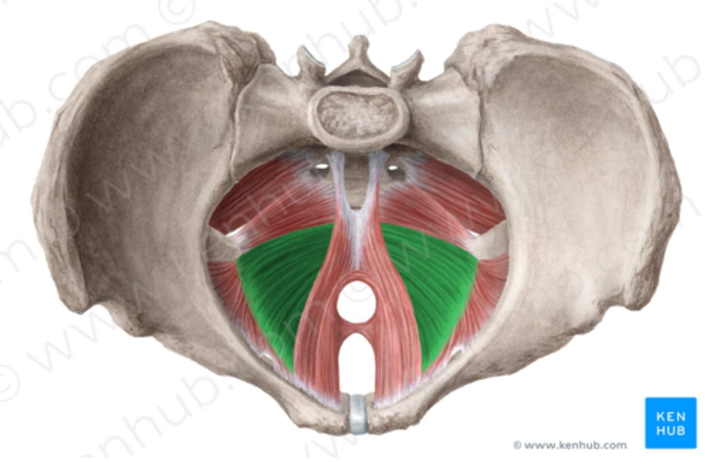
Iliococcygeus (image)
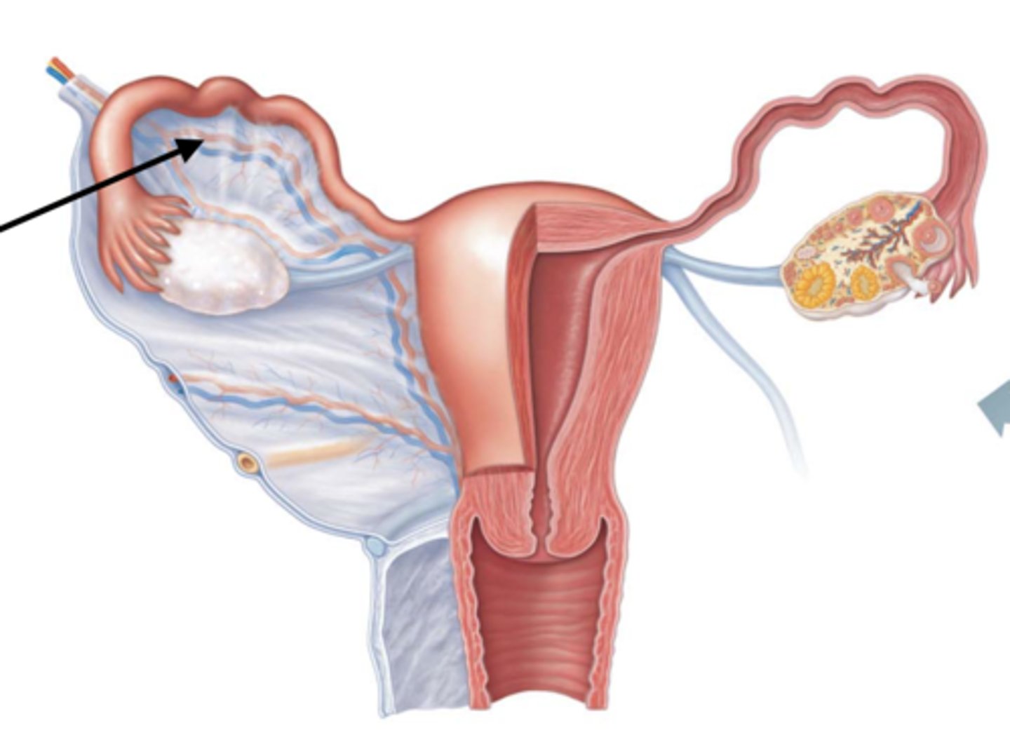
extends from sides of the uterus to the lateral wall and floor of the pelvis
holds the uterus in position
Broad Ligament
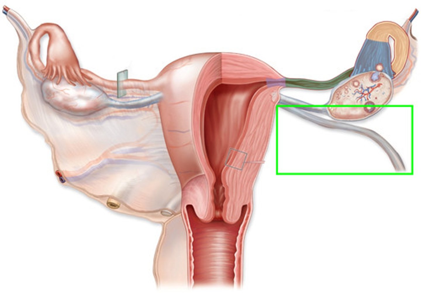
Binds the uterus to the anterior body wall
Round Ligament
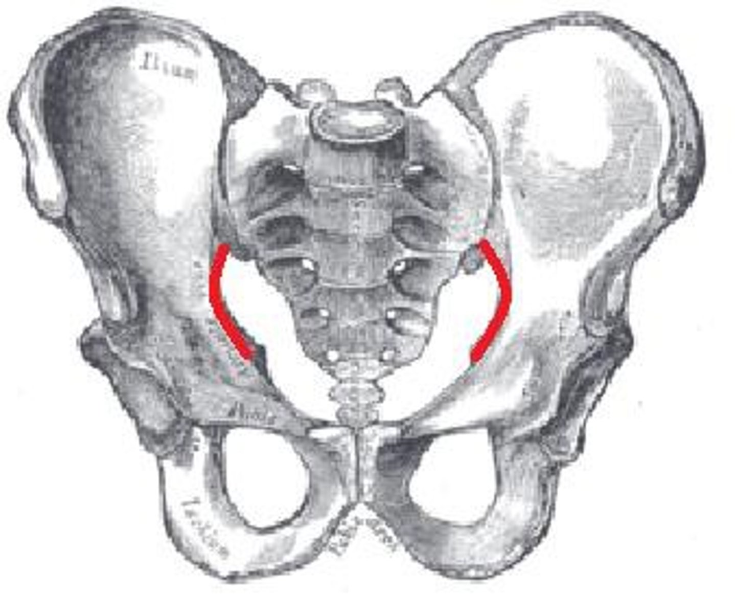
A ridge of bone that runs inferiorly and anteriorly from the auricular surface
Arcuate Line
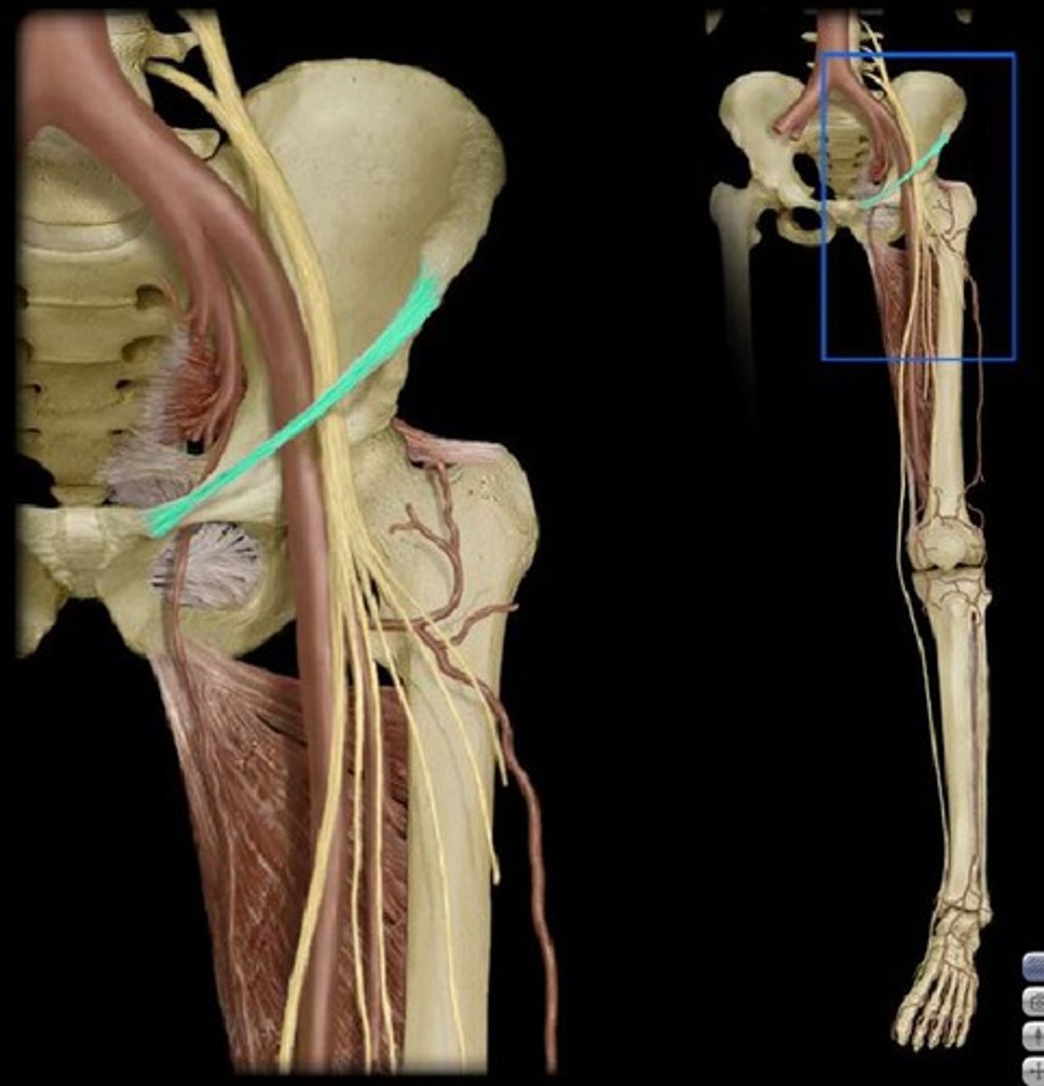
Inguinal Ligament
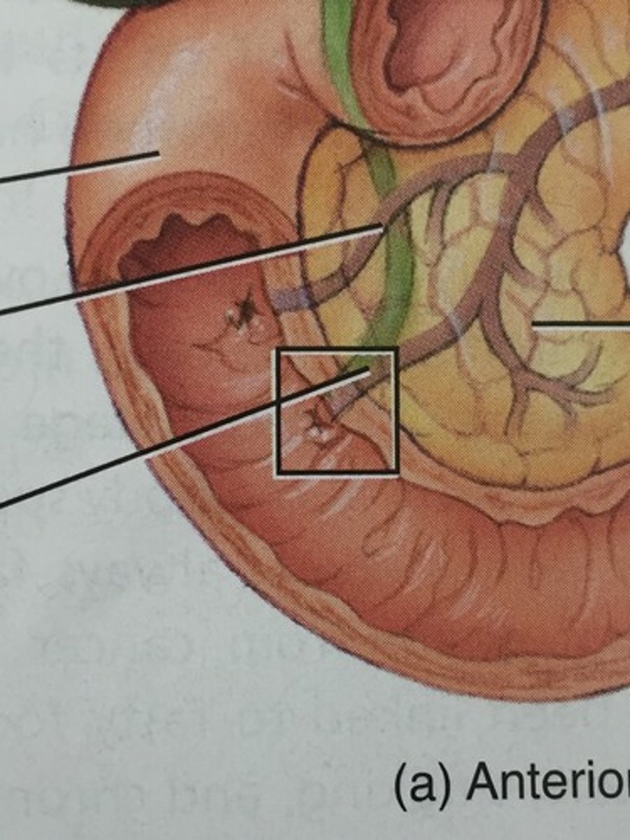
Formed by common bile duct and pancreatic duct
Heptopancreatic Ampulla
Portal Triad (image)
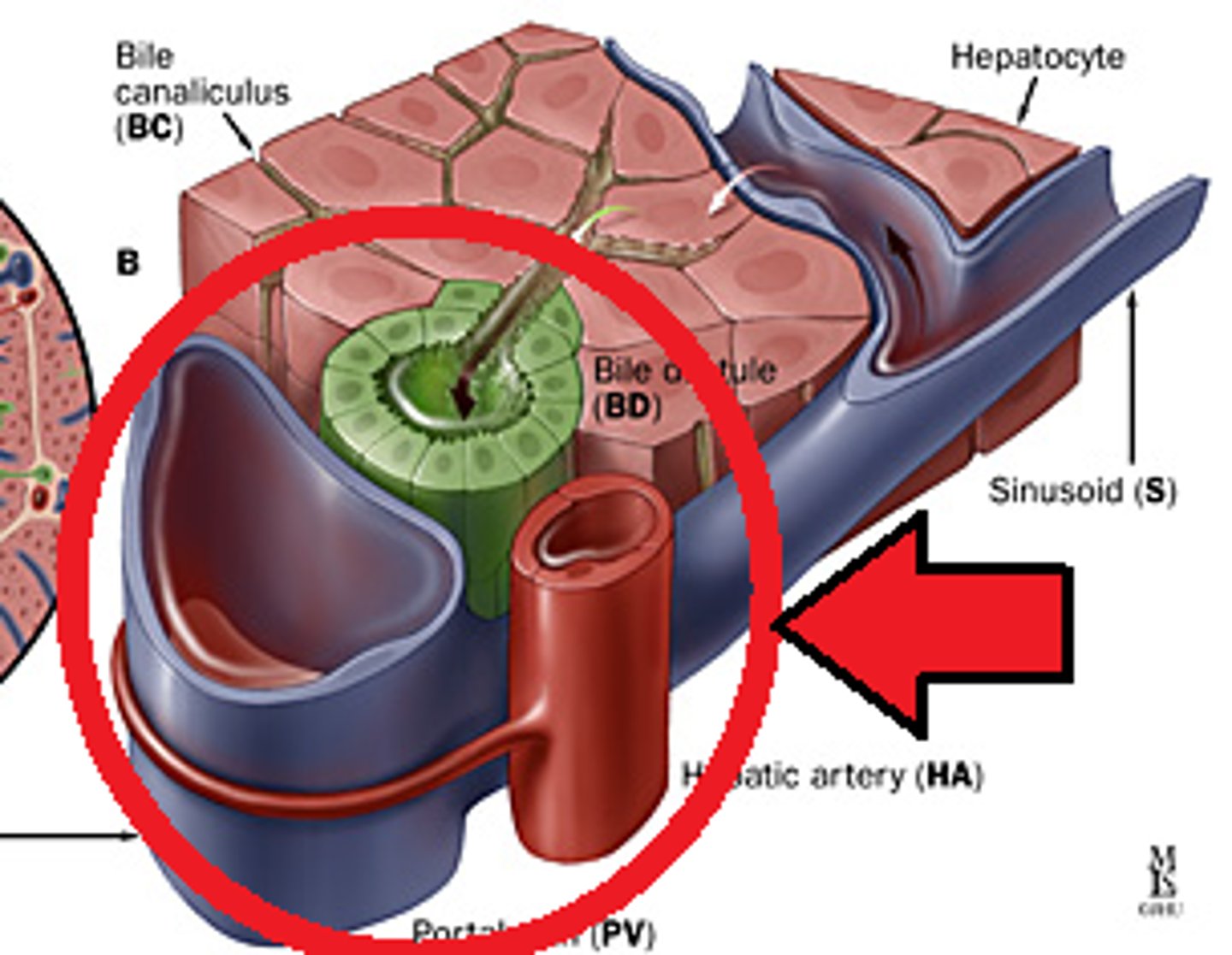
Made up of common hepatic artery, hepatic portal vein, and common bile duct
Portal Triad
Starts after the diaphragm ends
Abdominal Aorta (origin)
Ridges on the intestines
Hostra
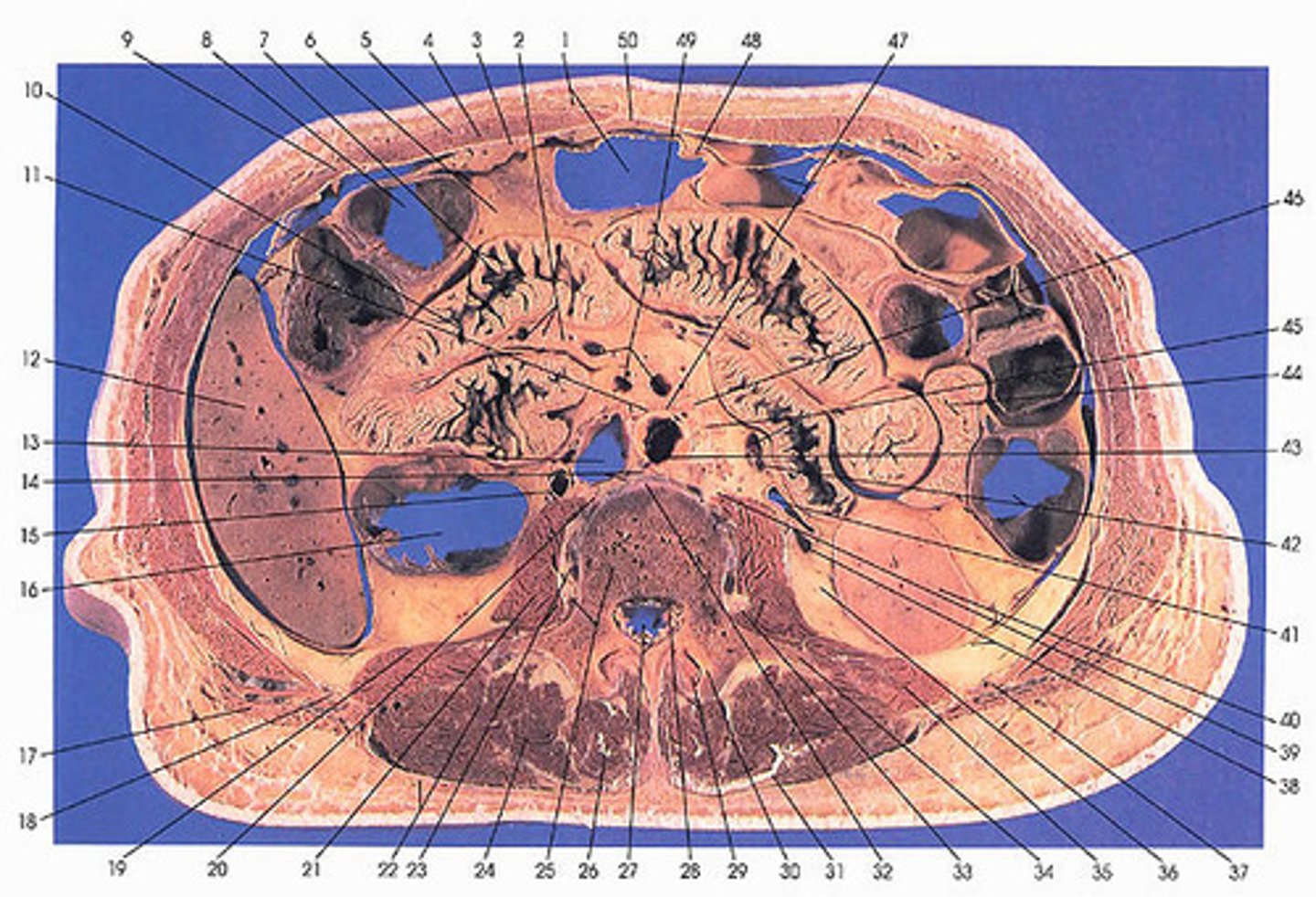
Liver
#10

Pulmonary Vein
#13

Inferior Vena Cava
#15
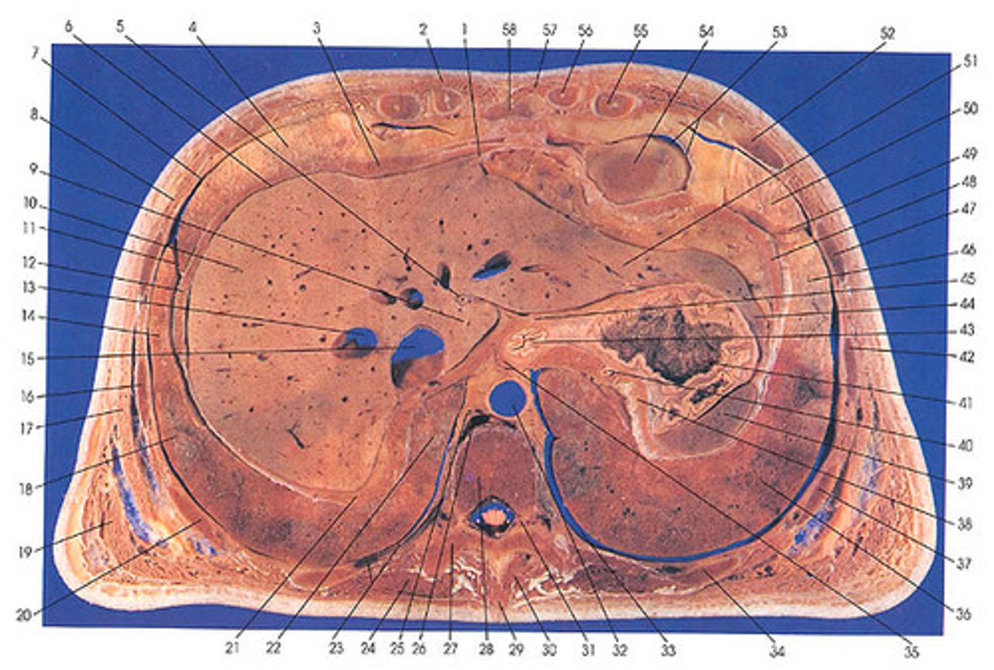
Descending Aorta
#32

Spleen
#39

Stomach
#41

Esophagus
#43

Transverse Colon
#46

Descending Colon
#41

Pancreas
#5

Gall Bladder
#14

Duodenum
#13
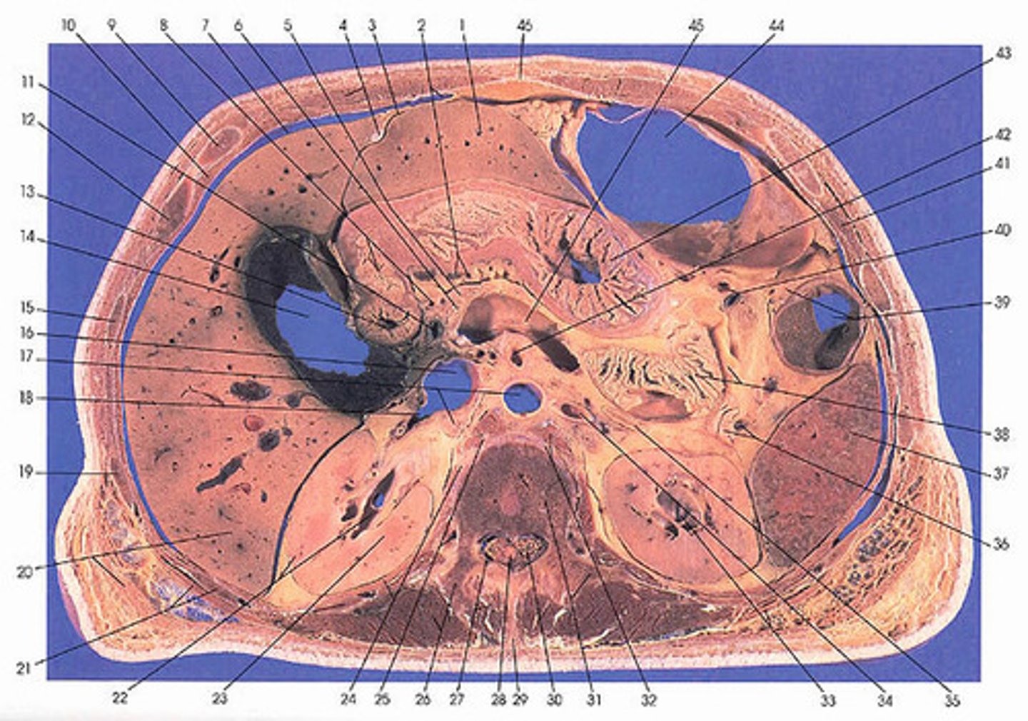
Ascending Colon
#9

Jejunum
#54 and #57
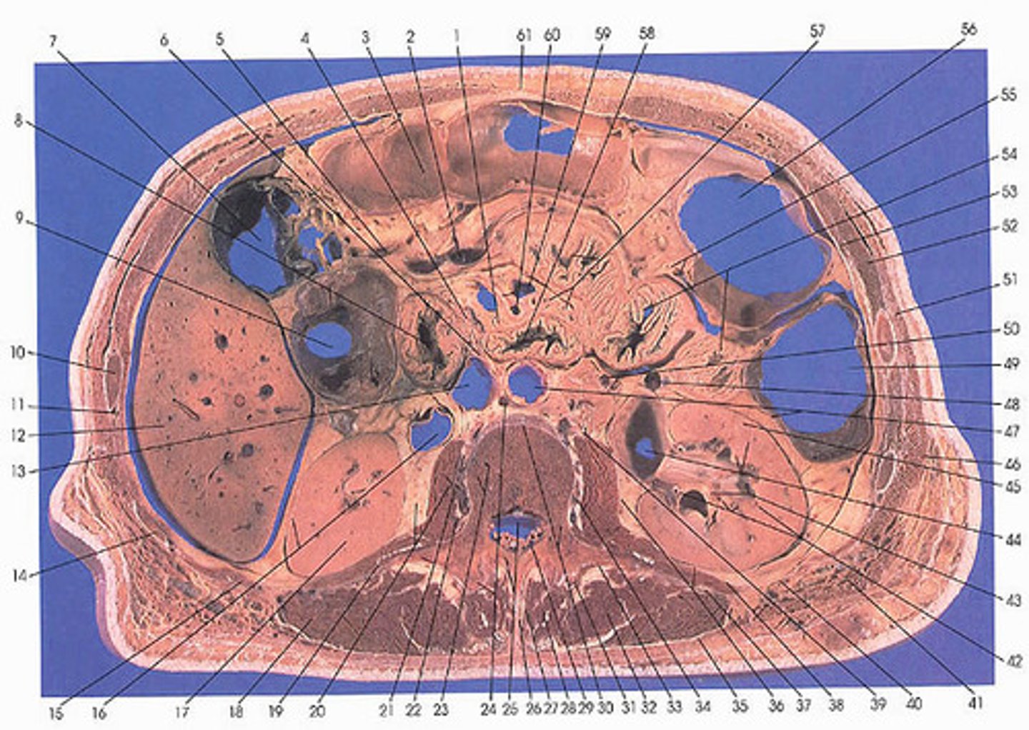
Ureter
#42 and #6

Urinary Bladder
#17
Sigmoid Colon
#62

Rectus Abdominis
#1

Rectus Sheath
#3

Linea Alba
#2
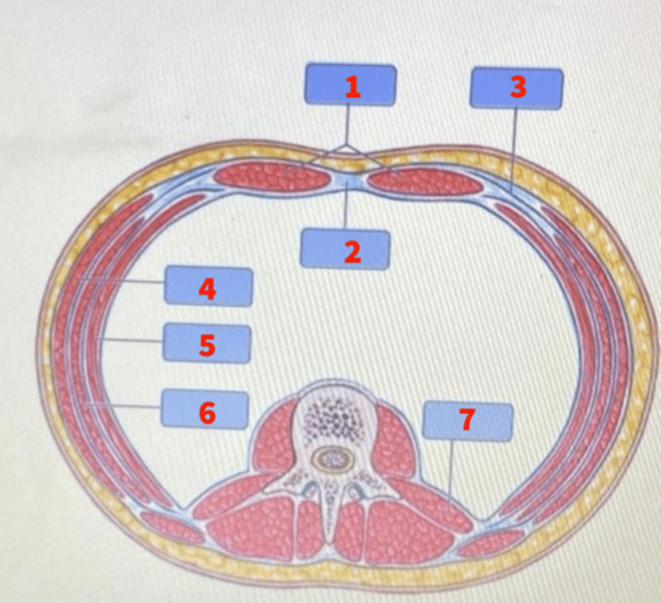
External Obliques
#4

Transverse Abdominis
#5

Internal Obliques
#6

Quadratus Lumborum
#7

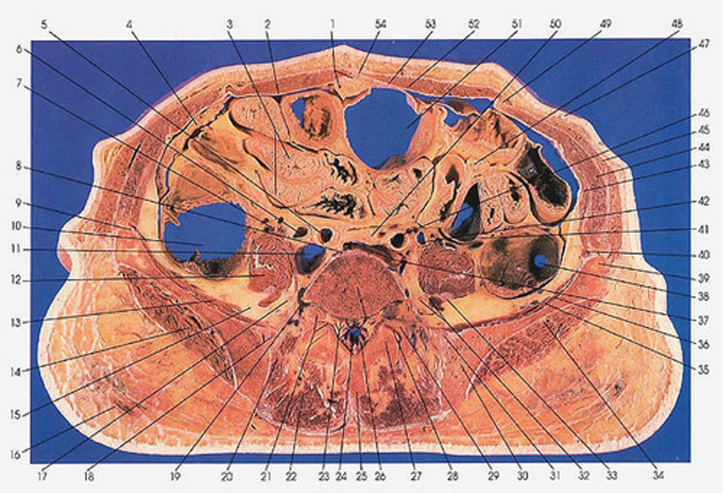
T10
Crossection

T12
Crossection
L1
Crossection

L3
Crossection
L5
Cross Section