Psychological Knowledge and Understanding - Psychology
1/62
There's no tags or description
Looks like no tags are added yet.
Name | Mastery | Learn | Test | Matching | Spaced | Call with Kai |
|---|
No analytics yet
Send a link to your students to track their progress
63 Terms
Two Components of the NS
Central Nervous System & Peripheral Nervous System
CNS - Control Nervous System
The control unit made of; brain, spinal cord, and brain stem
Carries sensory information up the spinal cord to the brain via sensory neurons.
PNS - Peripheral Nervous System
Is linked to the outside world; 2 divisions - automatic and somatic
Carries motor messages from the brain to organs and muscles in the body.
Autonomic NS
Involuntary bodily process, operates automatically
Carries messages from the brain to internal glands and organs via motor neurons.
Carries sensory messages to the brain about the activity level of glands and organs via sensory neurons.
Controls involuntary muscle movement therefore regulates internal organ function.
Regulates the function of glands.
Sympathetic and Parasympathetic
Somatic NS
Carriers sensory information from sensory neurons to CNS
Carries sensory information received by sensory receptor cells to the CNS via sensory neurons.
Carries motor messages from the CNS to skeletal muscles via motor neurons.
Controls voluntary skeletal muscle movement.
Controls involuntary skeletal muscle movement (reflex responses)
The 2 Divisions of the Sympathetic NS
Sympathetic and Parasympathetic
Sympathetic
Prepares the body for stress
fight, flight or freeze
increase heart rate
increase breathing rate
decrease digestion
regulates the glands and internal organ function to physically prepare the body for increased activity during heightened physical or emotional arousal
Parasympathetic
Calms the body and helps with energy conservation
reverses bodily functioning produced by the sympathetic NS by calming the body and maintaining an energy level suitable for normal bodily functioning
Neurons
Carry nerve impulses around the body
Neurons are the basic unit of the nervous system
They use electro-chemical means to conduct electrical impulses
Sensory Neurons
Detects a stimulus and sends an impulse to The CNS.
Motor Neurons
Carries an impulse from the CNS to a muscle or gland.
Interneurons
Connects sensory neurons to motor neurons in the spinal cord.
Cell Body
contains a nucleus that controls the activities of the neuron.
Myelin Sheath
Insulates the axon
Allows for rapid movement of the action potential along the axon
Dendrites
Extensions of the cell body
Receive neurotransmitters from pre-synaptic neurons
Converts them into electrical nerve impulses that are conducted towards the cell body.
Axon
The long projection of a neuron that conducts electrical nerve impulses and carries them away from the cell body.
Axon Terminal
the enlarged end points of axon branches that store neurotransmitters and release them into the synaptic cleft.
Role of Neurotransmitters
Are molecules found within the NS that act as chemical messages.
Neurotransmitters allow neurons to communicate by relaying information between them across the synapse, as well as from neurons to glands and muscle cells.
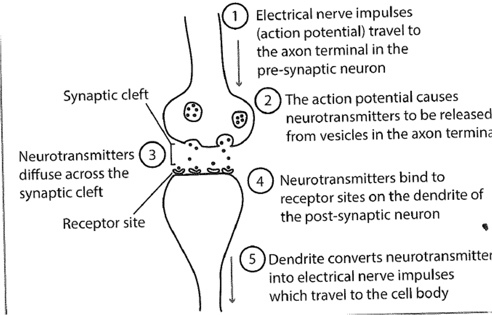
Steps of Neural Transmission
electrical impulses travel to the axon terminal in the pre-synaptic neurons
the action potential causes neurotransmitters to be released from vesicles in the axon terminal
neurotransmitters diffuse across the synaptic cleft
neurotransmitters bind to receptor sites on the dendrites of the post-synaptic neuron
dendrites convert neurotransmitters into electrical nerve impulses which travel to the cell body
Electro-chemical Signal
an electrical nerve impulse (action potential) whcih travels through the neuron
neurotransmitters travel between the synapse of communicating neurons.
The electrical nerves impulses are the electro component, and the neurotransmitters are the chemical component of the signal.
Direction of Transmission
from the dendrites to the cell body, then along the axon to the axon terminal
it causes the release of neurotransmitters into the synaptic cleft
myelin increases the transmission speed of the nerve impulses trough the axons
The Role of the Synapse
The synapse allows neural transmission to occur by converting the electrical nerve impulse from one neuron into a chemical signal and then back again into an electrical nerve impulse in another neuron.
The Brain
Receives, processes and interprets sensory information, integrates the information and coordinates a response
3 Regions of the Brain
Hindbrain, Midbrain, Forebrain
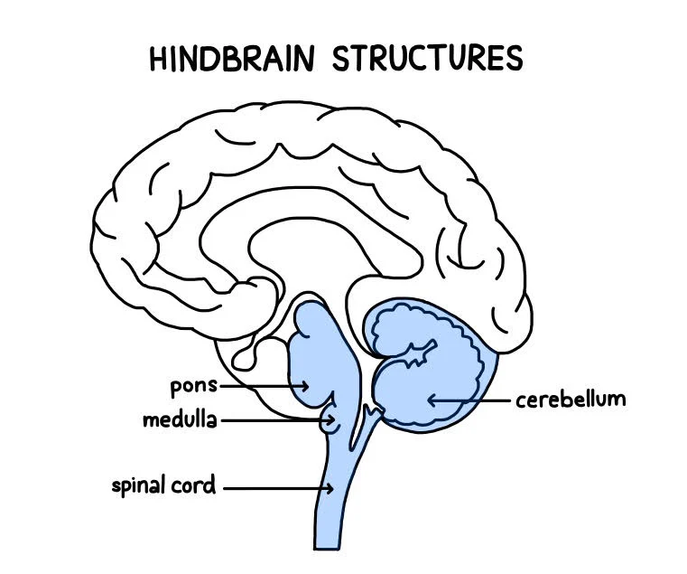
Hindbrain
Base of the brain and is responsible for lower-brain functions that occur with no conscious effort
controls basic autonomic functions e.g heart rate, breathing, sleep
coordinates voluntary muscle movements, balance, posture, and reflexes
Consists of pons, medulla, and cerebellum
Pons
a group of nerve that connect the cerebral cortex with the medulla
assists in transfer of neural messages between various parts of the brain & spinal cord
involved in dreaming, sleeping, arousal, coordination of some muscle movements
Medulla
lowest part of the brain connects the brain and spinal cord
relays information between brain and spinal cord
regulates involuntarily bodily functions, communicates with autonomic NS
Cerebellum
‘Little Brain’ - wrinkly similar to the outer layer of the brain
helps coordinate voluntary movement and balance by relaying motor information to and from the cerebral cortex
receives information from spinal cord, sensory neurons and brain to adjust posture and coordination
fine motor skills are controlled
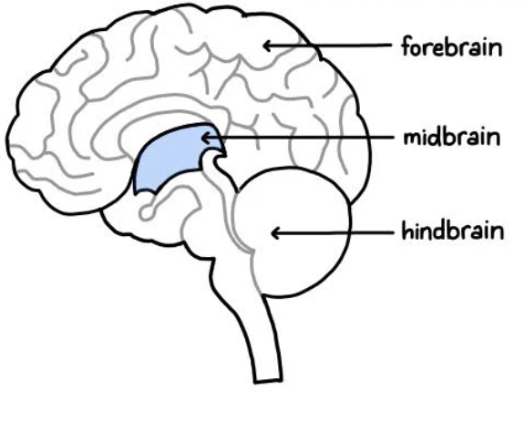
Midbrain
Connects the hindbrain and forebrain, below the cerebral cortex at the top of the hindbrain
processes information related to hearing, vision, movement, pain, sleep, arousal
helps keep us awake and alert
Consists of reticular formation
Reticular Formation
network of neurons that coordinates the function of vital brain systems
extends through SC to Midbrain
directs and filters information
arouses cortex to alertness
regulates autonomic NS function and sleep-wake cycle
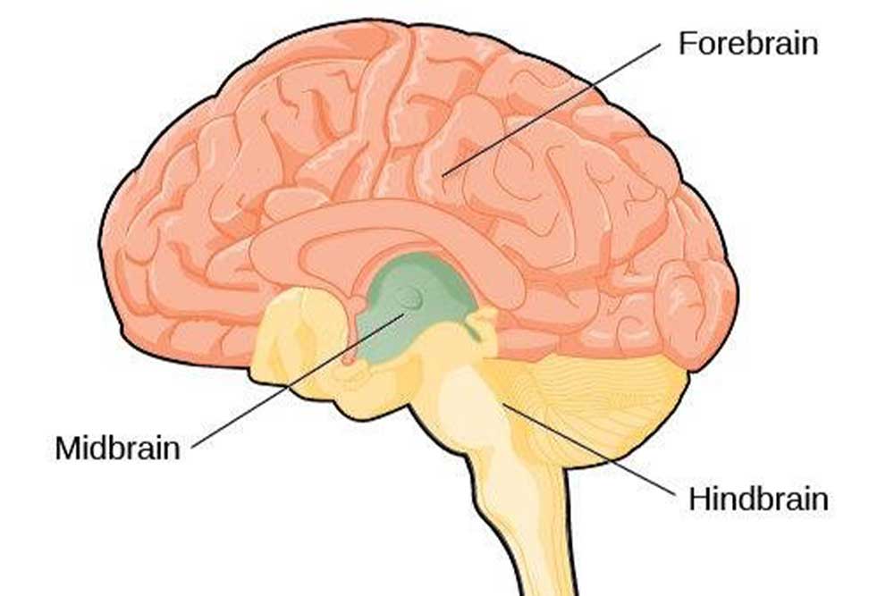
The Forebrain
located above the midbrain towards the top of the brain
most developed and complex region
responsible for emotions, motivations, sensations, perceptions, learning, memory, and reasoning
Consists of; hypothalamus, thalamus, cerebrum
Hypothalamus
Below the thalamus
maintains homeostasis
regulates endocrine system and the release of hormones
influences basic biological needs e.g hunger, thirst
controls ‘internal clock’ and sleep-wake cycle
Thalamus
2 egg-shaped structures joined together
relay system for sensory messages
conducts motor signals - brain stem to cortex
coordinates shift in consciousness
works with RF to focus attention
Cerebrum
the largest & most developed part of the brain
top of the forebrain
responsible for most conscious actions
cerebral cortex: the outer layer which is responsible for higher cognitive functions, voluntary movements, emotions and personality
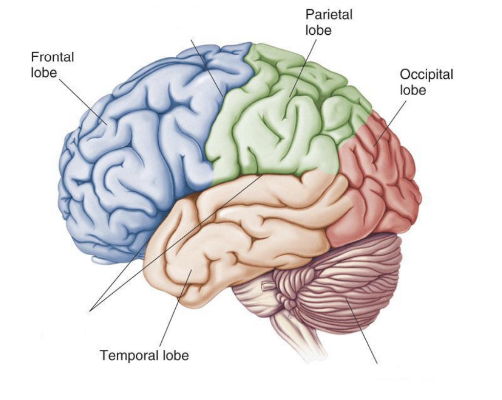
Lobes of the Brain
frontal, parietal, temporal, occipital
Left and Right Hemispheres
Left Hemisphere - analytics, logic, ideas, facts, maths
Right Hemisphere - creativity, intuition, arts, creation, feeling , imagination
They have contralateral control of the body
Specialised Functions of the Left Hemispheres
Receives sensations from the right side of the body and controls voluntary movements of the right side & is responsible for language and analytical function
Left Hemisphere - Language Functions
Broca’s and Wernicke’s Area
Speaking, writing, reading, and understanding language
Left Hemisphere - Analytical Functions
Breaks down information to process sequentially (ordering)
focuses on small details
logical and sequencing used in maths
judging time and rhythm
Specialised Functions of the Right Hemisphere
Receives sensations from the left side, controls voluntary movements of the left side, is responsible for non-verbal & information processing
Right Hemisphere - Information Processing
Processes information simultaneously and holistically
assembles pieces of information into a coherent image, by identifying patterns and general connections
Right Hemisphere - Non-verbal communications
Uses non-verbal responses to answer questions e.g pointing or nodding
understands context
understands sarcasm, jokes and irony
dominant in detecting and expressing emotion non-verbally
Spatial Skills
recognising patters, faces, melodies; puzzles, reading maps
Corpus Callosum
Tract of nerve fibres connecting left & right hemispheres
deep in the centre of the brain
Functions: physically connects the hemispheres, transmits informations registered in one to the other, exchanges and integrates information
Cerebral Cortex
Outer layer of the brain
most developed structure
contains 70% of NS neurons
wrinkled to increase surface area, therefore more neurons and neural connections
ultimate control and processing centre
Functions:
initiates, plans and controls voluntary movements
receive, process and integrate sensory information
Cerebral Cortex Example
Choosing a new blanket for bed
coordinate voluntary movement to touch the blanket
receive and process sensory information from hand about the texture
apply cognitive though process to visualise the blanket on the bed
decide if the colour will match decor
process sensory information to decide if it will be warm enough
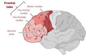
Frontal Lobe
Largest of the lobes
Consists of:
Pre-frontal Cortex
primary motor cortex
broca’s area
Frontal Lobe - Pre-frontal cortex
forward part of the frontal lobes
responsible fro advanced cognitive and executive functions
contributes to personality, intelligence, social skills
cognitive processes e.g decision making
responsibility to predict outcomes
emotion control and impulse control
Frontal Lobe - Primary Motor Cortex
controls voluntary movement - directs skeletal movement
Frontal Lobe - Broca’s Area
formulates the structure of sentences & analysis grammar
control the production of speech
Parietal Lobe
Controls bodily (somatic) sensations
receive and integrates information
determines the location of the body and objects
Consists of:
primary somatosensory cortex
Parietal Lobe - Primary Somatosensory Cortex
neurons at the front of the lobe
registers and processes sensations detected by the body
Temporal Lobes
Involved in hearing, language skills, social understanding
consists of:
Primary Auditory Cortex
Wernicke’s Area
Temporal Lobes - Primary Auditory Cortex
Registers & processes auditory information
right temporal lobe - non-verbal sounds
left temporal lobe - verbal sounds
Temporal Lobe - Wernicke’s Area
Controls comprehension and identifies words as sounds
Occipital Lobe
Registers and processes visual information
Consists of:
Primary visual cortex
Occipital Lobe - Primary Visual Lobe
information is transmitted from retinas
has specialised neurons to respond to specific visual ques
Phineas Gage
An explosive detonated prior to his expectations, resulting a in a 42 inch long 1.2 in wide metal rode to be blown into his skull, into the frontal lobe
He surprisingly had no pain, no loss of consciousness, normal pulse, breathing and, vision
But long term effects were major personality changes, reasoning and capacity to understand and follow social norms was diminished. He became vulgar and an irresponsible vagent
Roger Sperry
The role of the corpus callosum using split-brain experiments
Deduced that the two hemispheres worked independently and when the corpus callosum was severed the connected between the two was lost
Done to originally treat epilepsy
The information received from the optic nerves of each eye was still processed contra-laterally
If the word/image was flashed to the right of the dot the participant could say what they saw but not if the word/image was on the left
but the image/word shown on the left was able to be drawn with the participants left hand as the left hand is controlled by the right side (creative) an the word/image flashed on the left is processed by the right
Walter Freeman
Role of the pre-frontal cortex using frontal lobotomy
He believed that mental issues sprung from self-awareness and overactive emotions, so by severing the connection between the thalamus and pre-frontal cortex would eliminate excessive emotion
done under local anaesthesia do the patient can provide feedback and feelings
The knife inserted cut the nerve fibres that connect the thalamus and pre-frontal cortex
his goal was to reduce patient agitation - but in most cases apathy, decreased concentration, and emotional numbness was produced
Electroencephalograph (EEG)
External Recording - records the movement of electricity across the scalp
indicates neuronal activity, the changed in activity indicate underlying functions
ADVANTAGES: cheap, transportable, silent, no radiation, non-invasive
DISADVANTAGES: low spatial and temporal resolution, poor cortex measures, can’t locate specific areas
Computed Tomography (CT)
Still Imaging - series of x-ray images, 2D slices, can show th extent of a lesion and contrast dye can be used
ADVANTAGES: better than 1 x-ray, quicker than MRI, metal doesn’t matter, can be reconstructed to make 3D, good spatial resolution
DISADVANTAGES: higher radiation levels, cost, doesn’t show functions, poor temporal resolution
Magnetic Resonance Imaging (MRI)
Still Imaging - magnet causes hydrogen in H2O in cells to orient in a single direction, pulse of radiation waves causes atoms to spin at a certain frequency and direction
ADVANTAGES: no ionising fields, good spatial resolution, goods contrast between white and grey matter
DISADVANTAGES: strong magnetic field, poor temporal resolution, cost, size of machine
Functional Magnetic Resonance Imaging (fMRI)
Dynamic Imaging - shows the function of NS and diagnose metabolic disorders
sensors detect a change in oxygen in an area of heightened neural activity
if high neural activity haemoglobin has less O2 as it is being used
Haemoglobin w/oxygen reacts different in a magnetic field than haemoglobin w/ out
ADVANTAGES: satisfactory temporal and spatial resolution, no ionising radiation
DISADVANTAGES: strong magnetic field