Brain and Behavior - University of Texas at Arlington - Chapter Three
1/118
There's no tags or description
Looks like no tags are added yet.
Name | Mastery | Learn | Test | Matching | Spaced |
|---|
No study sessions yet.
119 Terms
Central Nervous System
Includes brain and spinal cord.
Peripheral Nervous System
Connects the brain and spinal cord to the rest of the body. Made up of the Somatic Nervous System and Autonomic Nervous System.
Somatic Nervous System
Consists of afferent axons carrying messages from the sensory organs to the central nervous system and efferent axons carrying messages from the central nervous system to its targets (i.e., muscles and glands).
Autonomic Nervous System
Controls visceral organs (heart, blood vessels, intestines, and others). Made up of the sympathetic and parasympathetic nervous systems.
Sympathetic Nervous System
A network of nerves that prepare the organs for rigorous activity. Consists of chains of ganglia (neuronal cell bodies in the PNS) just to the left and right of the spinal cord’s central regions (thoracic and lumbar areas). Prepare the organs for “fight or flight” (increasing breathing and heart rate, decreasing digestive activity). They are closely linked and thus tend to act “in sympathy” with one another (hence the name), although certain events activate more parts than others. Sweat glands, adrenal glands, muscles that constrict blood vessels, and muscles that erect hairs of the skin have sympathetic input but no parasympathetic input. Mainly releases norepinephrine though some sweat glands use acetylcholine.
Parasympathetic Nervous System
System of nerves that facilitate vegetative, non-emergency responses by the body’s organs. Parasympathetic activities are related to and usually opposite of sympathetic activities. Decreases heart rate, increases digestive activity, promotes sexual arousal, and in general conserves energy. Also known as the craniosacral system because it consists of nerves in the cranial and sacral parts of the spinal cord. Consists of long pregangionic axons that extend from the spinal cord to parasympathetic ganglia close to each organ which connect to shorter postganglionic fibers that extend to the organs themselves. Mainly uses acetylcholine for functions.
Dorsal
For the brain, referring to the top half of the brain close to the top of the head (same for four-legged animals). For the spinal cord, referring to the part toward the back, away from the stomach.
Ventral
For the brain, referring to the bottom half of the brain, close to the neck (same for four-legged animals). For the spinal cord, referring to the part toward the front, toward the stomach.
Anterior
To the front. Rostral for animals.
Posterior
To the back. Caudal for animals.
Superior
Above another part
Inferior
Below another part
Lateral
Towards the side, away from the middle
Medial
Toward the middle, away from the side
Proximal
Located close (approximate) to the point of origin or attachment
Distal
Located more distant from the point of origin or attachment
Ipsilateral
On the same side of the body
Contralateral
On the opposite side of the body
Coronal Plane (Frontal Plane)
A plane that shows brain structures as seen from the front
Sagittal Plane
A plane that shows brain structures as seen from the side
Horizontal Plane (Transverse Plane)
A plane that shows brain structures as seen from above
Lamina (Neuronal Lamina)
A row or layer of cell bodies separated from other cell bodies by a layer of axons and dendrites
Column
A set of cells perpendicular to the surface of the cortex, with similar properties.
Tract
Neural pathways (set of axons) within the CNS, also known as a projection. When a presynaptic axon in the CNS comunicates with a postsynaptic axon in the CNS, information is “projected” from one axon to the other.
Nerve
A set of axons in the periphery, either leading from the CNS to a muscle or gland or from a sensory organ to the CNS
Nucleus
A cluster of neuron cell bodies within the CNS
Ganglion
A cluster of neuron cell bodies, usually outside the CNS (as in the peripheral nervous system)
Gyrus (pl.:gyri)
A protuberance on the surface of the brain, part of the cerebral cortex. Part of the “wrinkles” that go outward.
Sulcus (pl.:sulci)
A fold or groove that separates one gyrus from another. Part of the cerebral cortex. Part of the “wrinkles” that go inward.
Fissure
A long, deep sulcus. Separates left and right half of cerebral cortex and bottom and top half of cerebral cortex.
Spinal Cord
Communicates with the sensory organs and muscles, except those of the head. Composed of grey matter and white matter.
Grey Matter
Located in the center of the spinal cord and densely packed with soma and dendrites. Includes the dorsal horn and the ventral horn.
White Matter
Composed mostly of myelinated axons that carry information from the grey matter to the brain (ascending) or to other areas of the spinal cord and target organs (descending).
Bell-Magendie Law
States that ventral roots transmit motor impulses while dorsal roots transmit sensory impulses.
Dorsal Roots
Afferent axons entering the spinal cord in the dorsal horn of the gray matter and carrying sensory information from sensory organs
Dorsal Root Ganglia (DRG)
Belong to the PNS, clusters of cell bodies of the sensory neurons located outside the spinal cord
Ventral Roots
Efferent axons, exit the spinal cord from the ventral horn carrying motor command to mucles and glands
Spinal Segments
There are 31 spinal segments in total which are capable of sending information to peripheral organs and receiving information from peripheral tissues. The 31 segments are divided into five groups: Cervical, Thoracic, Lumbar, Sacrum, and Coccyx
Cervical
The eight segments at the top of the spine, roughly located from bottom of ears to middle of shoulder blades
Thoracic
The twelve segments in the middle of the spine, roughly located from middle of shoulder blades to bottom of ribs
Lumbar
The five segments near the bottom of the spine, roughly located from bottom of ribs to top of pelvis
Sacrum
The five segments at the bottom of the spine, roughly located from top to bottom of pelvis
Coccyx
Very last segment of spine, roughly located near bottom of pelvis
Cervical Enlargement
Wider area in the cervical segments of the spine, helps with top limb movement
Lumbo-Sacral Enlargement
Wider area in the Lumbo-Sacral area of the spine, helps with bottom limb movement
Enteric Nervous System
Special system only in the digestive organs, controls/regulates digestion semi-independently of CNS signaling
Dual Innervation
Refers to organs which receive signaling from both sympathetic and parasympathetic systems, excludes sweat glands, adrenal glands, hair follicle muscles (arrector pili), and smooth muscles that constrict blood vessels.
The Hindbrain (Rhombencephaon)
Posterior (caudal for animals) to the midbrain. Consists of medulla, pons, and cerebellum.
Brainstem
Referring to the medulla, pons, and midbrain
The Medulla (Myelencephalon)
Located rostral to the spinal cord. Contains several vital centers that regulate/control vital reflexes such as breathing, heart rate, vomiting, salivation, coughing, and sneezing. The head and the organs connect to the medulla and adjacent areas by 12 pairs of cranial nerves.
Pons (Metencephalon)
Lies on each side of the medulla and rostral to it (think of wearing a neck pillow backwards). Contains the reticular formation, raphe nuclei, locus coeruleus, and some cranial nerves. In it, axons from each half of the brain cross to the opposite side of the spinal cord so that the left hemisphere controls the right and vice versa.
The Reticular Formation
The descending portion is one of several brain areas that control the motor areas of the spinal cord. The ascending portions sends axons up to much of the cerebral cortex, selectively increasing arousal and attention.
The Raphe Nuclei and Locus Coeruleus
Sends axons to much of the forebrain, modifying the brain’s readiness to respond to stimuli (like arousal and attention), adjusting vigilance and mood levels and sends axons down to the spinal cord to modulate pain.
Cerebellum
Structured attached to the hindbrain with many deep folds to form many folia (singular folium); helps regulate motor movement, balance, and coordination. Also important for shifting attention between auditory and visual stimuli, coordinating with the need of movement.
The Midbrain (Mesencephalon)
Consists of the tectum, tegmentum, and substantia nigra.
Tectum
Regarded as the roof of the midbrain (posterior or dorsal portion). There are two swelling on either side of the tectum and they are the superior colliculus (important to visual processing) and inferior colliculus (contributes to hearing).
Tegmentum
Anterior or ventral portion of the midbrain. Contains nuclei for cranial nerves and part of the reticular formation. Controls arousal, pain, and motor control.
Substantia Nigra
Gives rise to the dopamine (DA) containing pathway facilitating readiness for movement. Clinically, DA neurons degenerate in Parkinson’s Disease.
Cranial Nerves
Consists of twelve pairs of nerves (one half of pair on right, one on left), most of which are from the brainstem (except the olfactory and optic nerves). These are afferent and efferent nerves that regulate and control sensory and motor activity of the head, neck, shoulders, and visceral organs.
Olfactory Nerve
A cranial nerve whose major function is smell.
Optic Nerve
A cranial nerve whose major function is vision.
Oculomotor Nerve
A cranial nerve whose major functions are eye movement and pupil constriction.
Trochlear Nerve and Abducens Nerve
Cranial nerves whose major function is eye movement.
Trigeminal
A cranial nerve whose major functions are skin sensations from most of the face and control of jaw muscles for chewing and swallowing.
Facial Nerve
A cranial nerve whose major function are taste from the anterior 2/3rds of the tongue, control of facial expressions, crying, salivation, and dilation of the head’s blood vessels.
Statoacoustic Nerve
A cranial nerves whose major functions are hearing and balance
Glossoparyngeal Nerve
A cranial nerve whose major functions are taste and other sensations from the throat and posterior third of the tongue, control of swallowing, salivation, and throat movements during speech.
Vagus Nerve
A cranial nerve whose major functions are sensations from neck and thorax, control of throat, esophagus, and larynx, and control of parasympathetic nerves to stomach, intestines, and other organs.
Accessory Nerve
A cranial nerve whose major function is control of neck and shoulder movements.
Hypoglossal Nerve
A cranial nerve whose major function is control of the muscles of the tongue
The Forebrain (Prosencephalon)
The most anterior and prominent part of the mammalian brain and consists of two cerebral hemispheres, the outer regions being called the cerebral cortex (telencephalon). Each side of the cortex receives sensory information and controls motor movement from the opposite (contralateral) side of the body. The contents underneath the cortex are called sub-cortical regions (diencephalon).
The Limbic System
Consists of both cortical and sub-cortical structures and other interlinked structures which form a border around the brainstem. The main structures include the cingulate gyrus of the cerebral cortex, the olfactory bulb, the hypothalamus, the hippocampus, and the amygdala. Involved in mediating emotional responses (like aggression or fear), regulating motivated behavior (like rewarding or drug abuse), modulating mood levels, and learning/memory, etc.
Sub-Cortical Regions (Diencephalon)
Includes the thalamus, hypothalamus, pituitary, basal ganglia, basal forebrain, the hippocampus, fornix, the ventricles, and meninges.
Thalamus
Also known as the “inner chamber”. Relays information from the sensory organs to the sensory cortex. Most sensory information, except olfactory, is processed there before ascending to the cortex.
Hypothalamus
A small area underneath the thalamus near the base of the brain. Contains up to 11 small sized nuclei. Conveys messages to the pituitary gland to alter the release of hormones and also regulates a number of behaviors which include: drinking, regulating salt-water balance, eating, sex and other motivated behaviors, and body temperature setting. These are known as the body’s “internal issues”.
Pituitary Gland
Contains endocrine (hormone-producing) glands at the base of the hypothalamus
Basal Ganglia
The exception for using the term ganglia when referring to a structure inside the CNS. Located lateral to the thalamus. Comprised of the caudate nucleus, the putamen, and the globus pallidus. Integrates motivational and emotional behavior to increase the vigor of motivated actions. Also critical for gradual learning of skills and habits. Mainly has to do with movement, emotional expression, and some aspects of memory.
Basal Forebrain
One of the sub-cortical structures that lie on the ventral (toward the stomach) surface of the forebrain. Consists of two main parts: The nucleus basalis and the nucleus accumbens.
Nucleus Basalis
Receives input from the hypothalamus and basal ganglia, after which it send axons that release acetylcholine (ACh) to the cerebral cortex (mainly the prefrontal cortex). This area is key to integrating arousal, wakefulness, and attention. Patients with Parkinson’s disease and Alzheimer’s disease have impairments of attention and intellect because of inactivity or deterioration in the nucleus basalis.
Nucleus Accumbens
Reward center involved in motivating and rewarding responses like drug abuse.
The Hippocampus
Located between the thalamus and cerebral cortex, beginning at the anterior portion of the forebrain. Critical for storing long-term memory, particularly processing learning/memorizing new events. Shaped like a seahorse and follows curve of basal ganglia. Vital in prospective memory (memory of things you plan to do).
Central Canal
Located in the center of the spinal cord and connected to the cerebral aqueduct, which connects the four ventricles together. Filled with cerebrospinal fluid.
Ventricles
Fluid filled cavities in the brain that enable flow of cerebrospinal fluid.
Cerebrospinal Fluid
A clear fluid found in the brain and spinal cord. Provides “cushioning” for the brain. Reservoir of hormones and nutrition for the brain and spinal cord.
Meninges
Membranes that surround the brain and spinal cord. CSF flows between brain and meninges. The brain tissue has no pain sensors but meninges do. The meninges contain many blood vessels. Swollen blood vessels in the meninges cause headaches.
The Cerebral Cortex (Telencephalon)
Most prominent part of the mammalian brain; consists of six cellular layers on the outer surface of the cerebral hemispheres. Divided into two hemispheres and joined by two inter-communicating bundles of axons known as the corpus callosum and anterior commisure. The inter-hemisphere communications are important for cognitive processes and evaluation, like language and thoughts
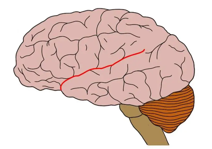
Lateral fissure

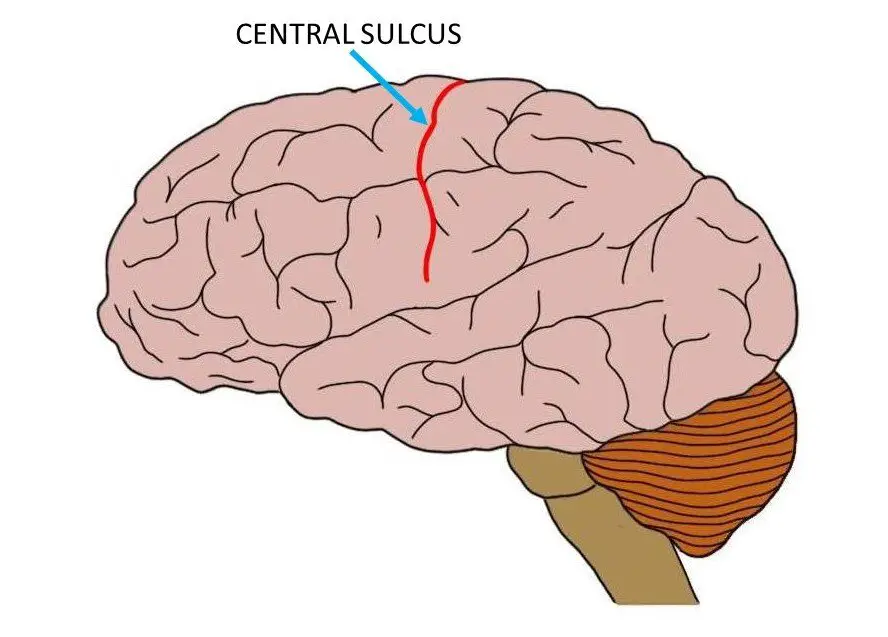
Central Sulcus

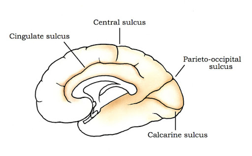
Parieto-Occipital Sulcus

Amygdala
Temporal lobe structure important for evaluating emotional information. Located superior to the hippocampus.
Primary Somato-Sensory Cortex (Post-Central Gyrus)
Dorsal to central sulcus. Contains point to point somatic representation areas for sensory regulation. Primary target for touch sensations (including pain) and information from muscle-stretch receptors and joint receptors.
Primary Somato-Motor Cortex (Pre-Central Gyrus)
Ventral to central sulcus. Contains point to point somatic representation areas for motor regulation. Controls fine motor movement.
Occipital Lobe
Located at the posterior (caudal) end of the cerebral cortex. The posterior pole of the lobe is known as the striate cortex (named for its striped look in cross section) or the primary visual cortex. Highly responsible for visual output, damage can result in cortical blindness (information goes in but nothing is processed; no visual thought or dreams).
Parietal Lobe
Lies between the occipital lobe and the central sulcus. Contains the post-central gyrus (Primary somato-sensory cortex). Monitors information about head, eye, and body positions and passes it on to brain areas that control movement. Handles spacial and numerical information.
Temporal Lobe
Located on the lateral portion of each hemisphere near the temples. The target for auditory information. The left side is involved in processing spoken language (comprehension). Responsible for complex actions of vision, such as moving vision, and some emotional and motivational behaviors, such as fear.
Temporoparietal Junction
Located where the parietal and temporal lobe meet, close to the occipital lobe. Conveniently located to receive input from vision, hearing, and body senses. Serves multiple functions, including attention, body awareness and social cognition. Damage to this area, especially to the right hemisphere, impairs the ability to imagine how something looks form a different perspective.
Frontal Lobe
Contains the prefrontal cortex and precentral gyrus. The prefrontal cortex is the integration center for all sensory information and information from other areas of the cortex. By receiving the information from all inputs, cognitive processing is integrated and performed by the prefrontal cortex, such as: abstract thinking, planning, decision making, and thoughts. Responsible for our ability to remember recent events and information (working memory). People with damage to the prefrontal cortex exhibit delayed responses to stimulus. Humans have a larger prefrontal cortex compared to other animals.
Prefrontal Cortex Regions
The posterior portion is mostly associated with movement. The middle zone pertains to cognitive control, emotional reactions, and certain aspects of memory. The anterior zone of the prefrontal cortex is important for making decisions, evaluating which of several courses of action is likely to achieve the best outcome.
Laminae
Six distinct layers of cell bodies parallel to the surface of the cortex. The first layer is known as the molecular layer and is made up of mostly dendrites and long axons. The second layer is known as the external granular layer and consists of small pyramidal cells. The third layer is known as the pyramidal cell layer and consists of pyramidal cells. The fourth layer is known as the internal granular layer and consists of cell smalls and serves as the main site for incoming sensory information. The fifth layer is known as the inner pyramidal layer and consists of large pyramidal cells and serves as the main source of motor input. The sixth layer is known as the multiform layer and consists of spindle cells.
Columns
Neural cells of the cortex that are divided based on similar properties. For example, if one cell in a column responds to touch on the palm of the left hand, then the other cells in that column do too. Perpendicular to laminae.