Module 3 - Lecture 4 to Lecture 8
1/62
There's no tags or description
Looks like no tags are added yet.
Name | Mastery | Learn | Test | Matching | Spaced |
|---|
No study sessions yet.
63 Terms
Module 3: Lecture 4, Action Potential
Dendrites
Receive inputs from other neurons
Cell body
Integrates all the inputs it receives
Axon
Sends output signals to the next
neuronGenerates and propagates action
potentials (aka impulse, spike)
→ How signals are sent around the
nervous system
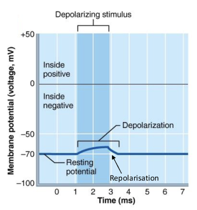
Depolarisation
Inside of membrane becomes more positive
→ Na+ coming into neuron
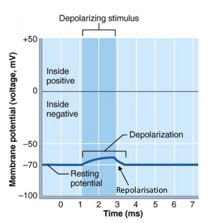
Repolarisation
Return to resting membrane potential from being depolarised
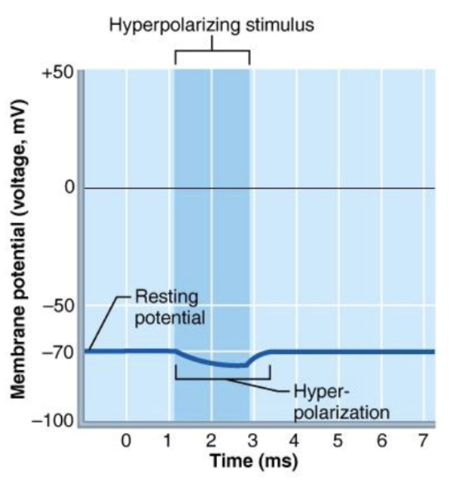
Hyperpolarisation
Inside becomes more negative
→ K+ leaving the neuron
Action potential
A rapid, brief change in membrane potential
Factors effecting speed of action potential
propagation
Axon diameter: Thicker axons propagate axons faster
Temperature: Reactions occur faster at higher
temperaturesMyelination: Insulates the membrane
and speeds up propagation
Module 3: Lecture 5, Synaptic Transmission
Synapse
A junction between two nerve cells, consisting of a minute gap across which impulses pass by diffusion of a neurotransmitter
Process of Synapse
Pre-synapse → Synaptic cleft → Post-synapse
Pre-synaptic neuron
Impulse happens at end of axon
Triggers opening of Ca+ channels
Neurotransmitter molecules are released
Synaptic cleft
Neurotransmitter molecules are released
Bind with receptors in post-synaptic cleft
Postsynaptic neuron
After neurotransmitter molecules bind into receptors
Results in another action potential down a neuron
Chemically gated channels
Responsible for triggering graded/local potentials at a
synapse
Voltaged-gated ion channels
Responsible for triggering action potentials in an axon
Excitatory synapses (EPSPs)
Depolarise the postsynaptic membrane
→ Closer to action potential initiation
threshold
Inhibitory synapses (IPSPs)
Hyperpolarize the postsynaptic membrane
→ Further from action potential initiation threshold
Summation of postsynaptic potentials
Determines whether the cell will reach action potential threshold
Temporal summation
Two EPSPs from the same presynaptic neuron occur close in time to depolarise the membrane to threshold
Spatial summation
Two EPSPs from different presynaptic neurons occur close together in time, to depolarise the
membrane to threshold.
EPSPs and IPSPs cancellation
No net change in membrane potential
Synaptic transmission dependency
The type of cell it is terminating on
The type of neurotransmitter the neuron releases
The types of neurotransmitter receptors on the postsynaptic membrane
Module 3: Lecture 6, Cell Communication & Receptors
Cell communication
Local and Long distance signalling
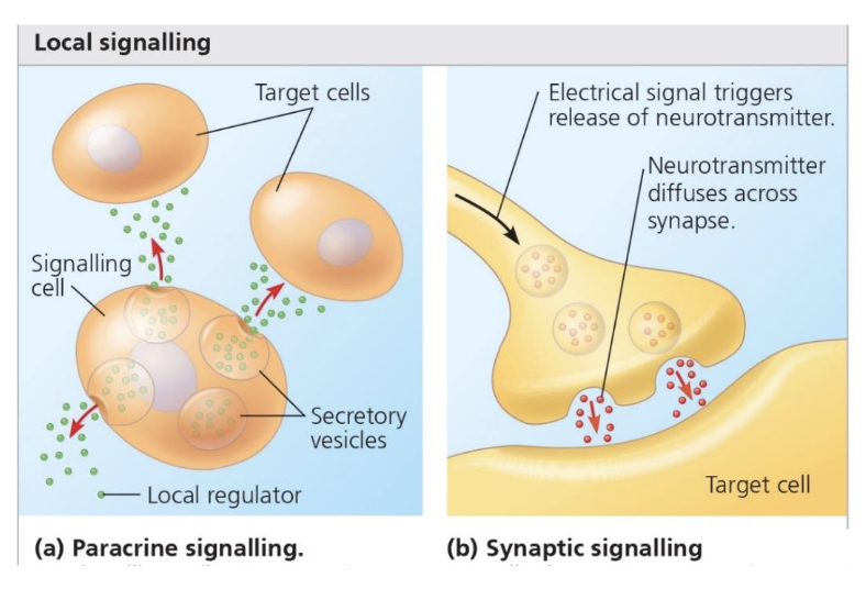
Local Signalling
Paracrine signalling: One cell releases a signalling
molecule that acts on nearby cells.Synaptic signalling (synaptic
transmission): Main communication between neurons.
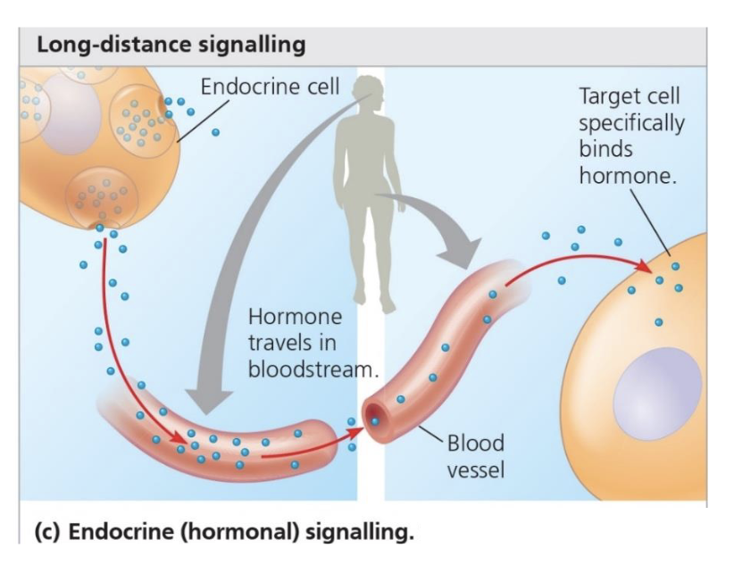
Long Distance Signalling
Signalling molecule is released from a
cell into the blood stream (Hormone)
3 stages of cell communication
Reception
Transduction
Response
Reception
The signalling molecule binds to a specific receptor
Transduction
A signalling pathway is initiated in the cytoplasm
Response
A cellular response is activated e.g. to activate an enzyme
Four families of receptors
1. Ligand-gated ion channels
2. G protein coupled receptors
3. Tyrosine kinase receptors
4. Steroid receptors
Receptors involved in neurotransmission
Ligand-gated ion channels
G protein coupled receptors
Ligand-gated ion channels
Fast neurotransmission
“Direct” neurotransmission
G protein coupled receptors
Slower neurotransmission
“Indirect” neurotransmission
Common neurotransmitters
Adrenaline: Fight or Flight
Dopamine: Pleasure
Noradrenaline: Concentration
Serotonin: Mood
Gaba: Calming
Acetylcholine: Learning
Glutamate: Memory
Endorphins: Euphoria
Tyrosine kinase receptors
Cell surface receptors
Respond to hormones only
Slower processes such as metabolism, growth
Steroid receptors
Intracellular receptors
Bind to steroid hormones and affect gene expression
Module 3: Lecture 7, Autonomic Nervous System
Motor (Efferent) Division
Motor nerve fibers
Conducts impulses from CNS to effectors (muscles and glands)
2 Types of Motor Division
Somatic/motor
Autonomic
Somatic/Motor Nervous System
Voluntary movement
Intervention and activation of skeletal muscle
Autonomic nervous system
Involuntary movement
Innervation of organs, glands,
smooth muscle
2 Types of Autonomic nervous system
• Sympathetic nervous
system
• Parasympathetic nervous
system
Sympathetic nervous system
“Fight or flight
Prepares the body for action
Parasympathetic nervous system
“Rest and digest”
Relaxes the body
Three differences between parasympathetic and sympathetic
Sites or origin
Relative lengths of fibers
Location of ganglia
Neurotransmitters of the ANS
Acetylcholine (ACh) → Produced by Parasympathetic
Noradrenaline (Ne) → Produced by Sympathetic
Module 3: Lecture 8, Reflexes
Types of movement
Reflex
Rhythmic
Voluntary
Reflex
Least complex, integrated at the spinal cord or brain stem
Rhythmic
Intermediate complexity, integrated in spinal cord with input from the brain
Voluntary
Most complex, integrated in cerebral cortex
Reflex
Rapid, automatic responses to stimuli
Classification of Neutral Reflex
Efferent division (Somatic/Automatic)
Integrating region (Spinal/Cranial)
Time (Innate/Learned)
Neurons (Monosynaptic/Polysynaptic)
Reflex Arc
Receptor
Sensory neuron
Integration centre
Motor neuron
Effector
Muscle stretch
Special sensory receptors called muscle spindles inside the muscle are activated.
Stretch Reflex
Stimulus: Muscle is stretched.
Receptor: Muscle spindle detects the stretch.
Afferent neuron (sensory): Sends signal from spindle to the spinal cord.
Synapse: Directly synapses (monosynaptic) with an alpha motor neuron in the spinal cord.
Efferent neuron (motor): Sends signal to the same muscle.
Response: Muscle contracts to resist further stretch.
➡ Fast, monosynaptic reflex.
Knee Jerk Reflex
Stimulus: Tap to the patellar tendon stretches the quadriceps muscle.
Receptor: Muscle spindle in quadriceps detects the stretch.
Afferent neuron: Sends signal to spinal cord (L2–L4).
Synapse: Monosynaptic connection with motor neuron.
Efferent neuron: Stimulates quadriceps to contract.
Response: Lower leg extends (kicks out).
➡ Simple, monosynaptic stretch reflex used in clinical testing.
Crossed Extensor Reflex
Stimulus: Painful stimulus (e.g., step on a nail).
Receptor: Pain receptors in foot activated.
Afferent neuron: Sends signal to spinal cord.
Synapse: Multiple interneurons involved (polysynaptic).
Ipsilateral side (same side): Activates motor neurons that contract flexors and inhibit extensors → leg withdraws.
Contralateral side (opposite leg): Activates extensors and inhibits flexors → leg extends to support the body.
Response: One leg flexes (withdrawal), other leg extends (support).
➡ Polysynaptic reflex involving coordination across both sides of the body.
Autonomic Nervous System Reflexes
Somatic reflexes: one motor neuron
Autonomic reflexes: 2 efferent neuron
Example of Autonomic NS reflexes
E.g. Need to urinate in response to a
full bladder stretching
E.g. Contraction of GI smooth muscle
in response to stretch from food
E.g. Changing blood pressure in
response to standing up
Marieb