Unit 1 Section 4
1/60
There's no tags or description
Looks like no tags are added yet.
Name | Mastery | Learn | Test | Matching | Spaced | Call with Kai |
|---|
No analytics yet
Send a link to your students to track their progress
61 Terms
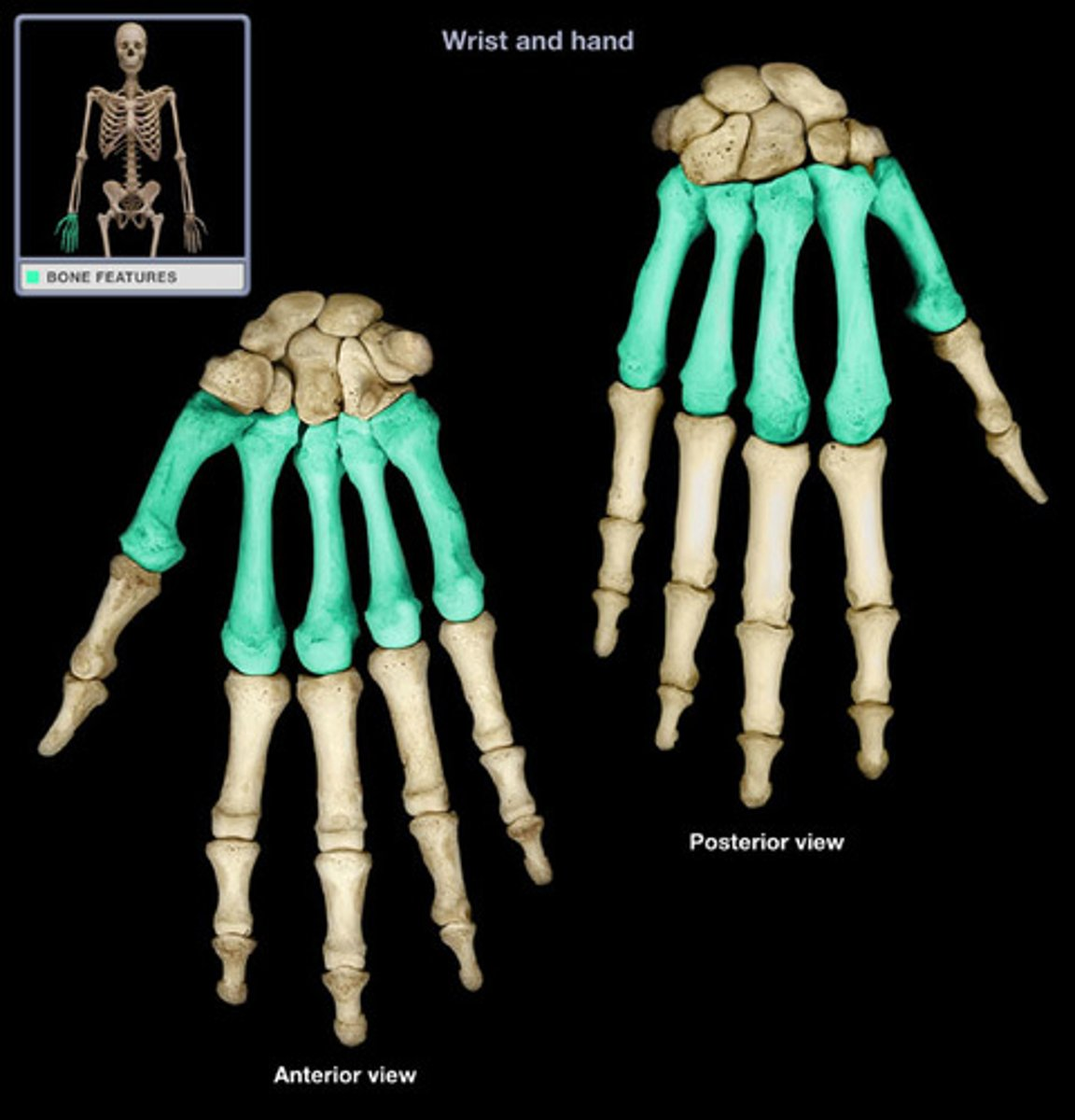
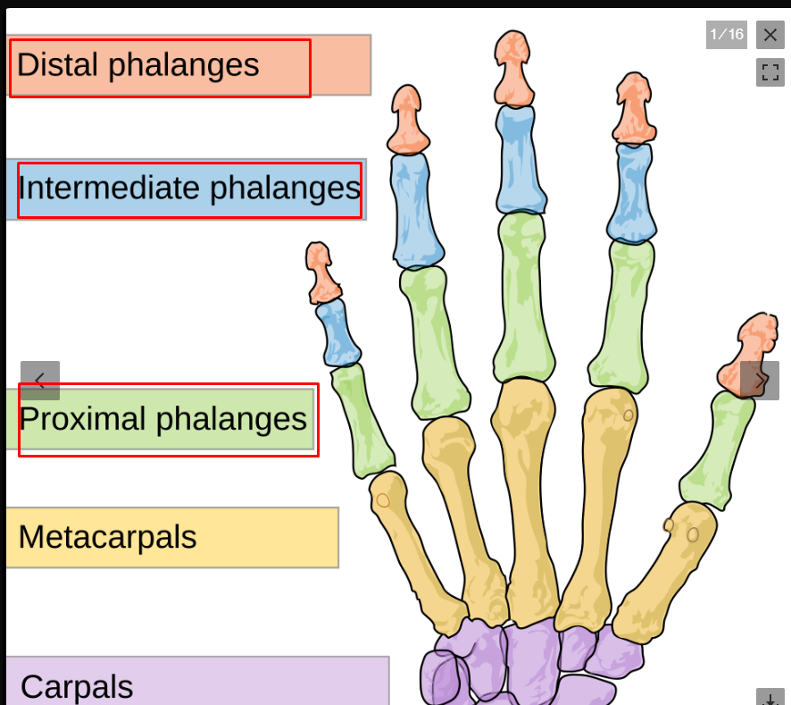
Metacarpal bones
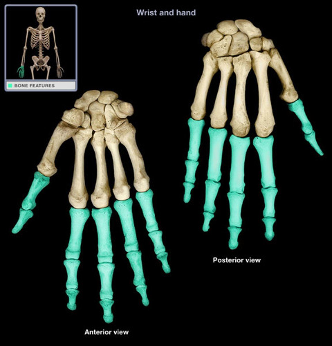
Phalanges (fingers)
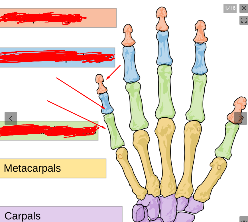
phalanges (green = proximal, blue = intermediate, red = distal)
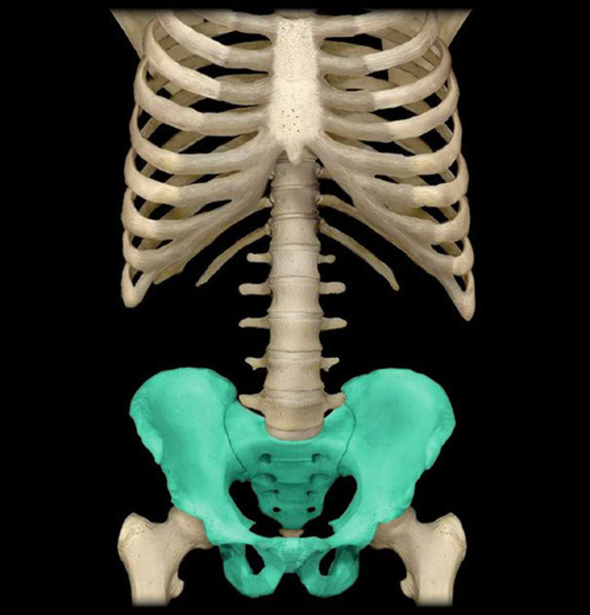
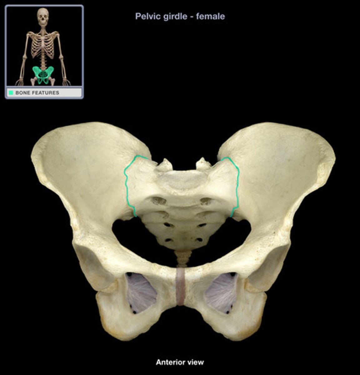
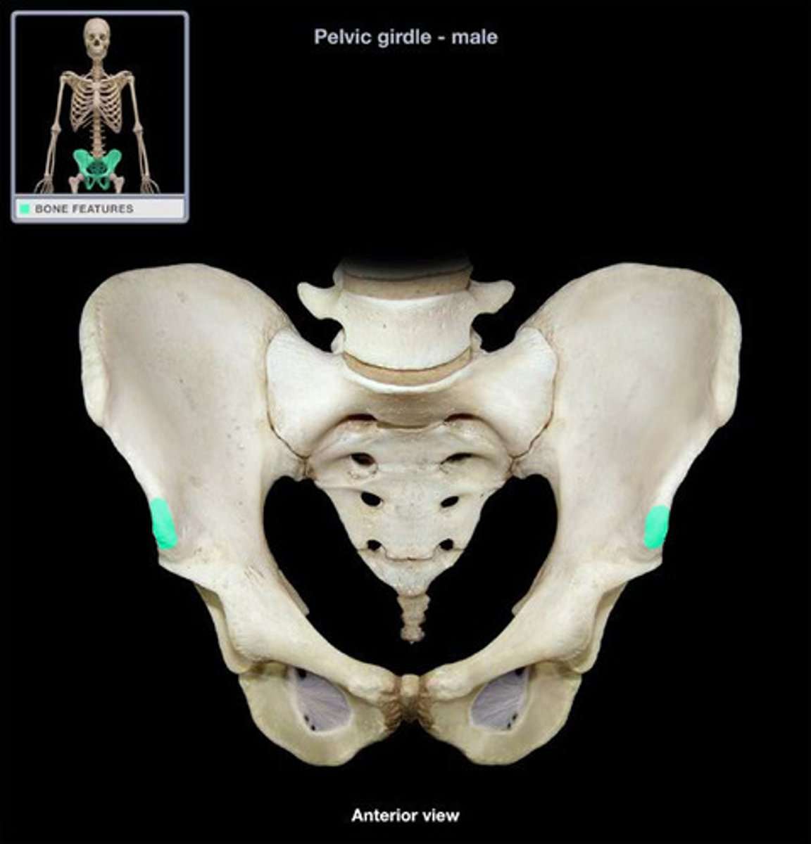
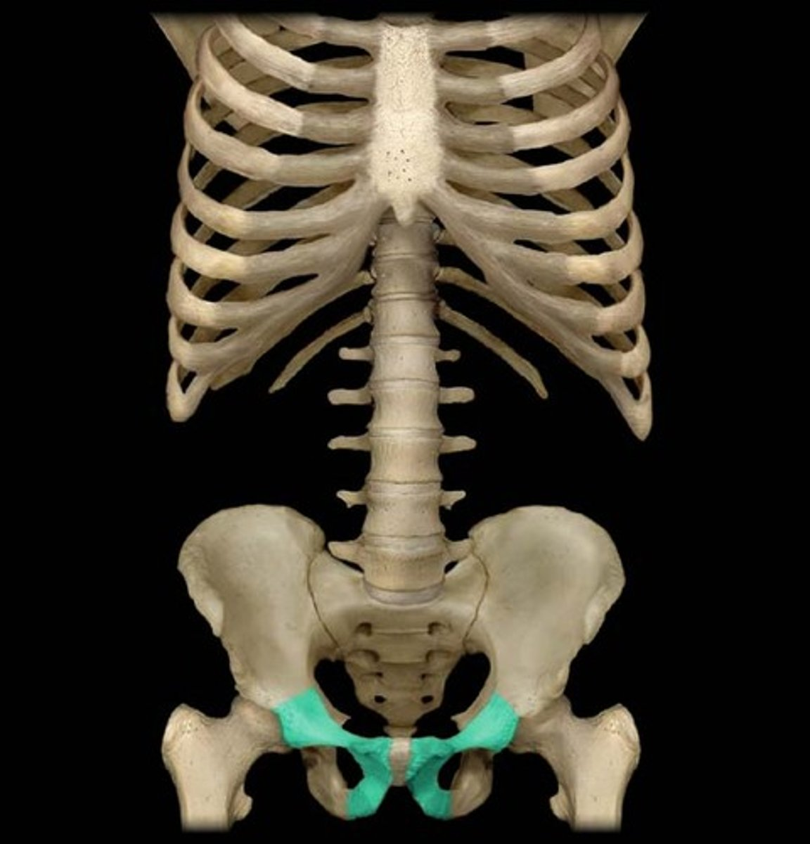
pelvis bone
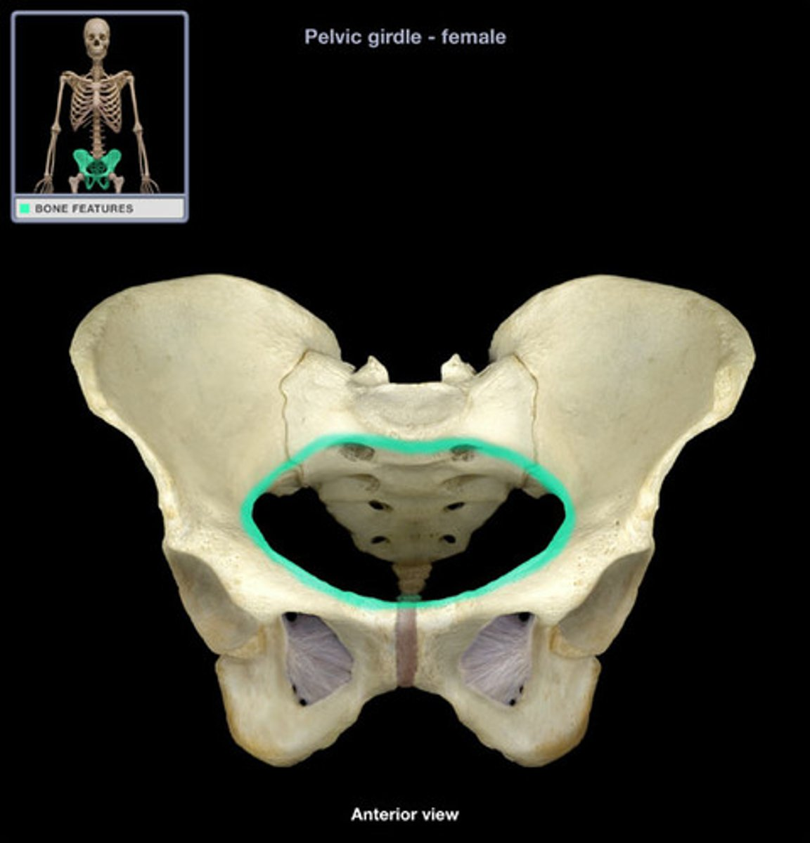
pelvic inlet - top rim of the true pelvis
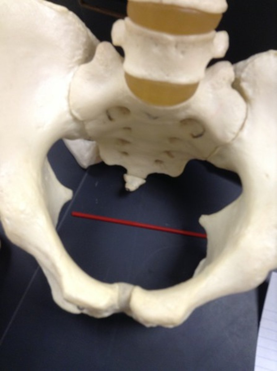
pelvic outlet - bottom hole or rim of the pelvis ; outlet = exit
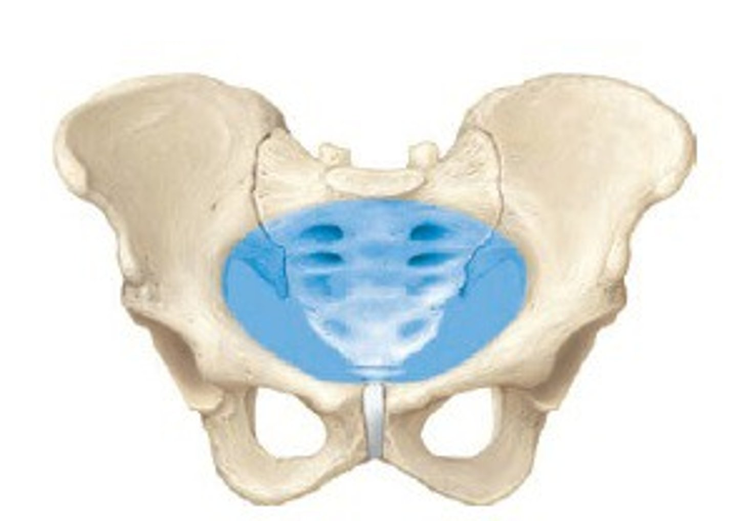
true pelvis - Inside / below the pelvic inlet ;
The pelvic inlet separates the false pelvis above from the true pelvis below.
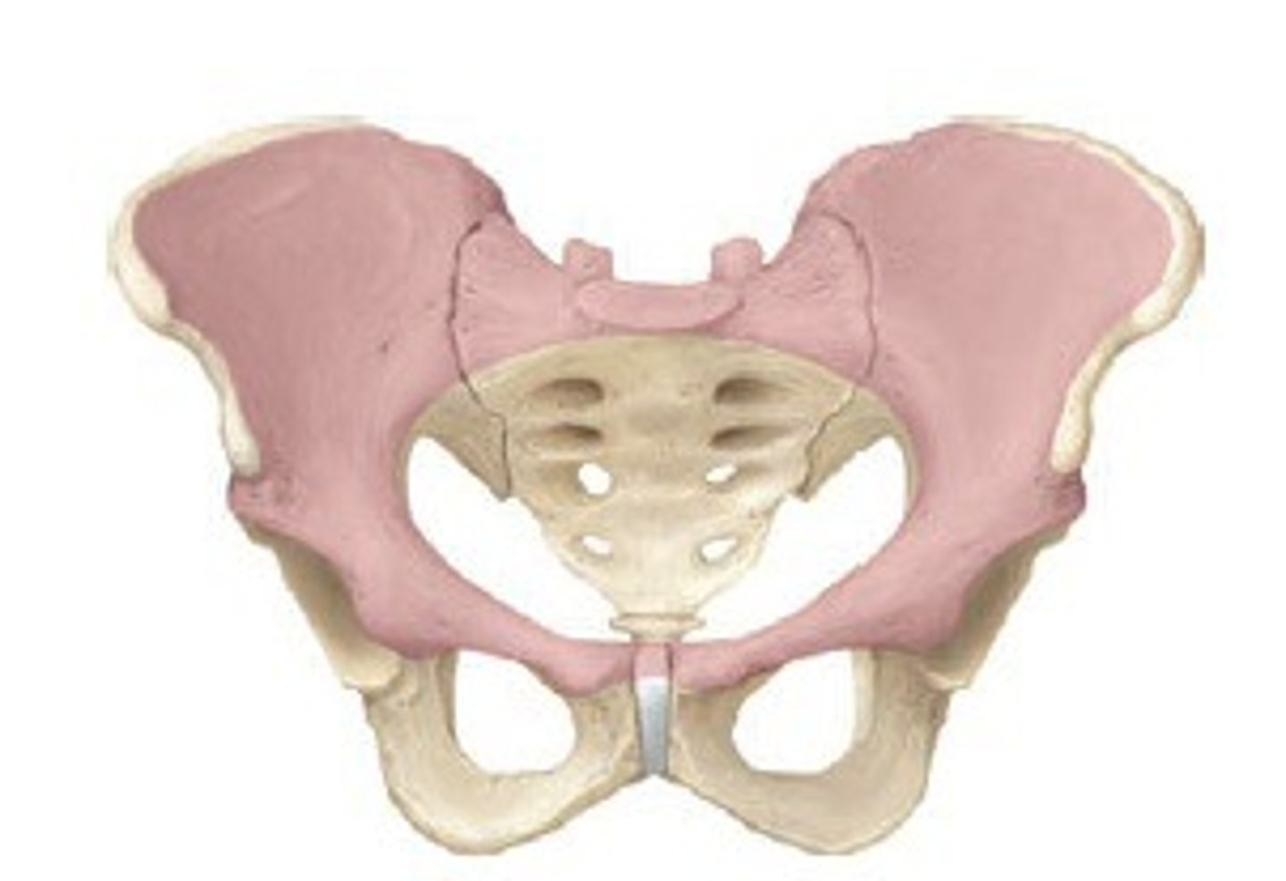
false pelvis - Above the pelvic inlet ; the greater pelvis
The pelvic inlet separates the false pelvis above from the true pelvis below.
sacroiliac joint - where the sacrum and the ilium connect
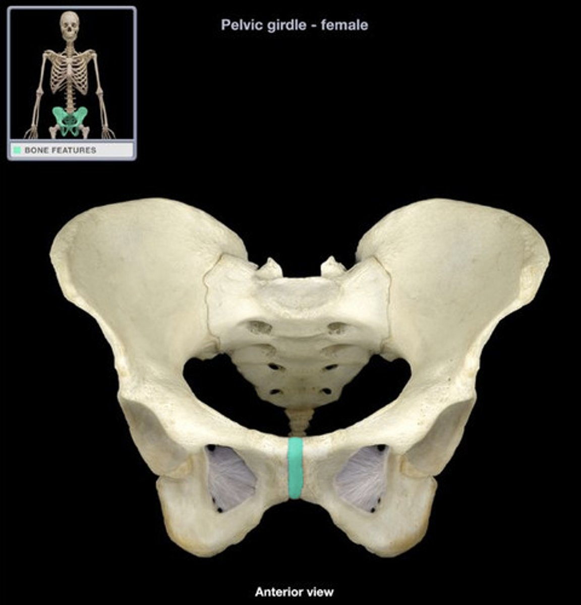
pubic symphysis - where the left and right pubic bones meet
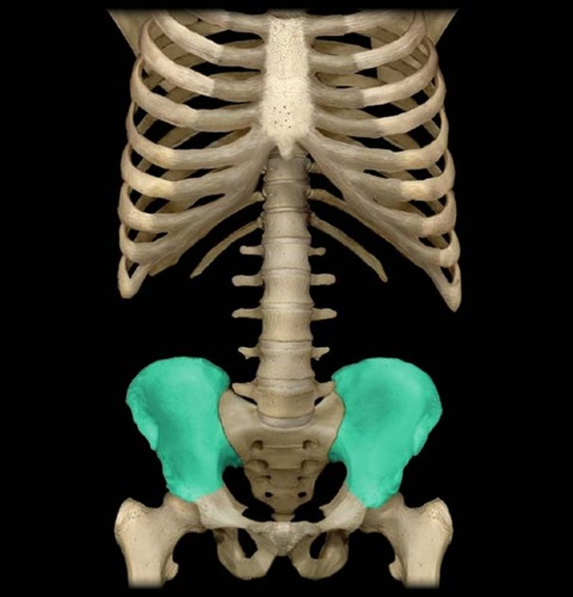
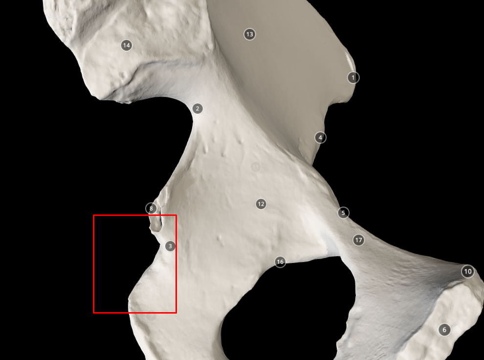
ilium bone
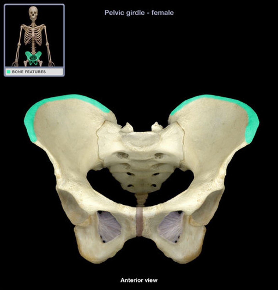
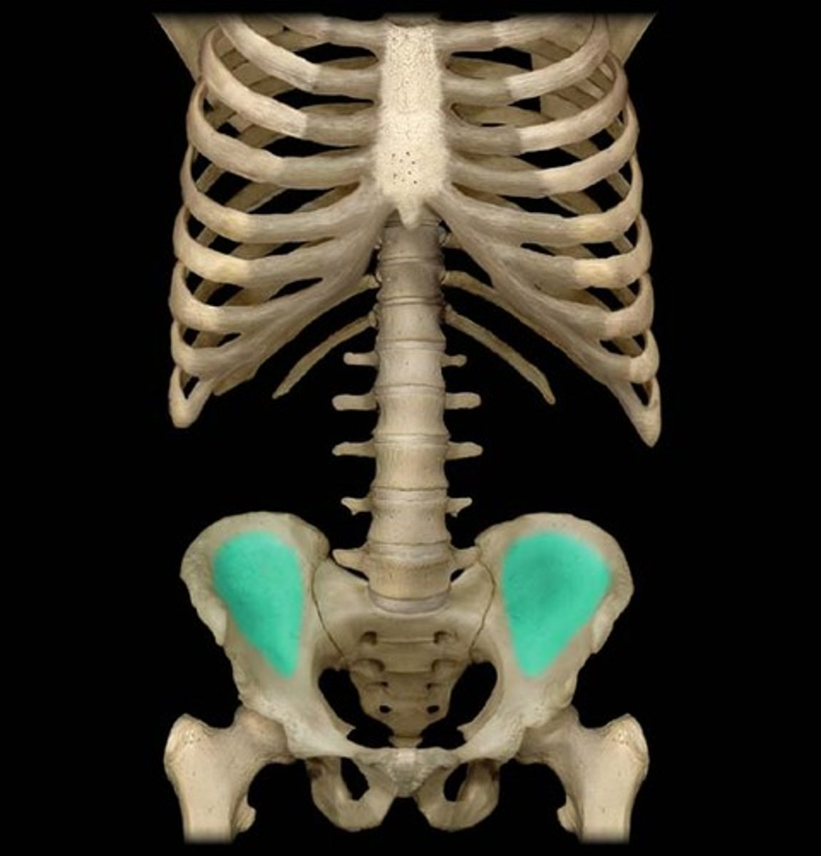
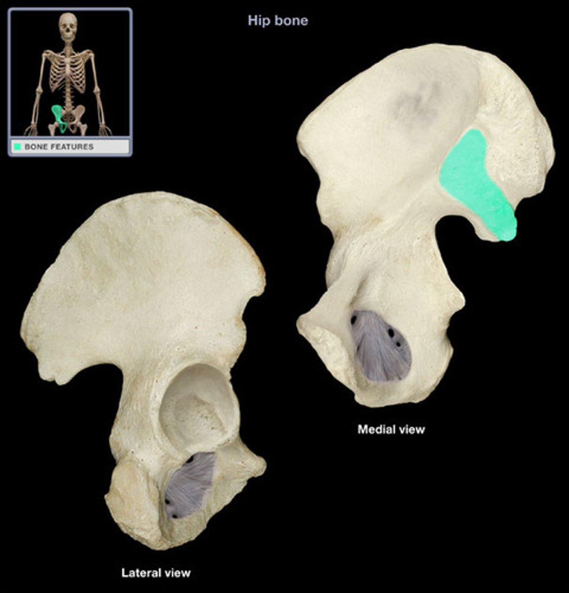
iliac crest - superior (top) edge or rim of the ilium bone
iliac fossa
auricular surface - the chunky, flat piece under the iliac tuberosity
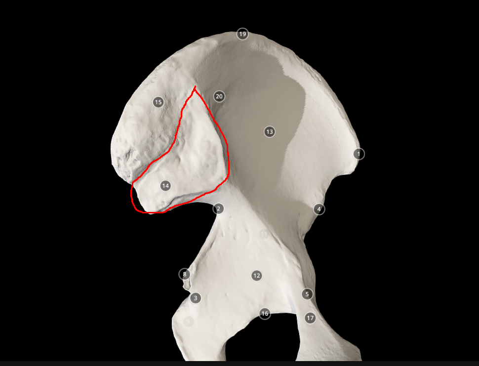
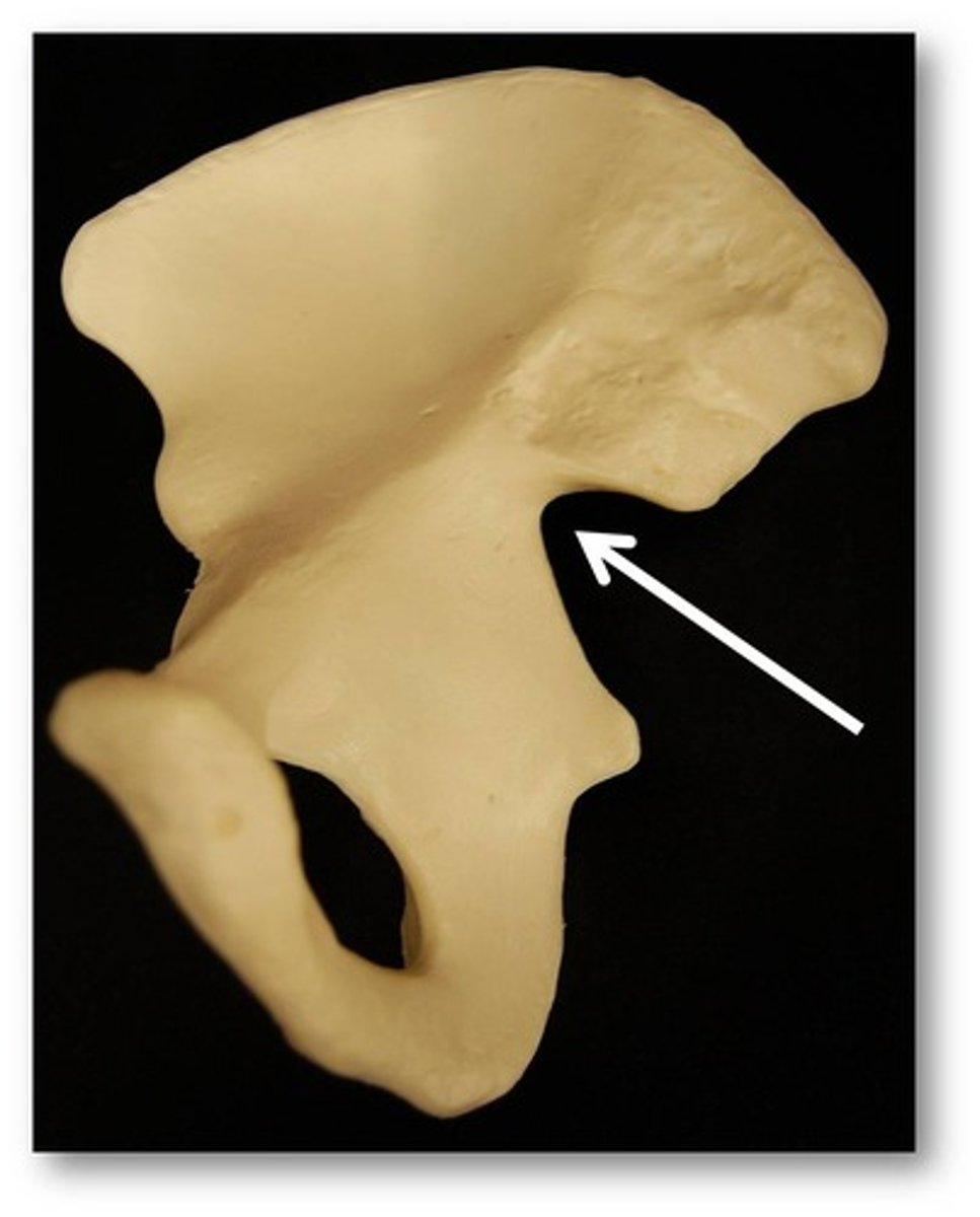
greater sciatic notch - the “hook” like hole in the ilium
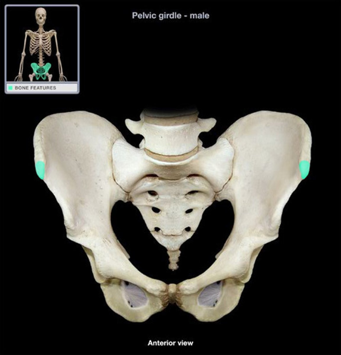
anterior superior iliac spine
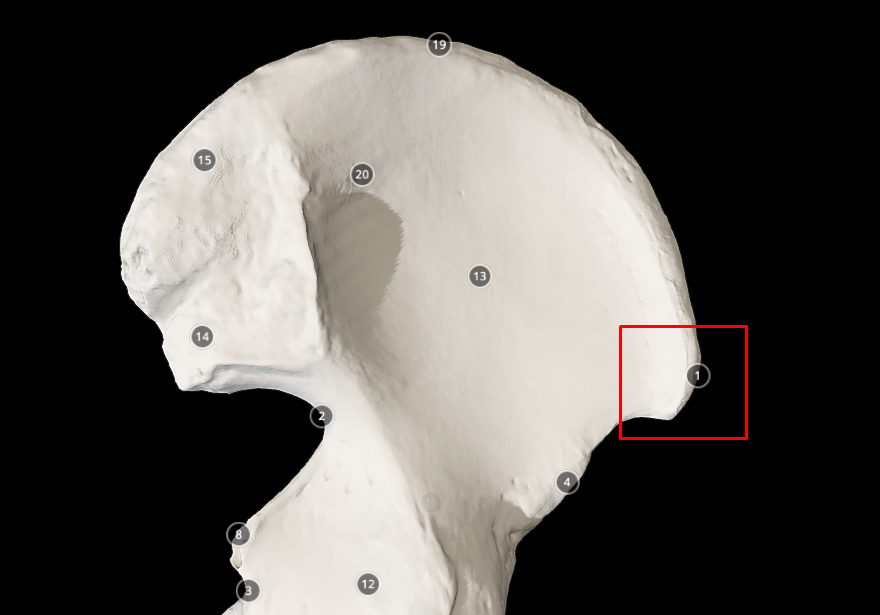
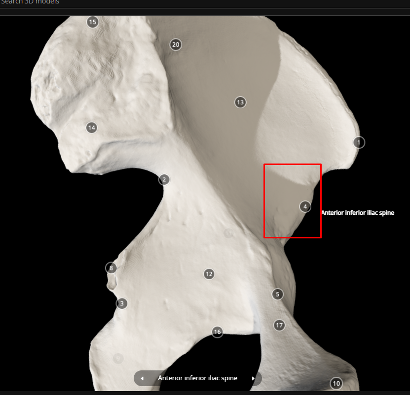
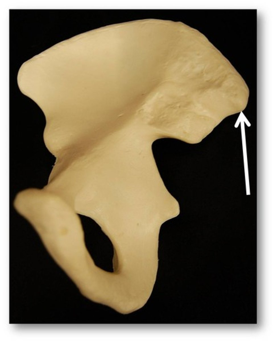
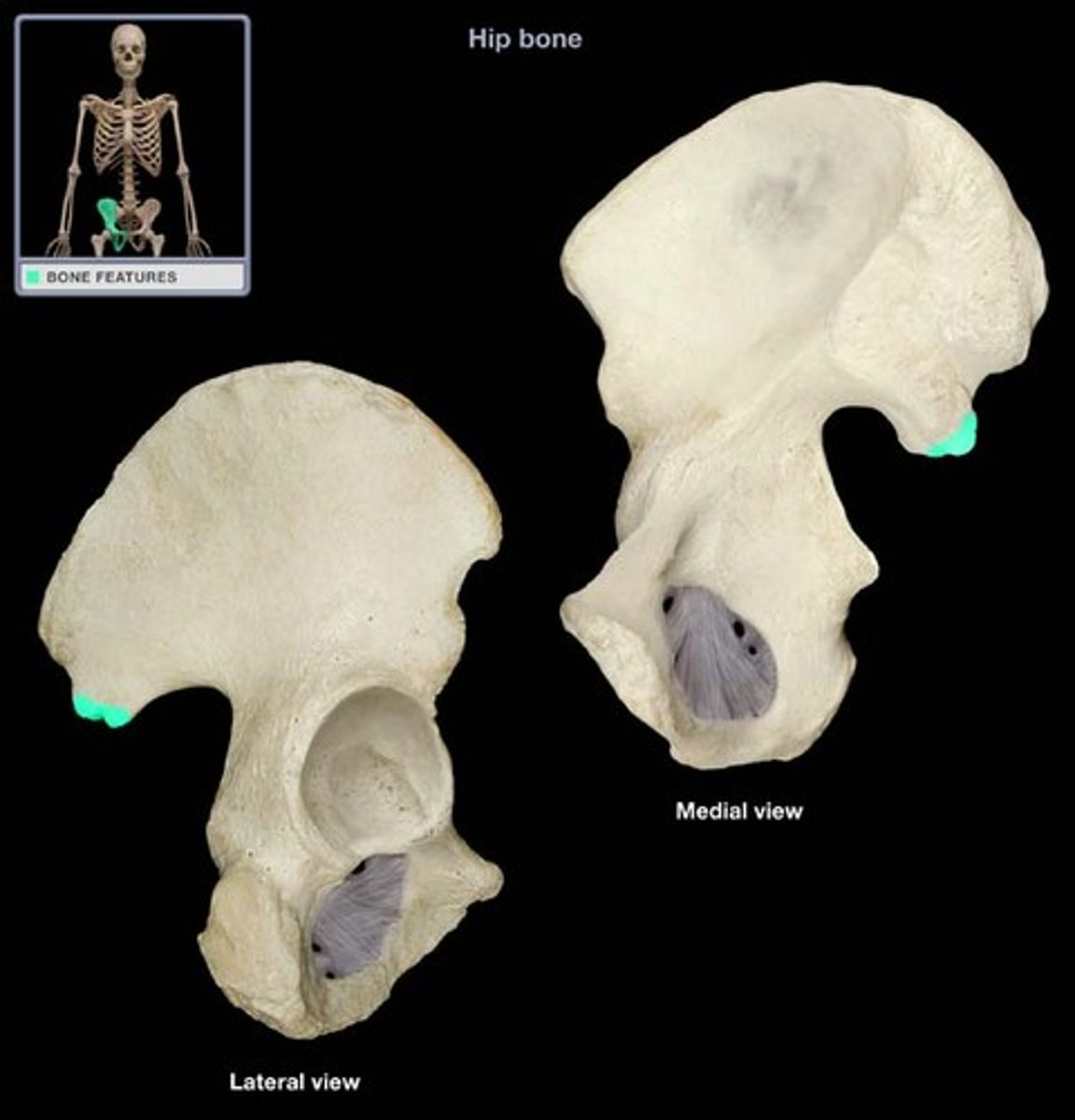
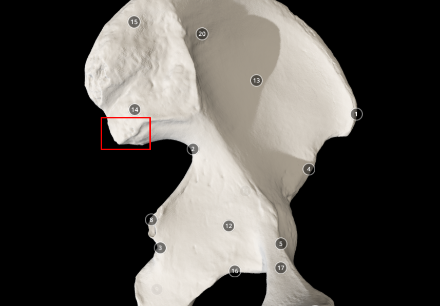
anterior inferior iliac spine
posterior superior iliac spine
posterior inferior iliac spine - point above the greater sciatic notch
ischium bone
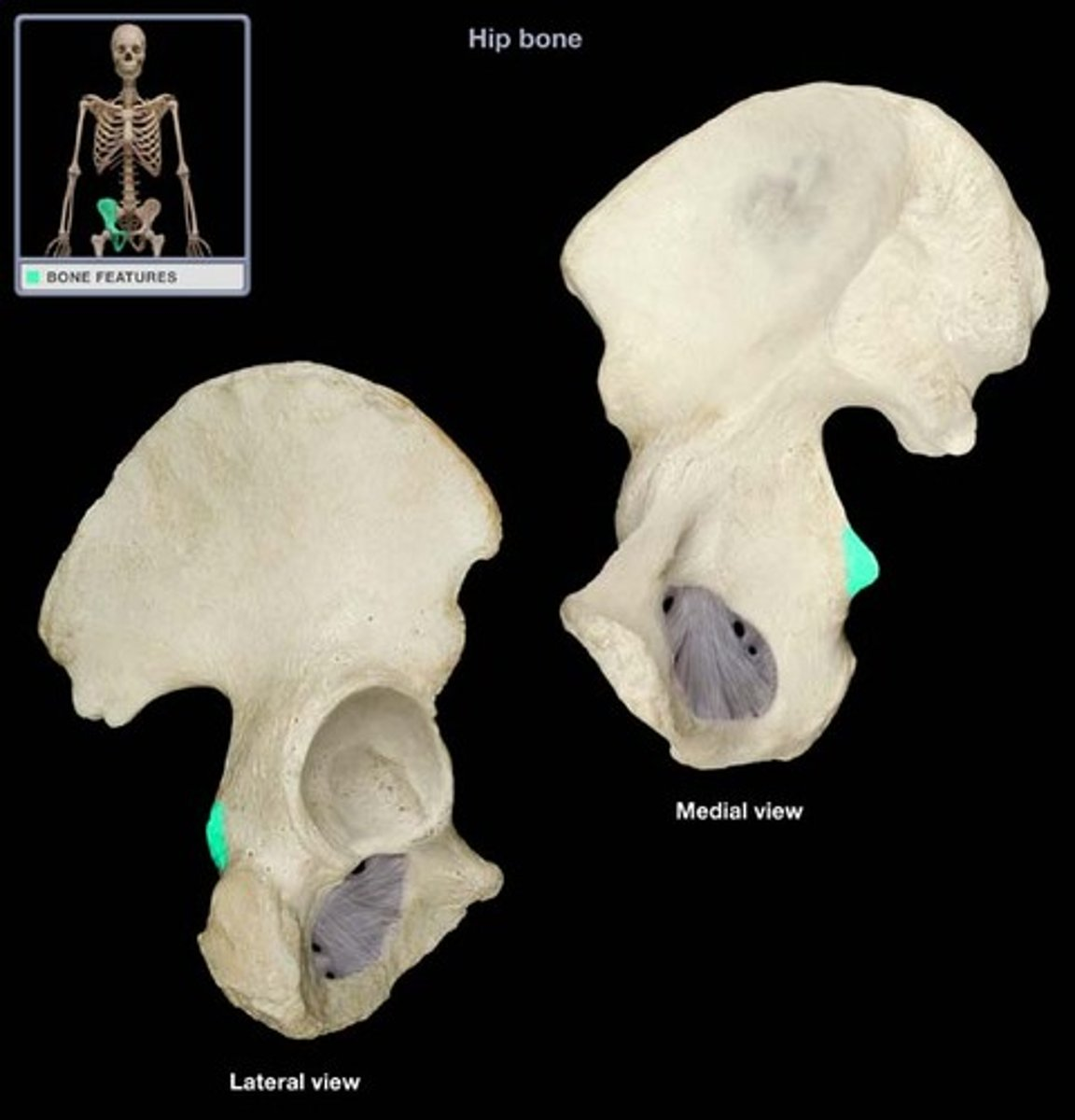
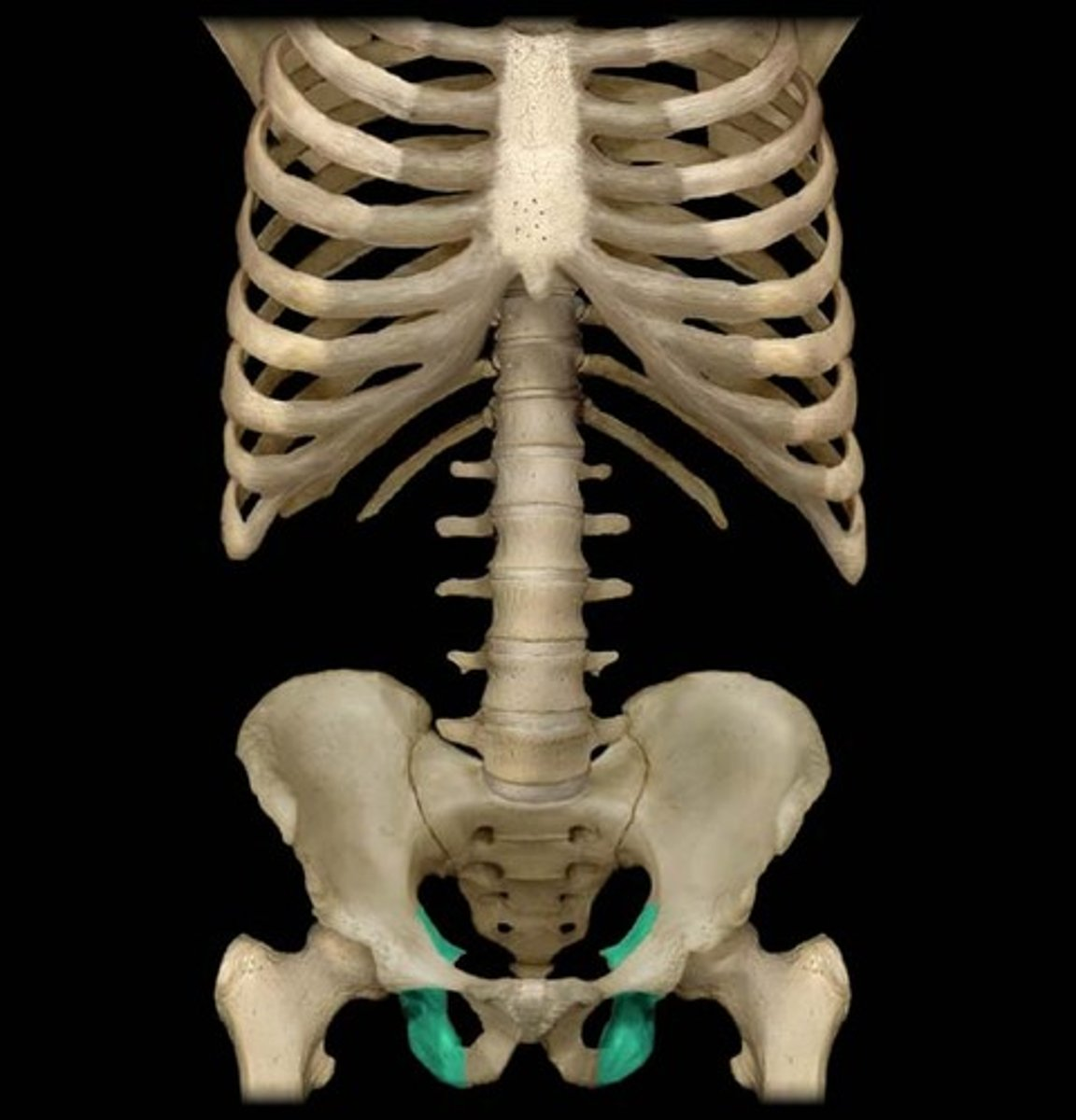
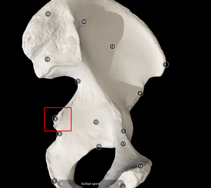
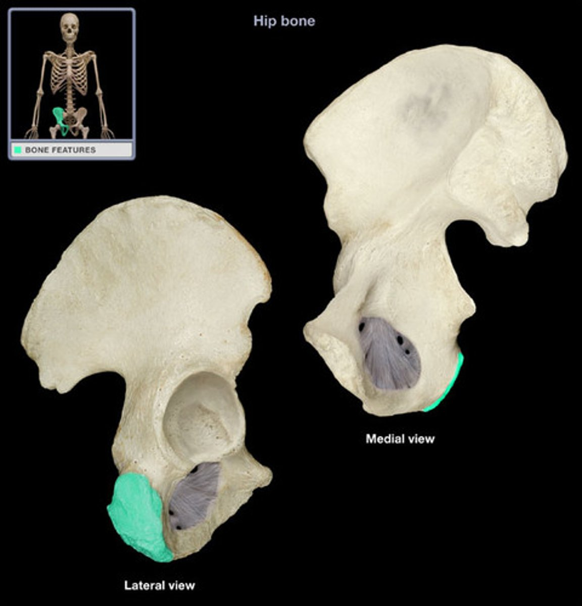
ischial spine - below the greater sciatic notch
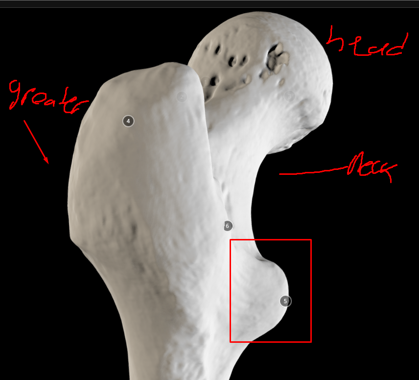
ischial tuberosity - big bump near the acetabulum fossa
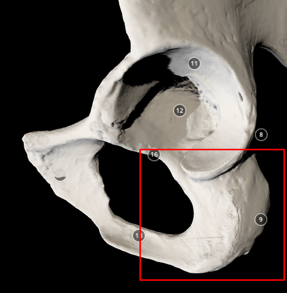

lesser sciatic notch
pubis bone
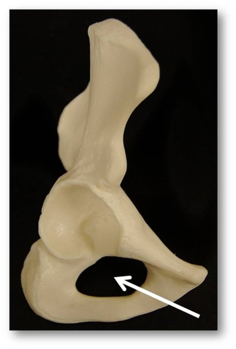
obturator foramen - big hole in the pubis and ischium
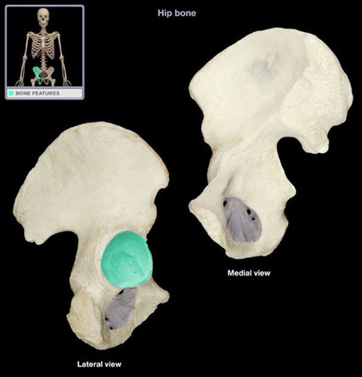
acetabulum - slot for head of femur
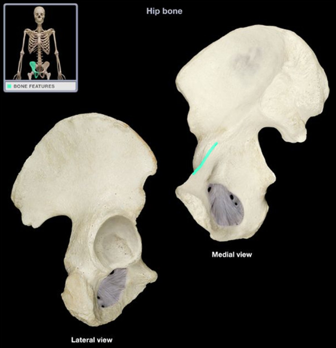
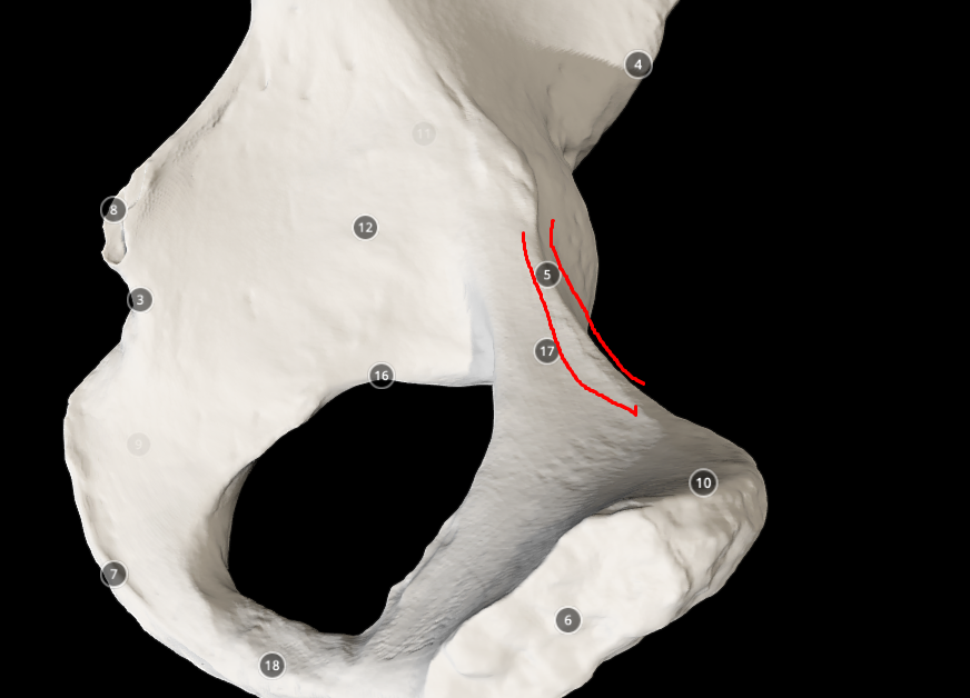
pectineal line - ridge like structure on the thinnest part
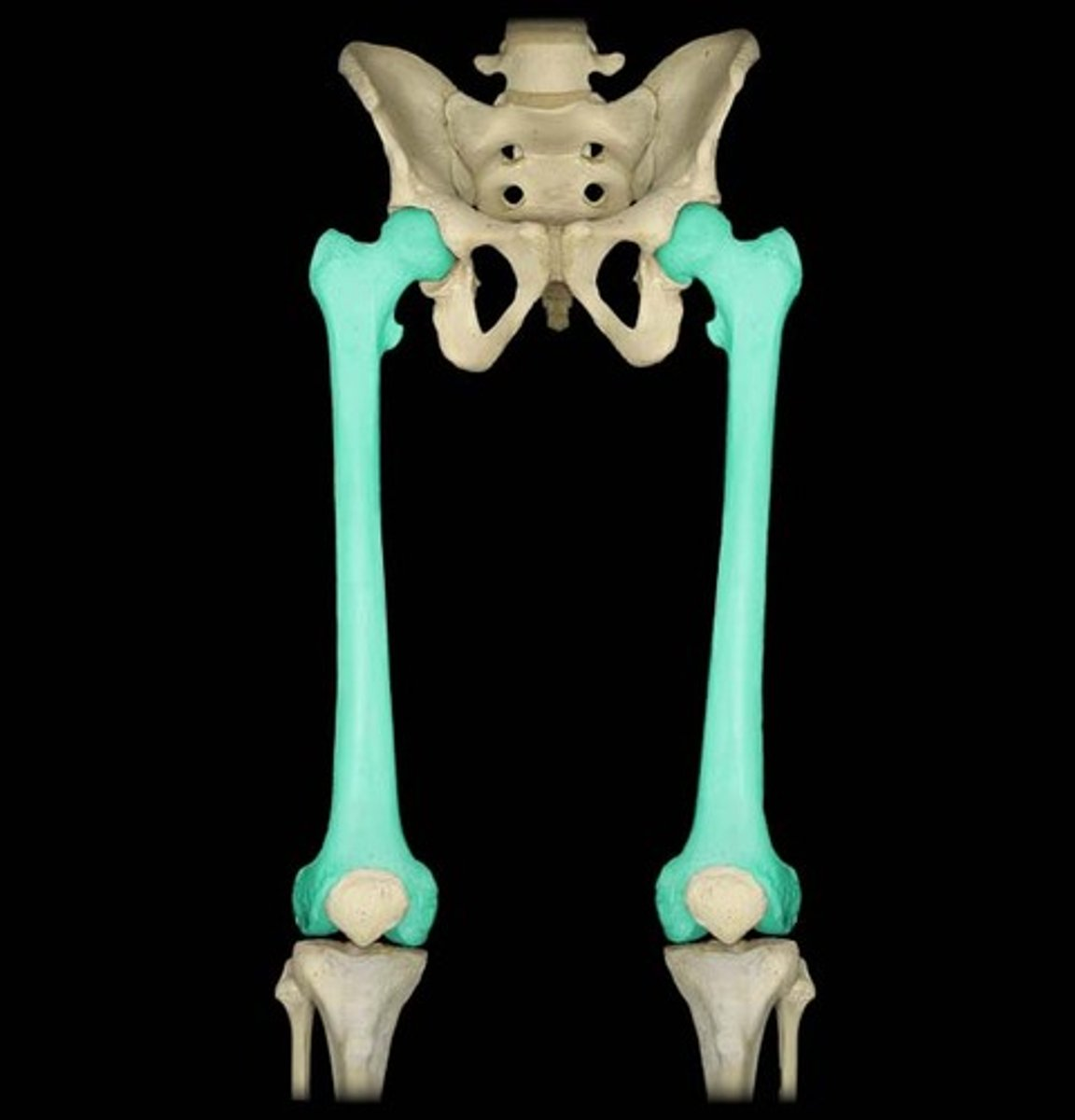
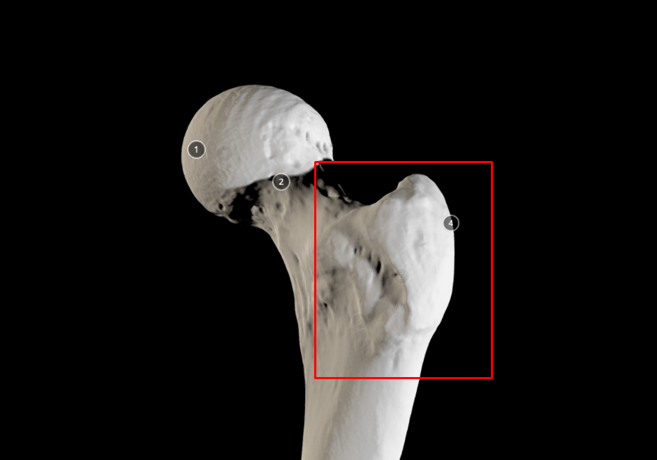
femur bone
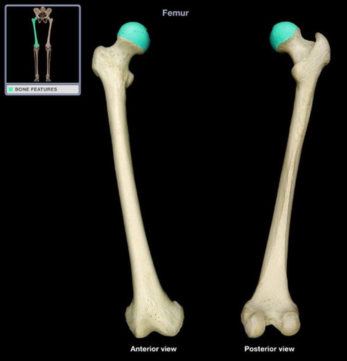
head of femur
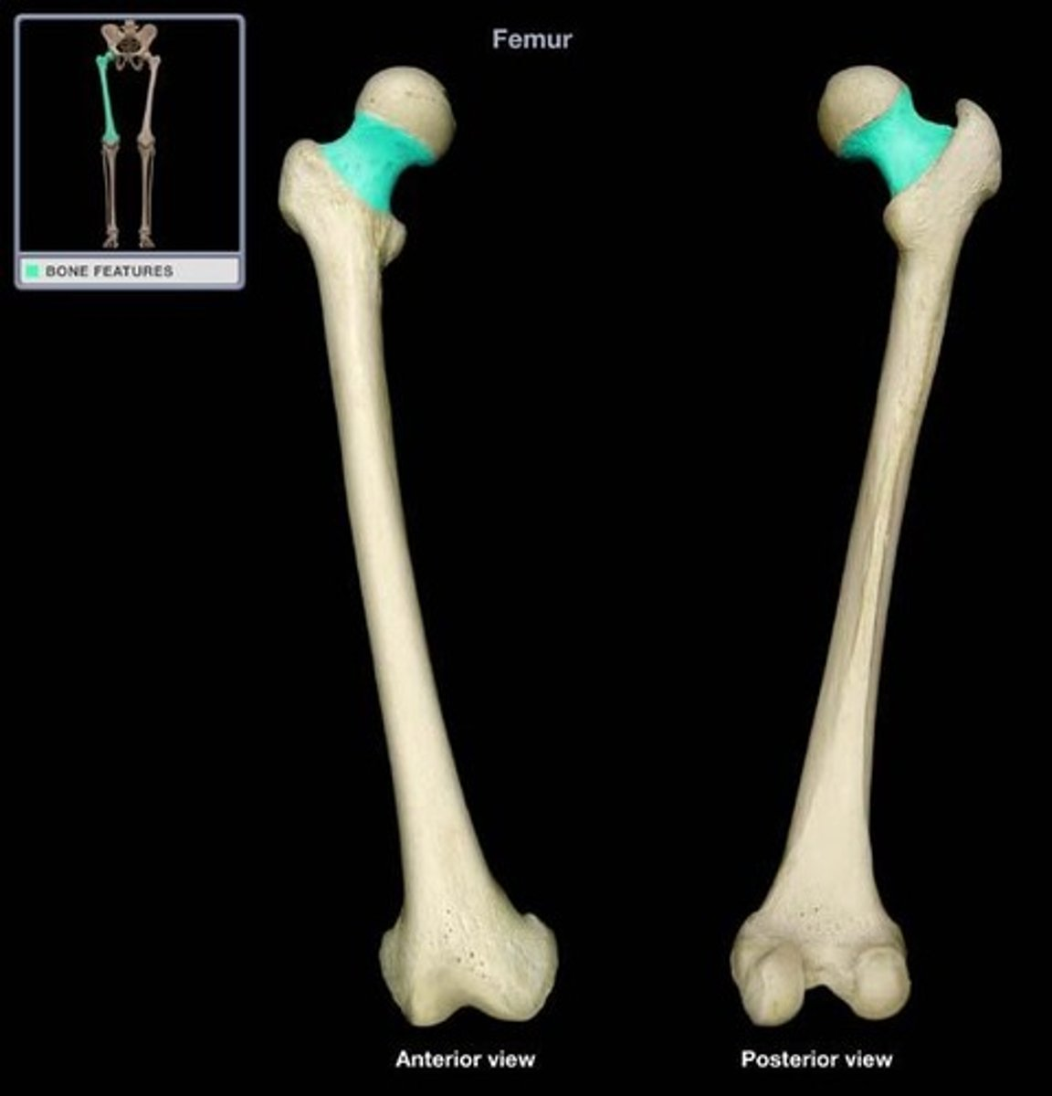
neck of femur
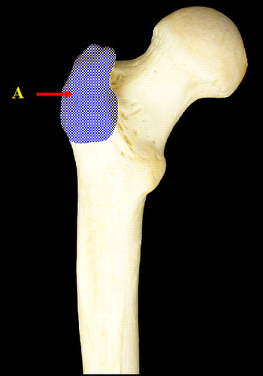
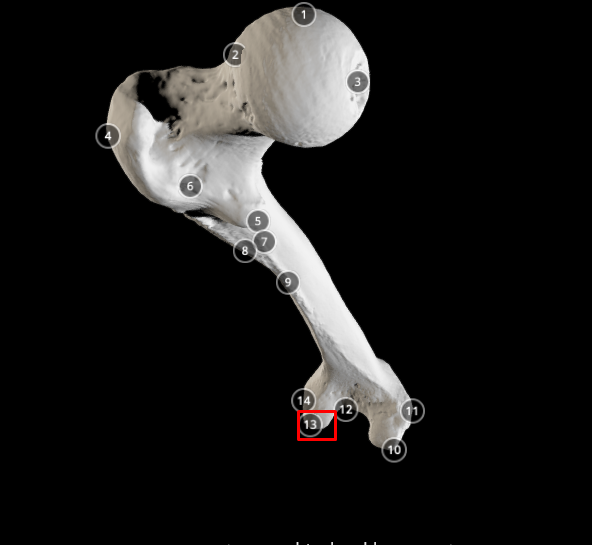
greater trochanter - big tip size structure next to the neck of femur
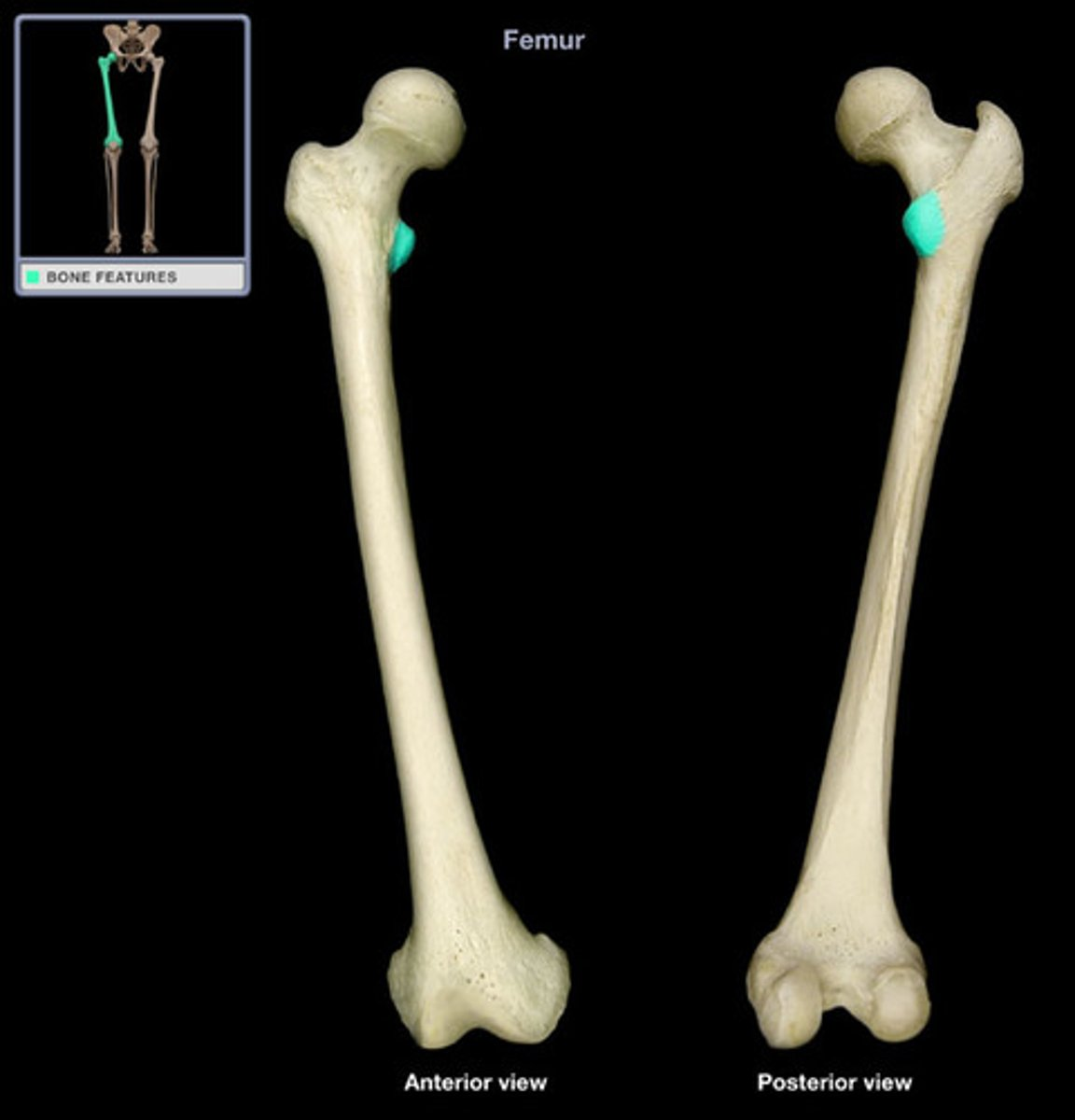
lesser trochanter - smaller bump at the bottom of the neck of femur
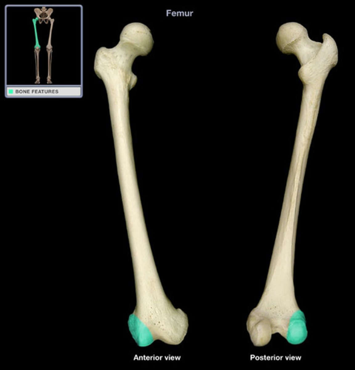
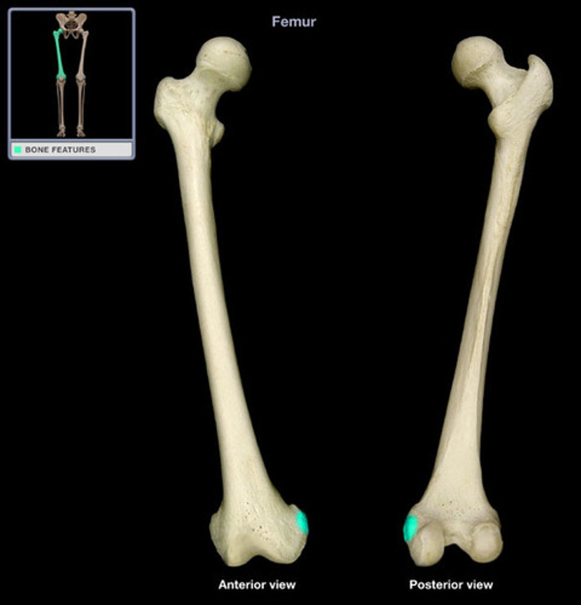
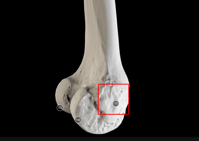
lateral condyle (of femur) - on the greater trochanter side
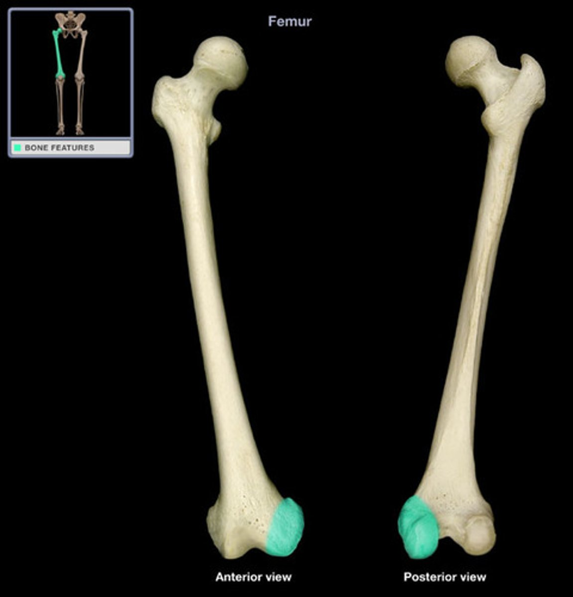
medial condyle (of femur) - on the head side
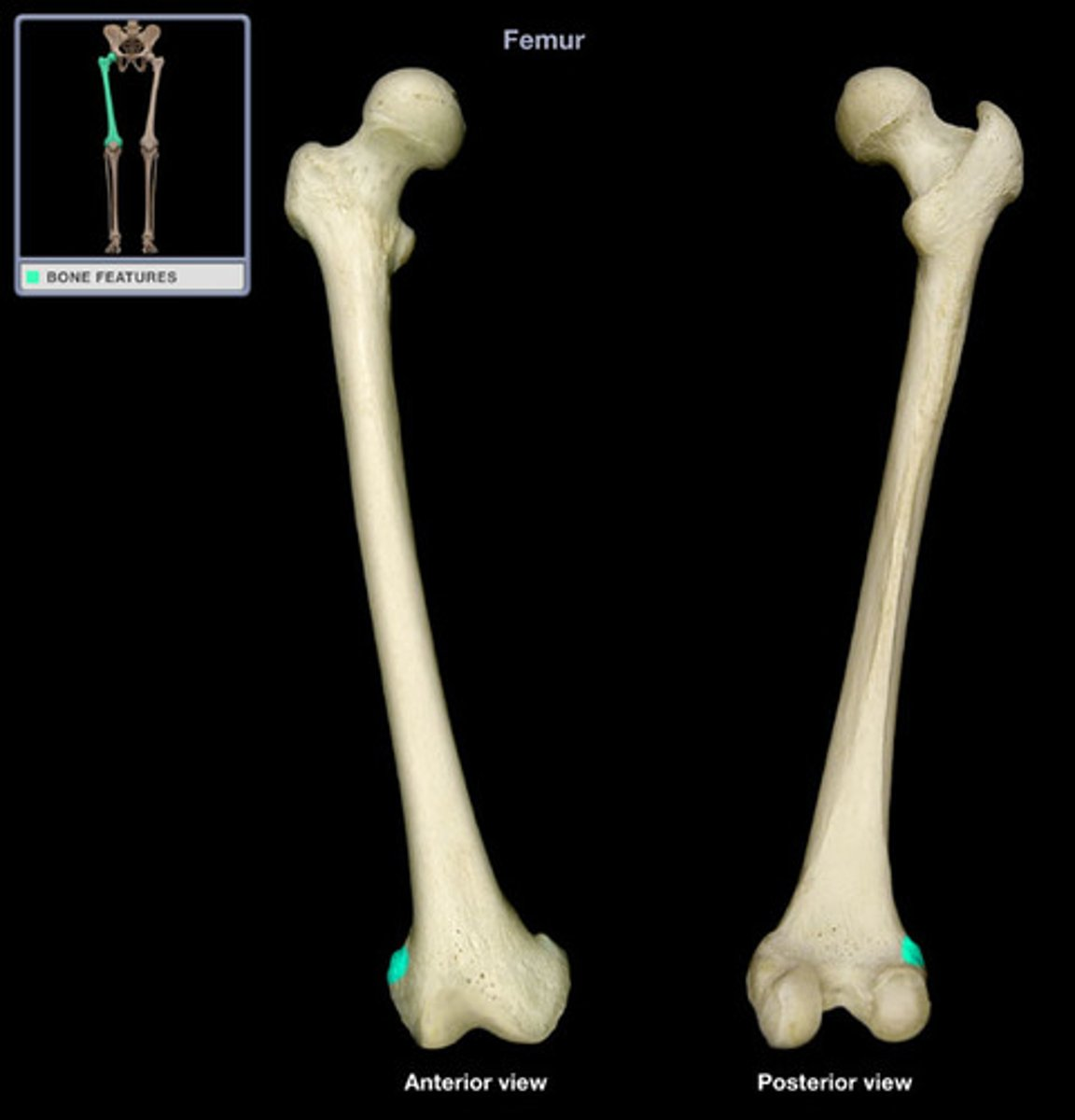
lateral epicondyle (of radius) - above the lateral condyle
medial epicondyle (of femur) - above medial condyle
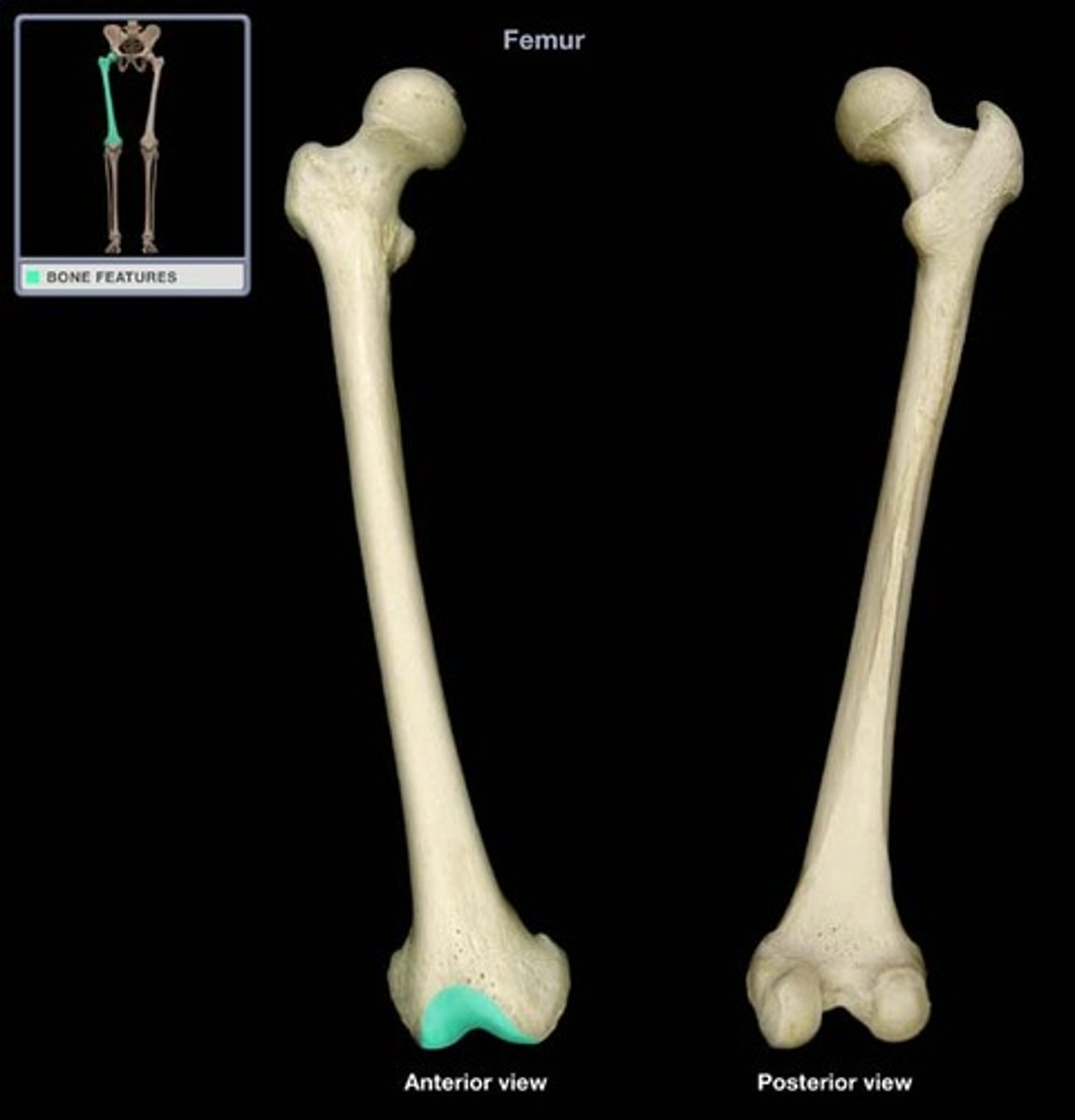
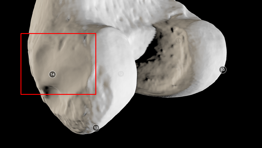
patellar surface - looks like an “n” ; also bones on one side, patellar surface on the other
popliteal surface - triangular groove

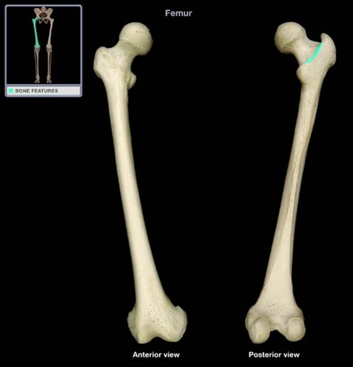
intertrochanteric line - boundary of between tranchanters and neck
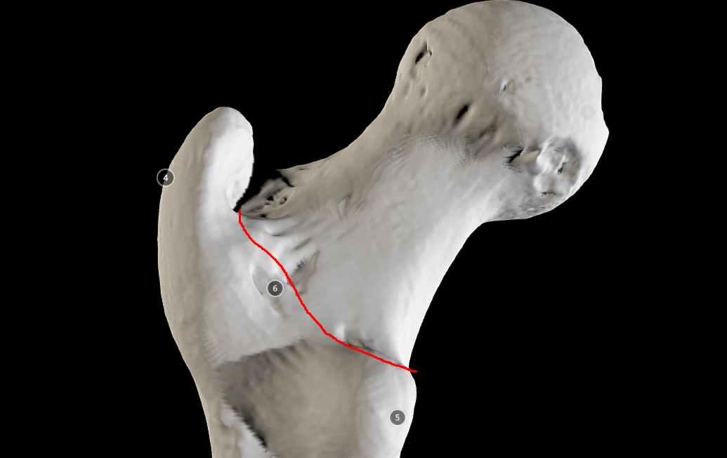
Intertrochanteric crest - ridge between greater and lesser trochanter
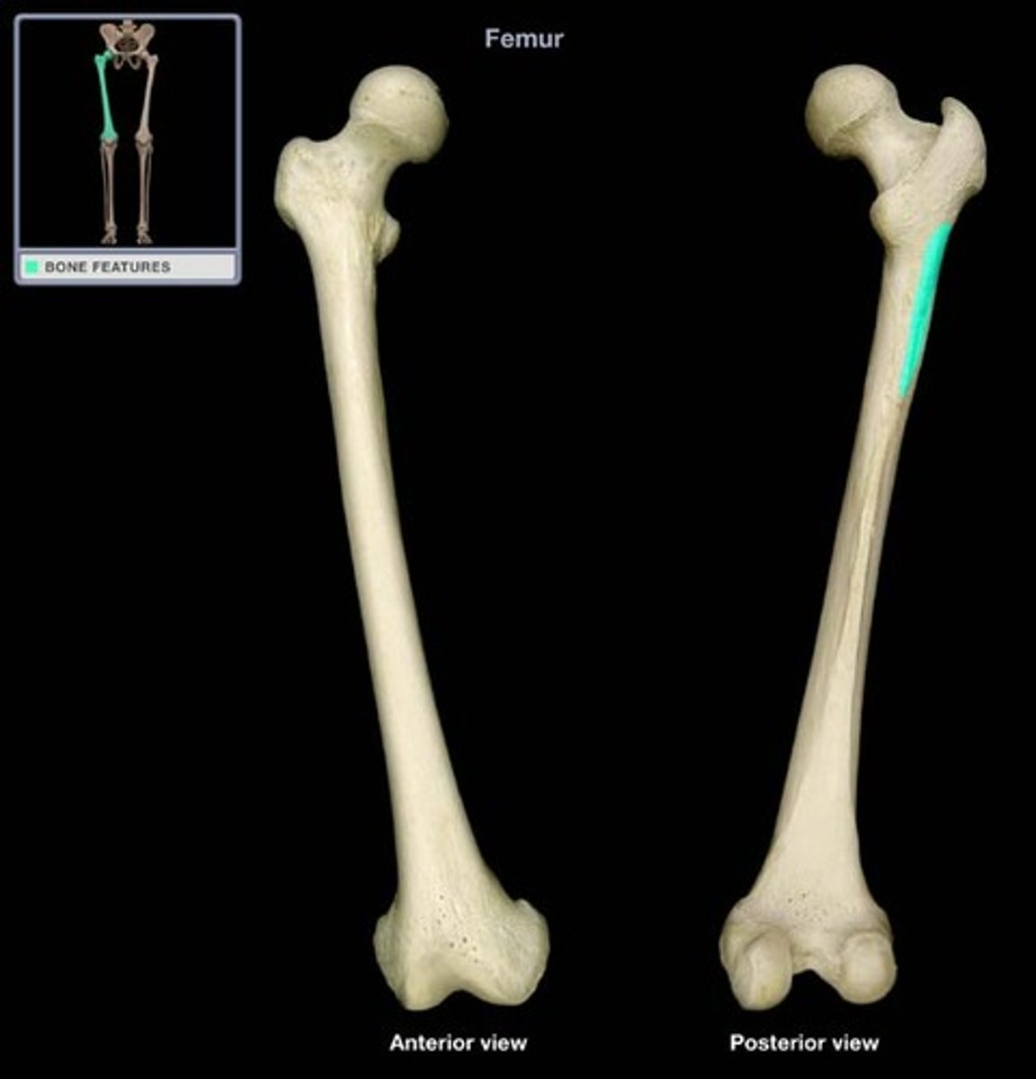
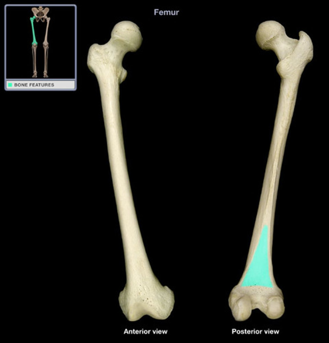
Gluteal tuberosity - buldge in line and below the greater trochanter
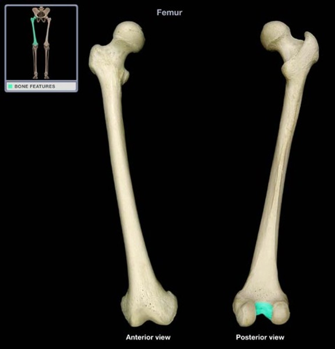
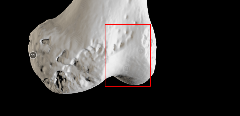
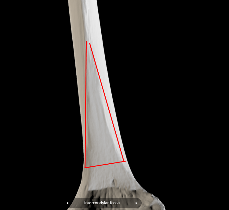
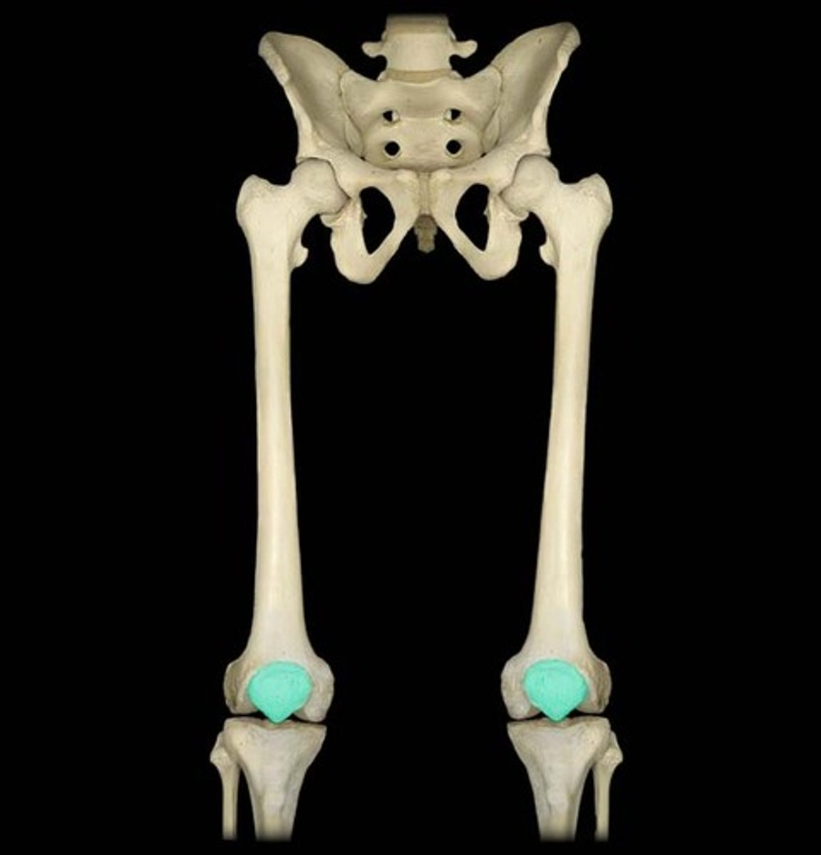
Intercondylar fossa - ditch between lateral and medial condyles
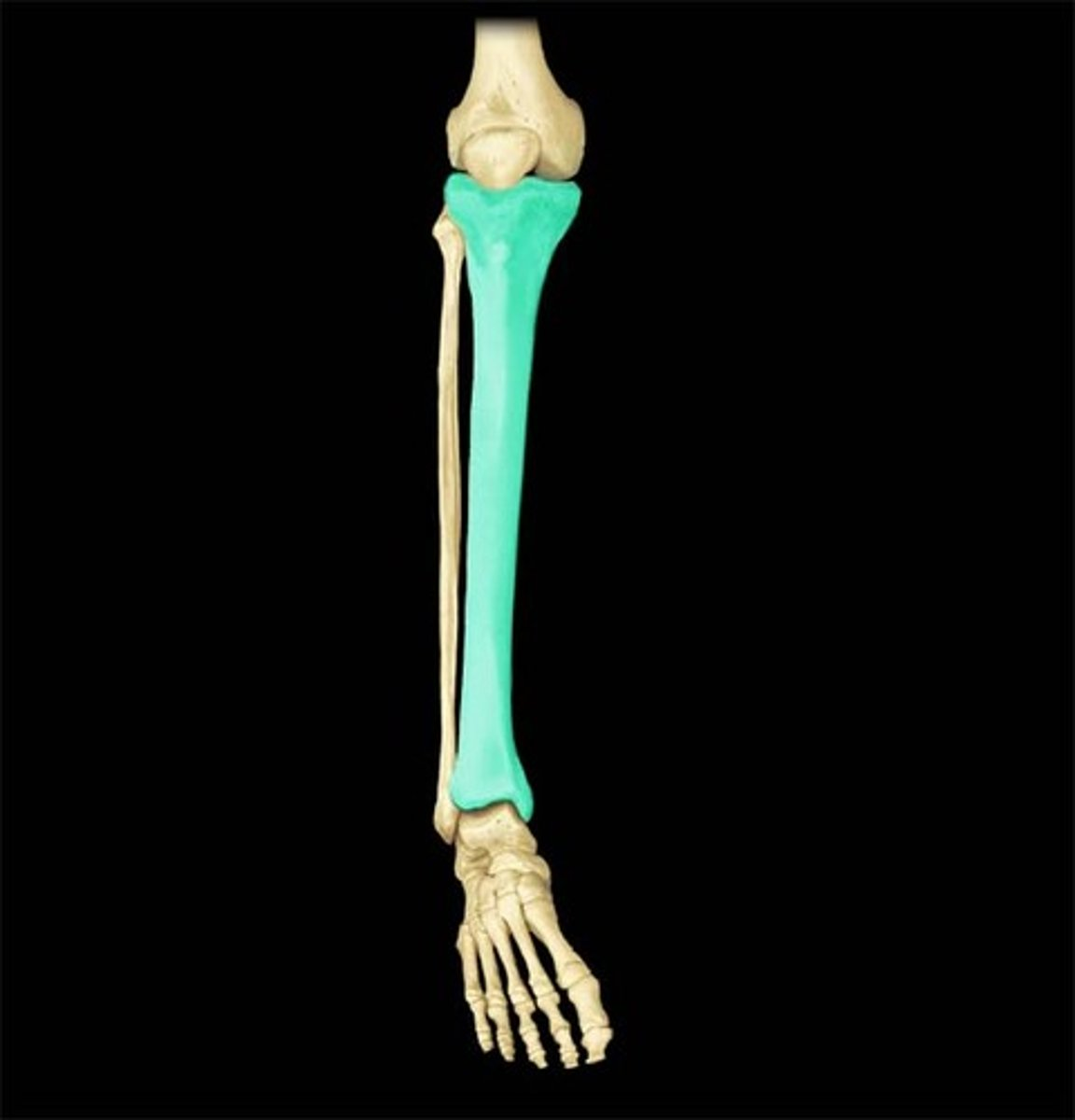
Tibia bone
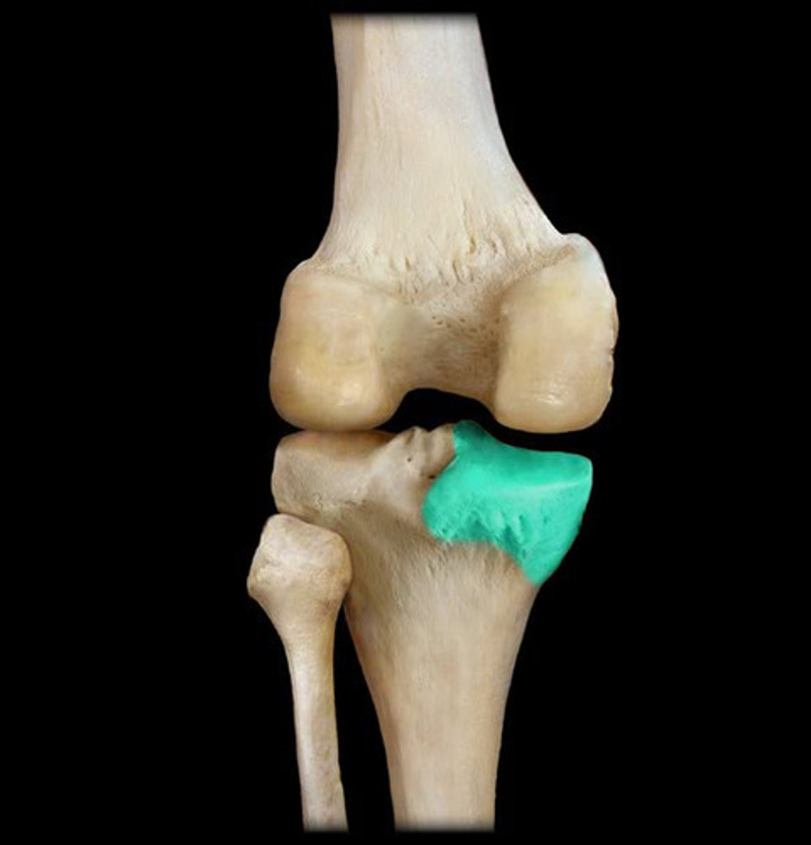
Medial condyle (of tibia) - connects with MC of femur (also head side of femur) ; ALSO LARGER
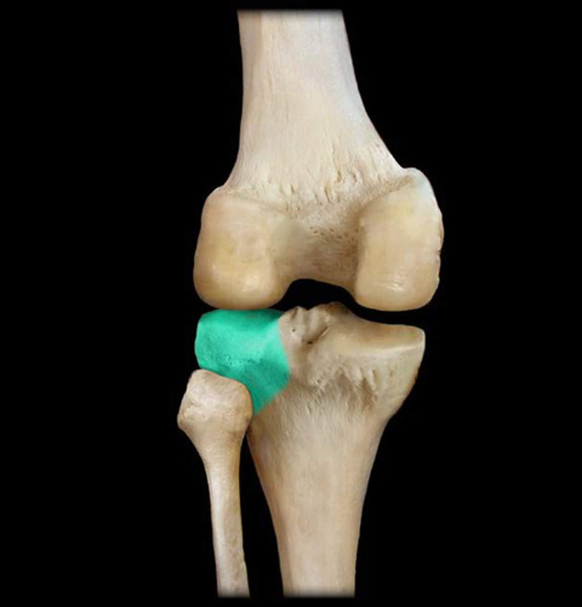
lateral condyle (of tibia) - connects with LC of femur and touches the fibula bone ; on the great trochanter side ; ALSO SMALLER than MEDIAL CONDYLE
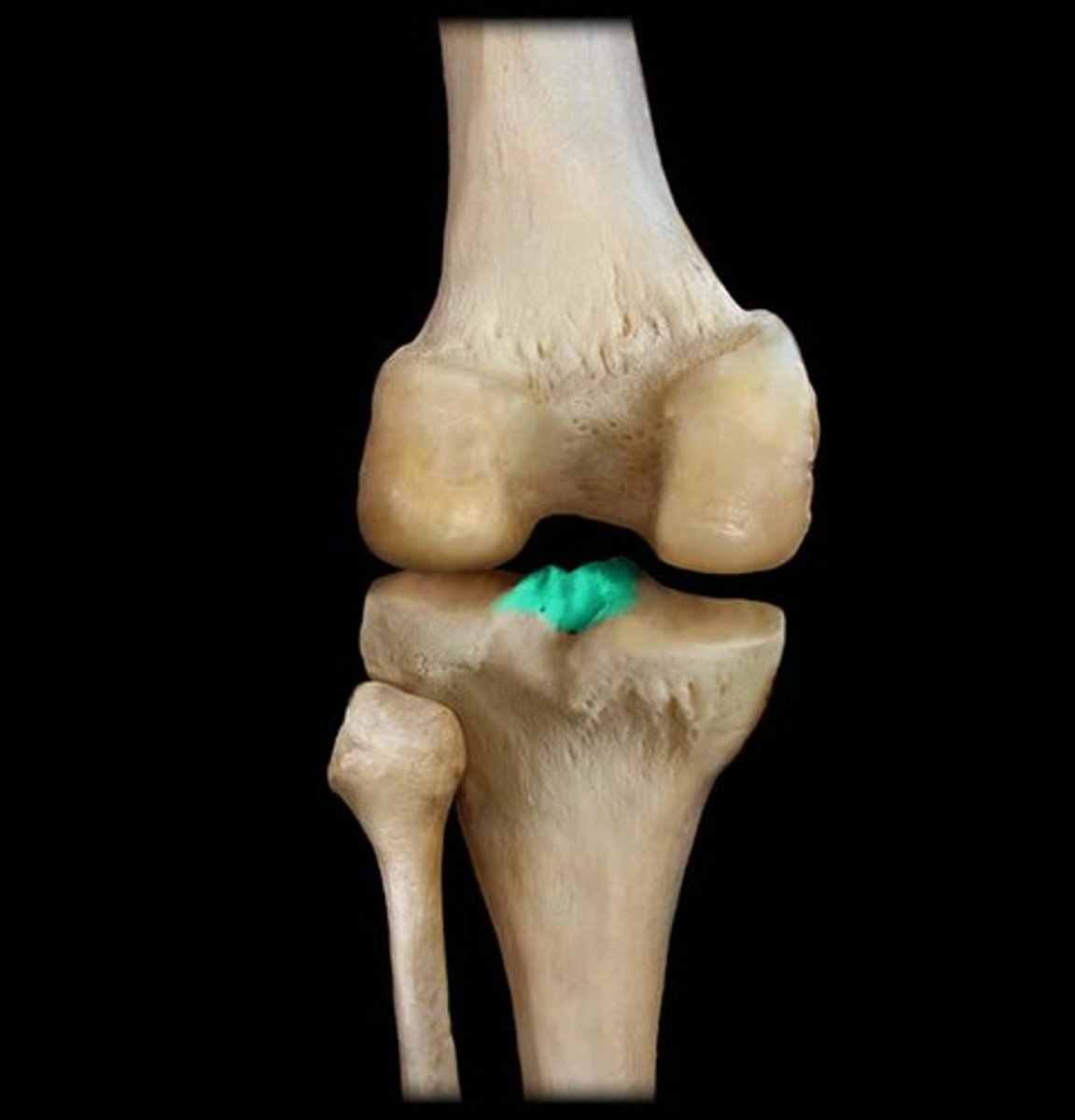
Intercondylar eminence - bump that lays between both condyles of the tibia
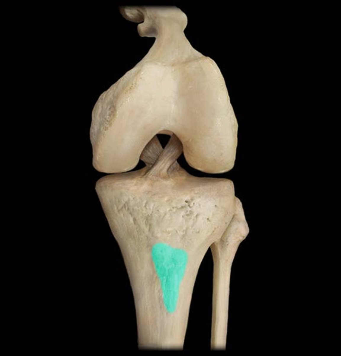
Tibial tuberosity - on the back side of the tibia
Medial malleolus - looks like the styloid process but on the tibia ; on the medial condyle side
FIBULA BONE
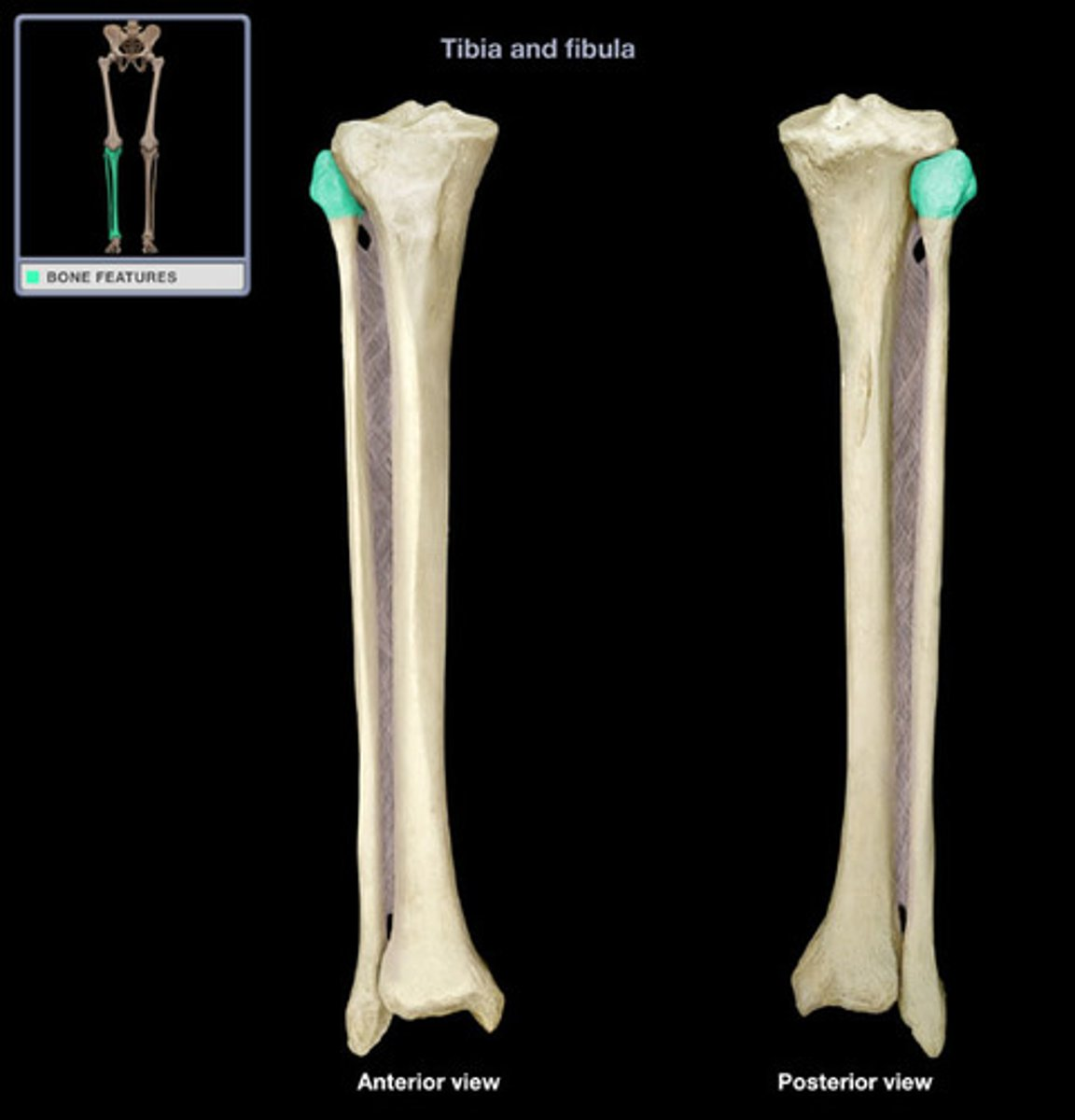
head of fibula
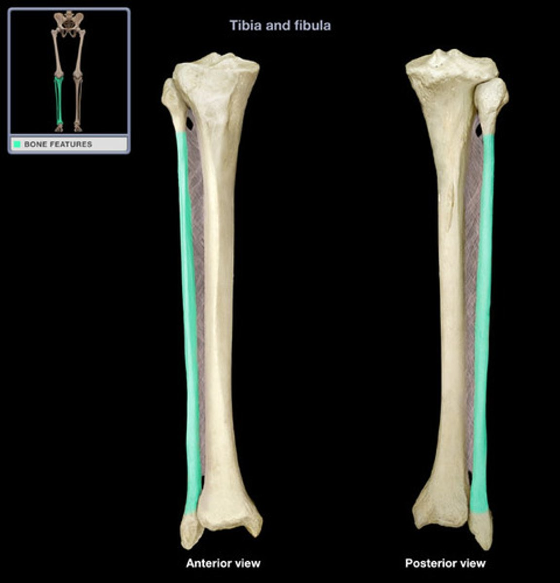
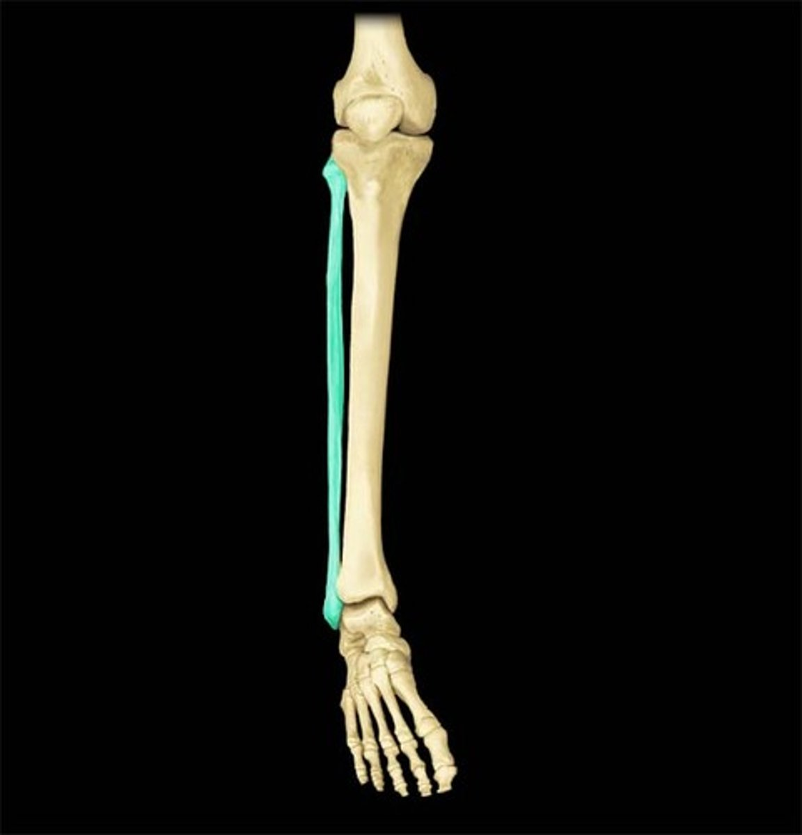
shaft of fibula
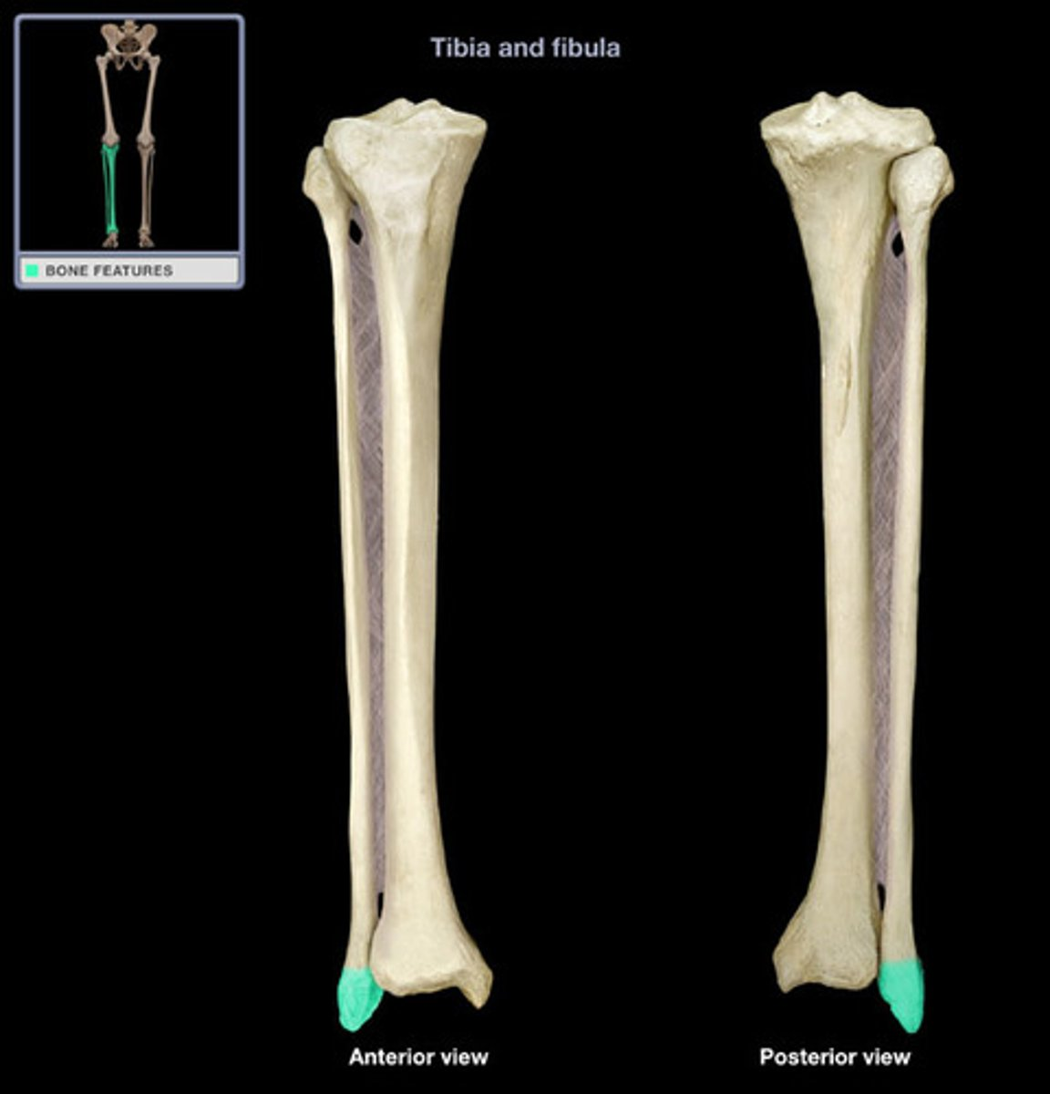
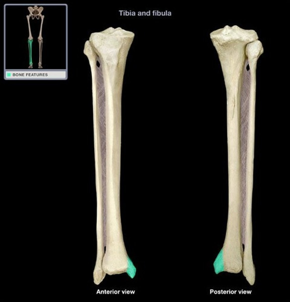
lateral malleolus
Patella bone
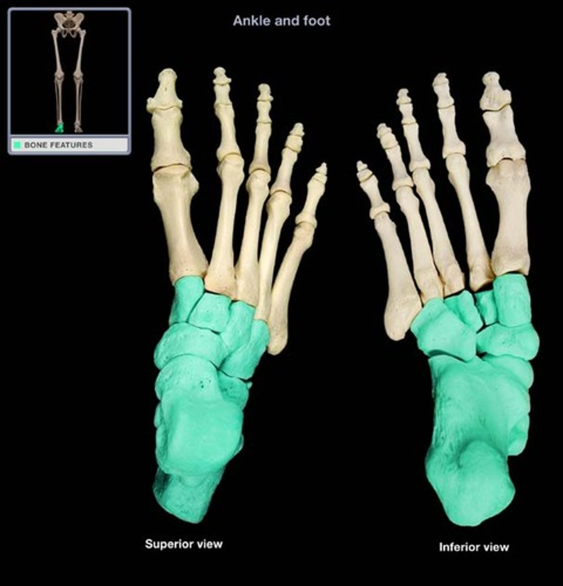
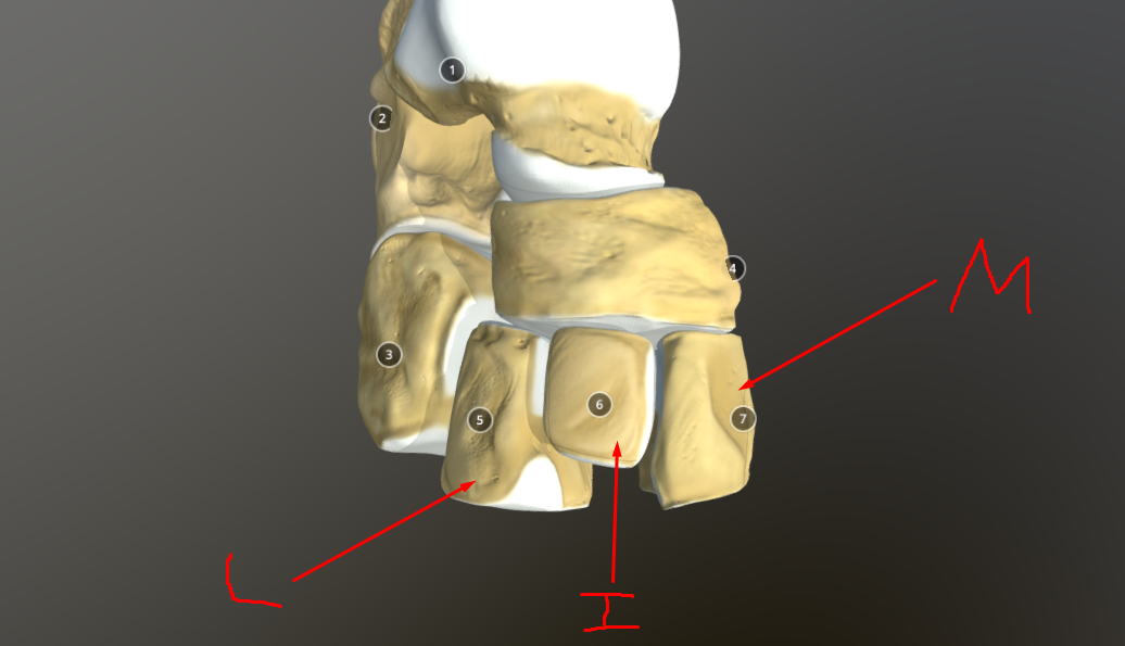
tarsal bones
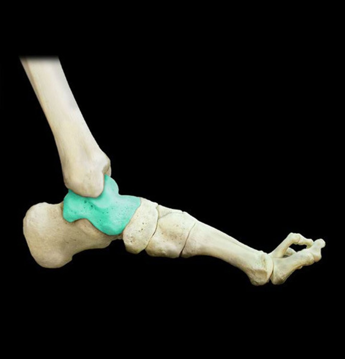
talus- on top of heel bone and connects to tibia
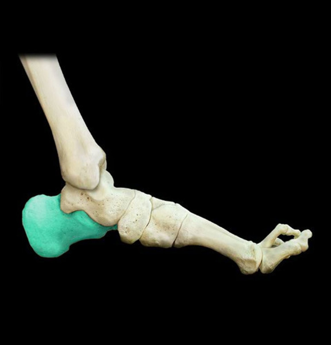
calcaneus
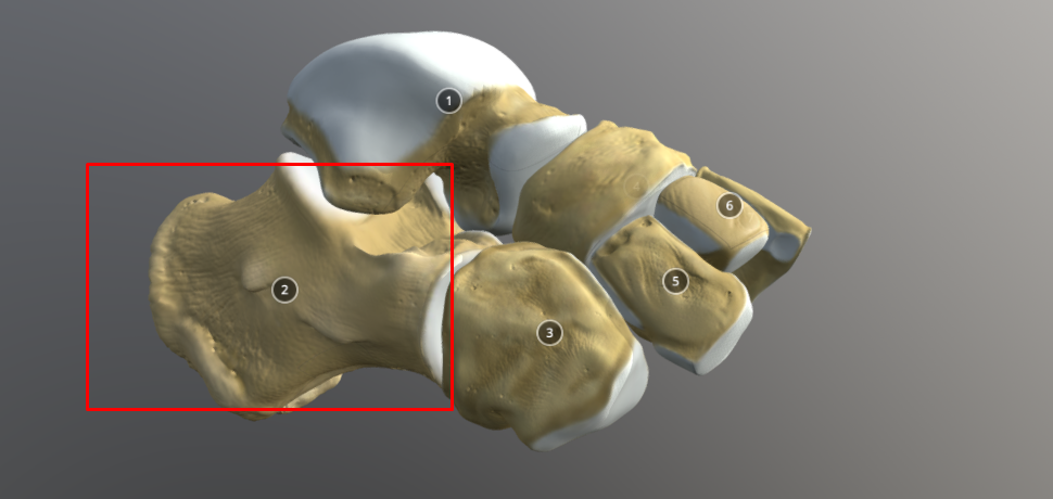
navicular - in front of talus
cuboid - in front of calcaneus
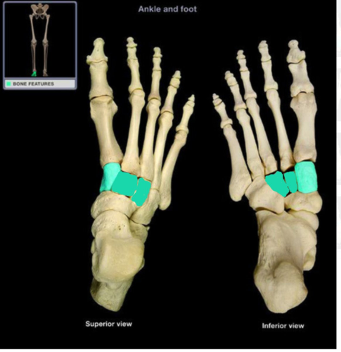
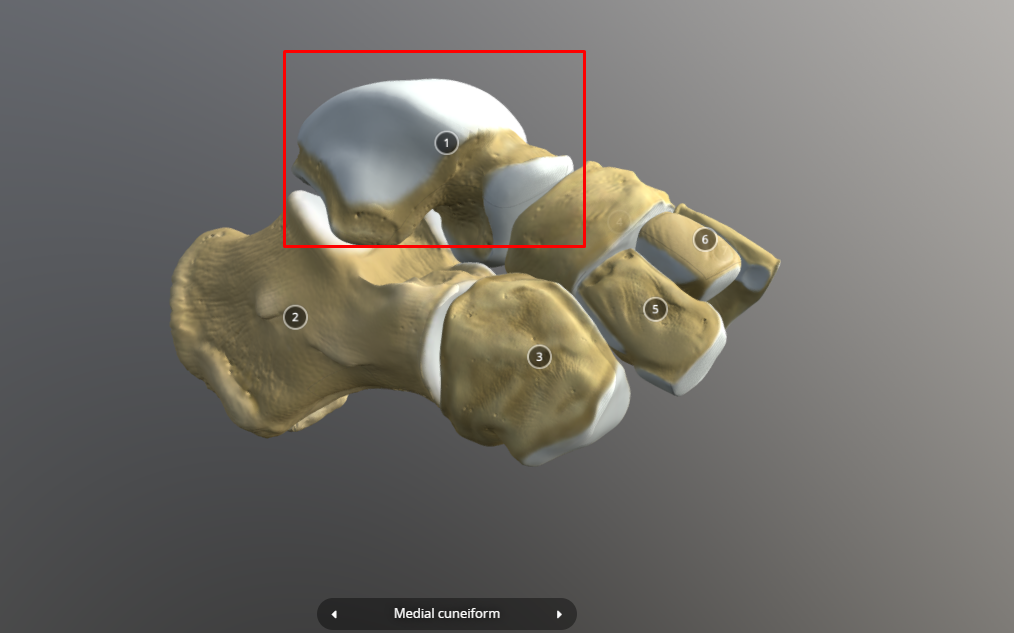
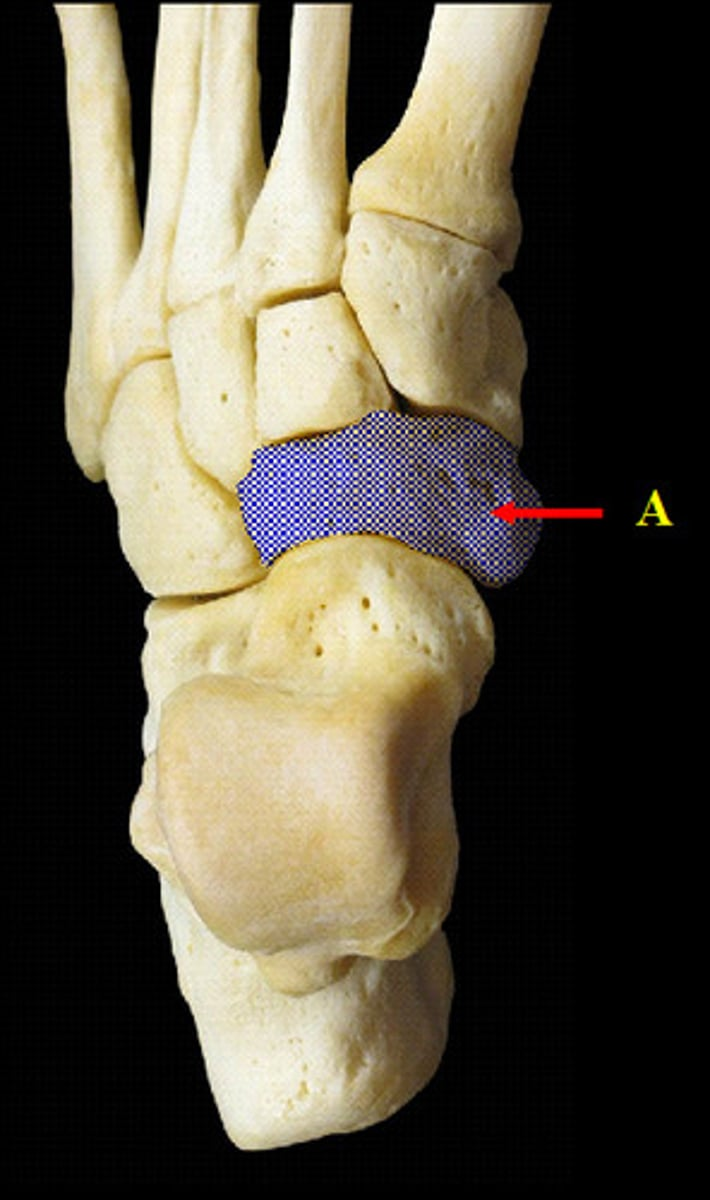
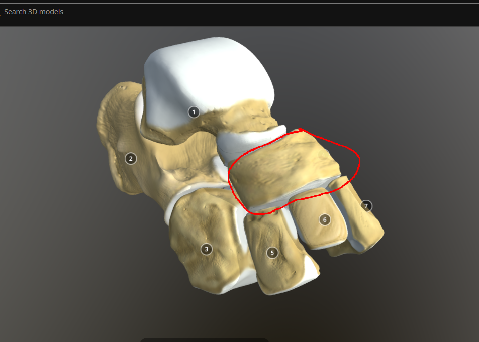
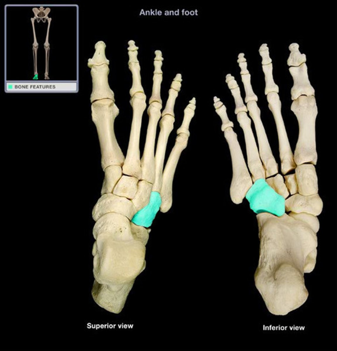
cuneiforms (medial, intermediate, lateral)
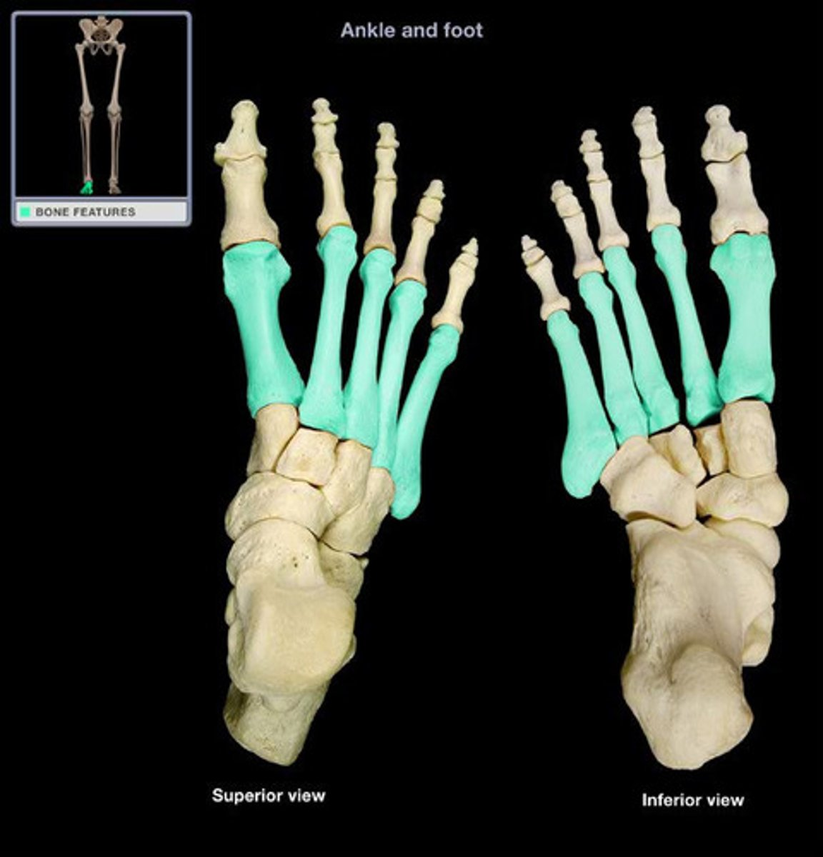
metatarsal bones
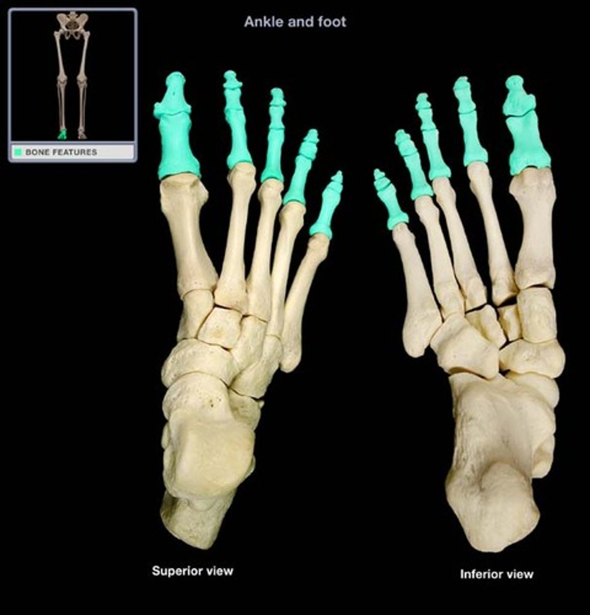
phalanges (toes)