Chapter 12 The Cell Cycle and Mitosis
1/54
Earn XP
Description and Tags
Use to study for the quiz tomorrow.
Name | Mastery | Learn | Test | Matching | Spaced | Call with Kai |
|---|
No analytics yet
Send a link to your students to track their progress
55 Terms
What are the three key role of cell division?
Asexual Reproduction
Growth and Development
Tissue Renewal|Repair
Give an example of asexual reproduction.
An amoeba, a single-celled eukaryote, divides into two cells, each an individual organism.
Give an example of growth and development.
A fertilized egg divides into a two-celled sand dollar embryo.
Give an example of tissue renewal or repair.
Dividing cells in bone marrow make new blood cells.
cell cycle
the cyclical process of a life of a cell from the time it is first formed from a dividing parent cell until its own division into two daughter cells or every cell forming from a preexisting cell
What does cell division result in?
Genetically identical daughter cells
genome
A cell’s endowment of DNA or genetic information
Contrast a human cell’s genome and a prokaryotic genome.
A prokaryotic genome is often a DNA molecule while eukaryotic genomes usually have multiple.
How many chromosomes are in a human somatic cell?
46
gamete
A reproductive cell that has had as many chromosomes as somatic cells
Name the two types of gametes.
Sperm and egg
How many chromosomes are in a human gamete?
23
chromatin
A complex of a DNA molecule and associated proteins that make up a chromosome
How many DNA molecules are in a human’s somatic cell?
46
What does a replicated chromosome with two sister chromatids look like?
An X
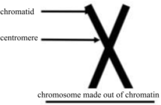

centromere
A region containing specific and repetitive chromosomal DNA sequences where the chromatid is attached most closely to its sister chromatid, the narrow waist on a chromosome

chromosome
A long, thin packaged gene-carrying structure consisting of chromatin

chromatid
A copy of a duplicated or original chromosome, a fibe
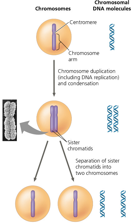
Summarize what occurs at the DNA level in each stage.
1. The uncondensed chromosomes in a eukaryotic cell are not duplicated. One chromosome has one chromatin containing one DNA molecule.
2. The chromosomes duplicate, becoming two sister chromatids, each containing the DNA molecule or a copy of it, from the original chromosome. They are connected along their length by sister chromatid cohesion.
3. Molecular and mechanical processes separate the sister chromatids into two chromosomes and distribute them to two daughter cells.
mitosis
the division of the genetic material in the nucleus or one nucleus into two genetically identical nuclei, usually followed by cytokinenis
How is mitosis different from cytokinesis?
Cytokinesis is the division of the cytoplasm, splitting the cell into two. Mitosis is the division of the genetic material.
What occurs in meiosis?
Gametes are produced through cell division, yielding non-identical daughter cells with one set of chromosomes, half as many as the parent cell.
How is the chromosome number of the daughter cells different from the parent cell?
The daughter cells have one set of chromosomes, half as many as the parent cell. In humans, the chromosome number goes from 46, two sets, to 23, one set.
By what process are the damaged cells in a would replaced?
Mitosis
By what process are eggs formed?
Meiosis
By what process does a zygote develop into a multicellular organism?
Mitosis
In what process are identical daughter cells produced?
Mitosis
What process reduces the chromosome number of daughter cells?
Meiosis
A hedgehog has ninety chromosomes in its somatic cells. How many chromosomes did the hedgehog inherit from each parent?
45
A hedgehog has ninety chromosomes in its somatic cells. How many chromosomes are in each of the hedgehod’s gametes?
45
A hedgehog has ninety chromosomes in its somatic cells. How many chromosomes will be in each somatic cell of the hedgehog’s offspring?
Ninety
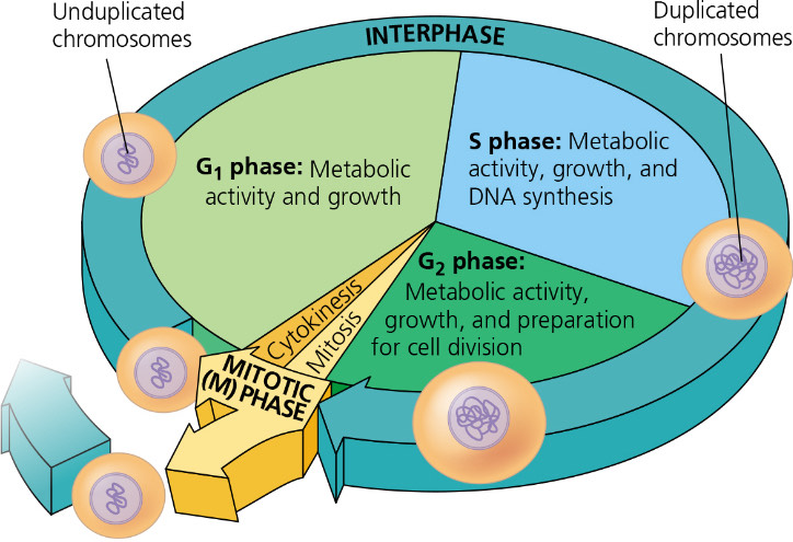
What does the mitotic phase alternate with?
Interphase in the cell cycle
Give a brief explanation of what happens in each phase or part of the cell cycle.
G1: A cell grows
S: A cell grows and copies its chromosomes
G2: A cell grows and prepares for cell division.
M: A cell divides.
What are the four components of the mitotic spindle?
Fibers that are made of microtubules and associated proteins
Asters
Centrosomes
What is the source of the components of the mitotic spindle?
Centrosome
In animal cells, the assembly of spindle microtubules starts at the centrosome. What is another name for a centrosome?
Microtubule-organizing center
What does a centrosome contain?
Two centrioles
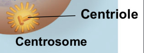
Describe what happens to a centrosome during interphase.
In animal cells, two centrosomes remain together near the nucleus.
kinetochore
a structure of specialized proteins assembled on specific sections of chromosomal DNA at each centromere
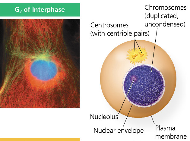
Interphase
During G2, a nuclear envelope encloses the nucleus, which contains one or more nucleoli. Two centrosomes, regions in animal cells that organize the microtubules of the spindle, form by the duplication of one centrosome. Each contains two centrioles.
During S Phase, chromosomes are duplicated, which cannot be individually distinguished because they are not condensed.
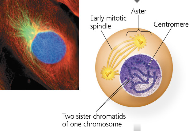
Prophase
Nucleoli disappear.
Chromatin fibers condense and coil tightly into discrete chromosomes observable with a LM. Each duplicated chromosome appears as two identical sister chromatids joined at their centromere and, in some species, all along their arms by cohesins or sister chromatid cohesion.
The mitotic spindle, named for its shape, starts to form. It is composed of microtubules that extend from the centrosomes. The radial array of shorter microtubules are asters or stars. The centrosomes move away from each other, propelled partly by the lengthening microtubules between them.
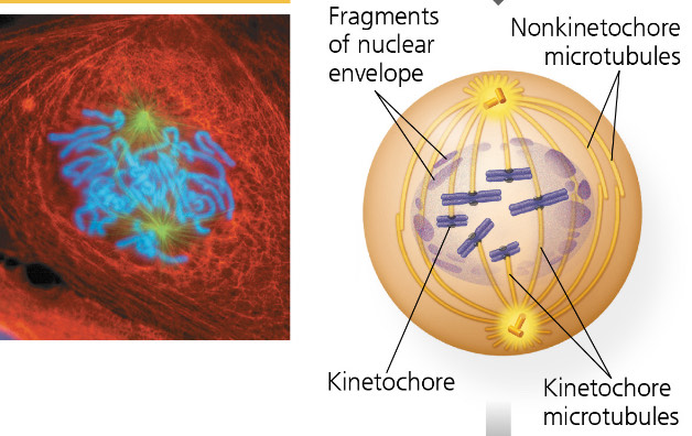
Prometaphase
The nuclear envelope fragments, allowing the microtubules to invade the nuclear area. Microtubules attach to kinetochores, becoming kinetochore microtubules, which jerk the chromosomes back and forth. Nonkinetochore microtubules interact with those from the opposite pole of the spindle, lengthening the cell.
Chromosomes condense. Each chromatid forms a kinetochore at its centromere, thus, there are two kinetochore per chromosome.

Metaphase
The centrosomes reach opposite poles of the cell.
The chromosomes arrive at the metaphase plate, a plane that is equidistant between the spindle’s two poles. The chromosomes’ centromeres lie at the plate. For each chromosome, the kinetochore microtubules of the sister chromatids come from opposite poles of the cell.
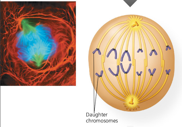
Anaphase
It is the shortest stage of Mitosis, often lasting a few minutes.
It starts when the cohesion proteins are cleaved, allowing each pair of sister chromatids to part suddenly. Each becomes a chromosome. The two daughter chromosomes move toward opposite ends of the cell as their kinetochore microtubules shorten. The centromeres move ahead of the arms at a rate of about 1µm|min because the microtubules are attached to that region.
The cell elongates as the nonkinetochore microtubules lengthen.
In the end, the two ends of the cell have equal and complete collections of chromosomes.
Telophase
Two daughter nuclei form. Two nuclear envelopes arise from the fragments of the original one and other parts of the endomembrane system. Nucleoli appear.
The chromosomes become less condensed.
The remaining spending microtubules are depolymerized.
Mitosis completes.

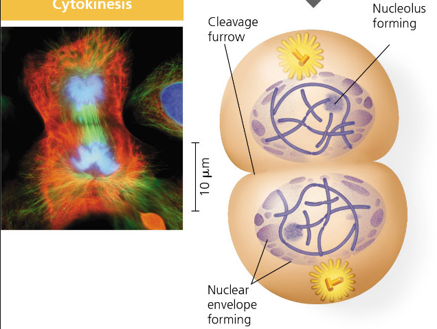
Cytokinesis
The cytoplasm divides, which is usually well under way by late telophase, causing two daughter cells to appear shortly after mitosis.
In animal cells, a cleavage furrow forms, pinching the cell in two.
Explain the difference between kinetochore and nonkinetochore microtubules.
Unlike nonkinetochore microtubules, kinetochore microtubules attach to the kinetochore.
What is the function of kinetochore microtubules?
One kinetochore is captured by microtubules, moving the chromosome towards the pole from which the microtubules extended. Microtubules from the opposite pole attaching to the kinetochore on the other chromatid stops this movement. The chromosomes move back and forth, until they settle on the metaphase plate, like a tug-of-war that ends in a draw.
What is the function of nonkinetochore microtubules?
They elongate, overlapping and interacting with other nonkinetochore microtubules from the opposite pole of the spindle, elongating the cell.
At which end do kinetochore microtubules shorten?
kinetochore
Explain the data that supports where kinetochore microtubule shortening occurs.
An experiment determined that motor proteins on the kinetochores walk the chromosomes along the microtubules, which depolymerize at their kinetochore ends after the motor proteins pass, releasing tubulin subunits. This is the Pacman mechanism because of its resemblance to the arcade game character that moved by eating all the dots in its path. As the chromosomes move poleward, the microtubule segments on the kinetochore side of a mark region on the kinetochore microtubules between a spindle pole and the chromosomes shortened, while those on the spindle pole side stayed the same.
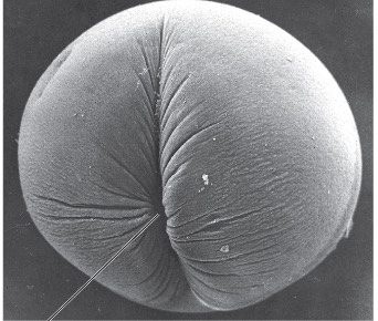
Describe cytokinesis in an animal cell.
It is a process called cleavage. The first sign is the cleavage furrow, a shallow groove on the cell surface near the old metaphase plate. On the cytoplasmic side of the furrow is a contractile ring of actin microfilaments associated with molecules of the protein myosin. The actin microfilaments interact with the myosin, contracting the ring. This is like the pulling of a drawstring. The cleavage furrow deepens until the cell is pinched in two, producing two seperate cells, each with a share of cytosol, organelles, and other subcellular structures.
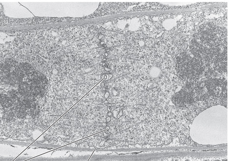
Describe cytokinesis in a plant cell.
Plants have cell walls. During telophase, a cell plate is formed. This enlarges until its surrounding membrane fuses with the plasma membrane along the perimeter of the parent cell. Two daughter cells result, each with its own plasma membrane. A new cell wall arises from the content of the cell plate between the daughter cells.
How is the cell plate formed?
Vesicles from the Golgi Apparatus move along microtubules to the middle of the cell, where they unite. Cell wall materials collect as the cell plate grows.
What is the source of the material for the cell plate?
Vesicles from the Golgi Apparatus