Microbiology Lab Exam 1
1/180
There's no tags or description
Looks like no tags are added yet.
Name | Mastery | Learn | Test | Matching | Spaced | Call with Kai |
|---|
No analytics yet
Send a link to your students to track their progress
181 Terms
Effective hand washing is important for minimizing direct person-to-person and indirect contact transmission of pathogens. It is critical for laboratory professionals to minimize transmission to ______, ________, and contamination of __________.
others, inoculation of oneself, and contamination of cultures
In which other professions would reducing the transmission of potential pathogens be considered important?
healthcare
food handlers
veterinary clinics
In the experimental results provided in this lab, which areas of the hands were typically the most difficult to clean, based on the individual’s hand-washing technique?
area under the fingernails
back of hands
between fingers
Lab 1-1 GLO Germ Hand Washing Exercise Overview
Goal: training aid for people to learn how to wash their hands more effectively
Steps: hands are covered with lotion (contains minute plastic particles), rub hands together to spread lotion, use UV to see spread, wash hands, use UV again to see remaining germs present
What is the single most effective way to prevent the spread of germs?
handwashing
How does the refractive index of immersion oil compare with that of glass?
Immersion oil has the exact same refractive index as glass
they have the same optical density meaning that the speed of light through immersion oil is equivalent to glass
this prevents the loss of light due to refraction
How do you calculate the total magnification of a specimen seen through a compound light microscope? Give an example using the 4X objective.
Total magnification = magnification of objective lens x magnification of ocular lens
ex: 4x objective lens → 4 × 10 = 40x total magnification
Differentiate between the following terms: parfocal, binocular, and compound microscope
Parfocal = the microscope’s ability to stay in focus when switching between objective lenses. requires only minor adjustments (fine focus knob and illumination).
Binocular = a microscope with 2 eye pieces; provides 3D vision.
Compound Microscope = uses more than 1 lens to magnify an object. has objective lenses & ocular lens to achieve higher magnification.
Differentiate between the terms magnification and resolution
Magnification = involves enlarging an object’s appearance.
Resolution = clarity of an image; improves with smaller limit of resolution.
Limit of resolution (resolving power) = measurement of how far apart 2 points must be for the microscope to view them as separate. determined by wavelength/numerical aperture of condenser & objective lens. the smaller the wavelength, the lower the limit of resolution and the higher the actual resolution.
Why is it advisable to start with the low power lens first when viewing a slide through the microscope?
To allow you to explore the slide and find + center the object that you are planning to study
also to ensure that the specimen is actually present
If you want to observe bacteria on a stained smear prep slide, at what point during the procedures would you add immersion oil to a microscope slide?
After focusing and centering the specimen under the 40x objective, but before switching to the 100x objective (oil immersion lens).
Lab 3-1 Introduction to the Light Microscope Overview
Purpose: To become familiar with the operations and limitations of a light microscope. Also, to examine two practice slides and learn about microscope functionality.
Steps: properly transport & clean microscope, turn on, start in 4x, move stage with mechanical slide holder, adjust diaphragm for light, coarse-focus knob, then 10x, coarse-focus, 40x, fine-focus, oil, 100x
when done, make sure to lower the stage and remove the slide
Refraction
The bending of light as it passes through the objective lens from the specimens that produces a magnified real image
What is the best limit of resolution achieved by a light microscope?
About 0.2 micrometers
Field of view
the visible area when looking through the microscope eyepiece at the specimen
Dark-field microscopy
special condenser used so light reflected off the specimen only enters the objective lens
brightly lit specimen against dark background
Phase contrast microscopy
special optical components to exploit subtle differences in the refractive indices of water & cytoplasmic components to produce contrast
light waves
Fluorescence Microscopy
fluorescent dye that emits fluorescence when illuminated with UV.
Ocular lenses
to remagnify (10x) the image produced by the objective lens
Revolving nosepiece
holds the objective lenses & allows for rotation to change power
Objective lenses
to magnify the specimen and produce a real image
4x = scanning power
10x = low power
40x = high dry power
100x = oil immersion power
Arm
attaches body of the microscope to its base
Stage Clip
holds the microscope slide in place on the stage and prevents it from moving while the specimen is being viewed
Coarse-Focus Knob
moves the stage up and down the focus the image
brings specimen into general focus
Fine-Focus Knob
slightly moves the stage up and down for focusing
handles small & precise movement of the specimen
Mechanical Stage Adjustment Knob
controls the movement of the stage and allows you to position the slide within the microscope’s field of view
Stage
flat platform where the microscope slide is placed for viewing
Lamp
light source so that the specimen can be viewed
Iris Diaphragm
controls the amount of light entering the condenser
Condenser Lens
concentrates the light and makes illumination of the specimen more uniform
What are the three domains? Identify whether each domain contains prokaryotes or eukaryotes.
Bacteria = prokaryotes
Archaea = prokaryotes
Eukarya = eukaryotes
What is the domain, classification group (protozoa, algae, or fungi), and mode of nutrition (heterotrophic or autotrophic) for paramecium?
domain = unicellular eukarya
classification group = protozoa (ciliates)
mode of nutrition = heterotrophic → searches for food in marine/freshwater
What is the domain, classification group (protozoa, algae, or fungi), and mode of nutrition (heterotrophic or autotrophic) for euglena?
domain = eukarya
classification group = algae
mode of nutrition = photosynthetic autotroph when light is available but heterotroph when light is not available. Known as a mixotroph.
What is the domain, classification group (protozoa, algae, or fungi), and mode of nutrition (heterotrophic or autotrophic) for spirogyra?
domain = eukarya
classification group = filamentous algae
mode of nutrition = photosynthetic autotroph
What is the domain, classification group (protozoa, algae, or fungi), and mode of nutrition (heterotrophic or autotrophic) for stentor?
domain = eukarya
classification group = protozoa (cilitates)
mode of nutrition = heterotrophic → in freshwater, can swim, but normally are anchored to a surface feeding
larger than paramecium
Identify the domain and kingdom for molds and yeasts. What are the differences between molds and yeast?
domain = nonmotile eukarya
kingdom = fungi
molds = multicellular; grow as hyphae
yeasts = unicellular; reproduce by budding
Differentiate between the terms hyphae and mycelia as they relate to fungi.
hyphae = the individual, thread-like filaments of cells that mold grow as
mycelia = interwoven masses of hyphae; the vegetative portion of a fungi
What are the four supergroups that eukaryotes are divided into?
Unikonta = closer to root of phylogenic tree; composed of heterotrophs & includes amoebas + fungi
Excavata = unicellular species that usually have a feeding groove excavated from one side of the cell and possess one or more flagella
Supergroup Archaeplastida = all descendants from an ancestor than engulfed cyanobacteria; all autotrophs with chloroplasts
SAR Supergroup = very diverse group joined because of strong DNA similarity of entire genome
Amoeba overview
two subgroups = gymnamoebas (classical amoebas) & entamoebas (some are parasitic)
move by forming pseudopods which also engulf their food
Fungi overview
nonmotile eukaryotes
cell wall is made of chitin
absorptive heterotrophs → secrete exoenzymes into the environment & absorb digested nutrients
Ciliates Overview
characterized by cilia covering their outer surface that provide motility when they sweep back and forth. cilia also lines the oral groove & sweeps food particles inward to be engulfed
paramecium & stentor
Lab 3-3 Microscopic Examination of Eukaryotic Microbes
Purpose: to observe representative samples of eukaryotes from the four subgroups and give students practice using the microscope, measuring specimens, and making wet-mount preparations.
Steps:
if solid media: add a loopful of water and mix a loopful of specimen on the slide
if liquid media: add a loopful of the broth directly to the slide
lower coverglass on slide at an angle so as not to trap bubbles
observe under microscope
Define the term pure culture
Pure culture = when a culture contains a single species.
What are the common types of media used to culture bacteria? What are the common tools used in a microbiology lab?
Media -
Broths = used to grow microbes when fresh cultures or large numbers of cells are required
Agar slants = used to grow stock cultures that can be refrigerated after incubation & maintained for several weeks
Plated media = used for obtaining isolation of species, differential testing, & quantifying bacterial densities
Tools -
inoculating loops / inoculating needles
pipettes
glass spreading rod
Give three reasons why it is important to practice aseptic technique in a microbiology lab
to prevent aerosol formation
to maintain a clean culture
to keep yourself safe
In what orientation should agar petri plates always be incubated? Why? Where on a petri plate should identification labels be written?
Agar petri plates should always be incubated upside down (lid facedown and agar up). This is so if condensation forms, it will fall onto the lid and not place moisture on the surface of the agar or interfere with microbial growth.
Identification labels should be written on the bottom of the petri dish, not on the lid. This is so that the label remains with the specimen even if the lid is lost also because the lid can rotate and ruin the orientation of the specimen on plate.
What does the term turbidity mean? Is this term used to describe the appearance of broth media, solid agar media, or both?
Turbidity = the cloudiness or opaqueness of a liquid caused by the presence of suspended particles.
the more turbid = the more microorganisms present
Turbidity is ONLY used in broth media
Culture
a medium that contains living microbes
Aseptic transfer
Transfer without contamination
What is the most important operational risk factor for aseptic transfers?
a procedure’s potential to release microorganisms into the air as aerosols and droplets
What practices should we adhere to during aseptic transfer for our safety?
minimize the potential of contamination (do the labs away from your book)
be organized
place all media tubes in a test tube rack when not in use whether they are sterile or not
take your time
never hold a tube culture by its cap
hold the inoculating loop or needle like a pencil in your dominant hand and relax
When do we use a fishtail inoculation?
We use a fishtail inoculation with an agar slant culture because we are seeding the agar in a wavy pattern resembling to movement of a fish tail.
Lab 1-4 Common Aseptic Transfers and Inoculation Methods
Purpose: to introduce and practice techniques for transferring microorganisms from one medium to another without contaminating the culture or the surrounding environment.
Steps:
broth → broth
label tube → grab new loop → flick specimen broth to ensure mixing → open cap of specimen with little finger and hold → grab one loopful of solution → replace cap → open cap of sterile broth → place inoculum in → replace cap → place inoculum in disinfectant
agar slant → agar slant
label tube → grab new loop → open cap of specimen with little finger and hold → grab a small amount of specimen in a fishtail method → replace cap → open cap of sterile slant → place inoculum using fishtail method → replace cap → place inoculum in disinfectant
same idea for a plate!
What does the term ubiquity mean?
Ubiquity = refers to the state of being present everywhere or in many places simultaneously
In microbiology, ubiquity often describes organisms that are widespread & found in various environments
Explain the terms mutualism, commensalism, and opportunistic pathogens
Mutualism = when a microbe actually benefits a host
Commensalism = when a microbe is benefitting but is having no significant effect on their host
Opportunistic pathogens = they are capable of producing a disease state if introduced into a suitable part of the body
What can you conclude from studying Figures 2.28 and 2.29 in your Leboffe lab manual, which feature the bacterium serratia marcescens?
The figures showcase the effect of incubation time on pigment production and the effect of incubation temperature on pigment production.
Serratia marcescens has increased pigment with a longer incubation period (ex: red after 48 hours) and has increased pigment at lower temperatures (ex: red at 25 degrees C)
Give some examples of the following categories used to describe the morphological characteristics of bacterial colonies growing on solid media → size
Size = the measurement of a colony’s dimensions
is the diameter if the organism is circular
is the length & width if the organism is shaped otherwise
Give some examples of the following categories used to describe the morphological characteristics of bacterial colonies growing on solid media → shape
Round (circular); Irregular; Punctiform (tiny pinpoint)

Give some examples of the following categories used to describe the morphological characteristics of bacterial colonies growing on solid media → margin
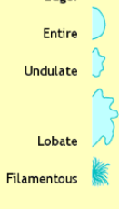
Entire (smooth); Undulate (wavy); lobate (lobed); filamentous; rhizoid (branched-like roots)
Give some examples of the following categories used to describe the morphological characteristics of bacterial colonies growing on solid media → surface
smooth; rough; wrinkled (rugose); shiny; dull
Give some examples of the following categories used to describe the morphological characteristics of bacterial colonies growing on solid media → texture
moist; mucoid (sticky); butyrous (buttery); dry
Give some examples of the following categories used to describe the morphological characteristics of bacterial colonies growing on solid media → elevation
flat; raised; convex; pulvinate (very convex); umbonate (raised in center)

Give some examples of the following categories used to describe the morphological characteristics of bacterial colonies growing on solid media → other features
color & optical properties → opaque (cannot see through) & translucent (light passes through)
What are free-living organisms?
Free-living = they do not reside on or in a specific plant or animal host and are not known to cause disease (nonpathogenic)
Saprophytes definition
decompose organic matter
What are pathogens?
If the microorganism causes damage to their host, that is, if they cause disease.
What is a reservoir?
Any area, including sites outside of the host organism, where a microbe resides and serves as a potential source of infection.
Lab 2-1 Ubiquity of Microorganisms
Purpose: To demonstrate the ubiquitous nature of microorganisms (that they are present nearly everywhere) and the ease with which many can be cultivated.
Steps: label the plates 1-8; conduct each petri dish as it pertains to the label (air, hair, vending machine, etc); if needed, use a cotton swab and gently streak the plate in a back and forth pattern; invert the plates and incubate them for 24-48 hours
What are the color, shape, and texture of microbial growth determined by?
the genetic makeup of the organism AND by environmental factors (nutrient availability, temperature, & incubation time)
How are colony morphological characteristics viewed?
With the naked eye, a hand lens, a stereo (dissecting) microscope, or a colony counter
Lab 2-2 Colony Morphology
Purpose: to observe, describe, and document the characteristics of microbial colonies growing on an agar plate.
Steps: look at cultures and compare the colonies present to the textbook. note their size, shape, margin, elevation, and texture.
Describe the basic types of morphologies and arrangements of bacterial cells
Arrangements -
single cells → all morphologies
diplo’s = pairs of cells → cocci & bacilli
strepto = chain of cells → cocci & bacilli
tetrads = group of 4 → cocci
sarcina = cuboidal → cocci
staphylo = irregular cluster → cocci
palisade & angular → bacilli
Morphologies -
cocci = sphere
bacilli = rod
spirilla = spirals
vibrios = curved rods
coccobacilli = short rods
spirochetes = flexible spirals
Describe two important reasons for heat fixation when preparing a smear
adherence to the slide → heat fixation ensures that the specimen is not wasted away during the staining process
preservation of morphology → heat fixation kills microbes and helps preserve their size and shape, which is vital for identification
List the three basic steps in the preparation of a smear
cells from a culture are spread in a thin film over a small area of a microscope slide
the slide with specimen is air dried
the specimen is fixed to the slide through heat fixation (5-7 min at 60 degrees C??) or other chemical fixations
Describe the differences in the preparation technique between smear prep slides made from a liquid broth culture versus a solid agar culture
Liquid broth culture = does not need water added since it is in liquid form and can adhere as is. 2 loopfuls of broth directly onto the slide.
Solid agar culture = requires a loopful of water to be added to the slide which the microorganism from the solid media is then mixed with
Describe the potential mistakes that a novice microbiology student might make with a bacterial smear preparation?
applying too much bacteria to the slide
forgetting to heat fix the bacteria before staining
not allowing the slide to dry completely before heat fixing (can burst cells or distort shape)
not using a sterile loop
What is the key difference between a simple stain and a differential stain?
Simple stain = the use of a single dye to provide a uniform color to all cells in order to observe the size, shape, and arrangement of cells.
Differential stain = the use of 2+ dyes to distinguish between different types of cells or structures
What is the purpose of a mordant? What mordant is used for the Gram stain technique?
Mordant = a substance used in staining procedures to bind a dye to a material, making the dye less soluble. In differential staining, a mordant ensures that the primary stain will adhere effectively to the cells.
Iodine is the mordant used for a gram stain technique
What makes mycobacterium smegmatis particularly resistant to staining? Differentiate between the two methods available for acid-fast staining.
Mycobacterium smegmatis is particularly difficult to stain because it contains 60% mycolic acid in their walls (waxy layer), making them resistant to water-soluble dyes.
Ziehl-Neeson method = uses heat as part of the staining process (carbolfuchsin stain enters wall through wax by steam heating); more sensitive
Kinyoun (K) method = “cold” stain; uses a more concentrated & lipid-soluble carbolfuchsin; less sensitive
Why is heat required in the endospore stain?
Heat is required to penetrate the tough outer covering made of keratin.
Why does the endospore stain require an older culture of bacillus? What would happen if a fresh 24-hour culture were used instead?
The endospore stain requires an older culture of bacillus because the older the culture, the more endospores present. Cultures form the endospores when exposed to stress and adverse conditions.
A fresh 24-hour culture would have very few, if any, endospores present. There would be primarily vegetative cells present and the sample would be all red/pink.
Summarize the steps in the Gram staining procedure, including the reagents used and the length of time each is used. Indicate which step is the most critical in Gram staining and explain why.
Steps -
prepare smear prep (including the air dry & heat fixation)
begin with up to 3 heat-fixed emulsions on one slide and place on the staining rack
cover the smear with crystal violet for 1 minute
grasp slide with slide holder on angle and rinse with distilled water over the staining rack
cover the smear with gram iodine for 1 minute
grasp slide with slide holder on angle and rinse with distilled water over the staining rack
hold slide on angle and decolorize with 95% ethanol until runoff is clear, about 5-10 seconds
once runoff is clear, immediately rinse with distilled water
counterstain the smear with gram safranin for 1 minute
grasp slide with slide holder on an angle and rinse with distilled water over the staining rack
gently blot dry in a tablet of bibulous paper
when dry, observe under oil immersion
The most critical step in gram staining is decolorizing because prolonged exposure can remove the stain from both types of bacteria (continues to decolorize as you reach for distilled water so account for this in application time)
What would happen if a student accidentally skipped iodine during a gram stain? Effect on gram-positive bacteria? Effect on gram-negative bacteria?
Effect on gram-positive = iodine is the mordant for crystal violet so without it, the cells would not be able to hold their purple color. WOULD APPEAR PINK.
Effect on gram-negative = would still appear pink.
What would happen if a student accidentally skipped gram’s decolorizer during a gram stain? Effect on gram-positive bacteria? Effect on gram-negative bacteria?
Effect on gram-positive = they would appear purple as they are retaining crystal violet
Effect on gram-negative = they would remain purple without the decolorizing step, instead of picking up the pink tones from safranin
What would happen if a student accidentally skipped safranin during a gram stain? Effect on gram-positive bacteria? Effect on gram-negative bacteria?
Effect on gram-positive = they would still appear purple
Effect on gram-negative = they would be colorless as the decolorizer would wash away the purple from the crystal violet and no pink would be added without safranin
What would happen if a student accidentally skipped heat-fixing during a gram stain? Effect on gram-positive bacteria? Effect on gram-negative bacteria?
Effect on gram-positive = loss of heat-fixing would lead all or most cells to be washed away so the stain would be lost or barely present
Effect on gram-negative = loss of heat-fixing would lead all or most cells to be washed away so the stain would be lost or barely present
Simple Stain → what reagents are used, what is the purpose of the stain technique, and what is an example of an organism used in lab that yields POSITIVE results for this stain
Reagents -
crystal violet = purple
methylene blue = blue
Purpose -
to enhance visibility of microorganisms & highlight their basic morphological features (size, shape, arrangement)
Example Organism -
Positive result for crystal violet = bacillus cereus, E. Coli, vibrio fischeri
Positive result for methylene blue = micrococcus luteus
Gram stain → what reagents are used, what is the purpose of the stain technique, and what is an example of an organism used in lab that yields POSITIVE results for this stain
Reagents -
Crystal violet = purple (gram-positive)
Iodine = mordant
95% ethanol decolorizer
Safranin = pink/red (gram-negative)
Purpose -
to classify bacteria into two main groups: gram-positive and gram-negative bacteria
Example Organism -
Gram-positive (purple) = micrococcus luteus, bacillus cereus, corynebacterium xerosis
Gram-negative (pink) = E. coli
Acid-Fast stain (Ziehl-Neeson) → what reagents are used, what is the purpose of the stain technique, and what is an example of an organism used in lab that yields POSITIVE results for this stain
Reagents -
carbolfuchsin = hot pink & positive
heat = mordant
acid alcohol = 95% ethanol & 3% HCL
methylene blue = blue & negative
Purpose -
to identify acid-fast bacteria such as mycobacterium or nocardia or to determine if the bacteria present contain the waxy, mycolic acid layer within their cell wall.
Example Organisms -
Pink (carbolfuchsin) = mycobacterium smegmatis
Blue (methylene blue) = micrococcus luteus
Schaeffer-Fulton spore stain → what reagents are used, what is the purpose of the stain technique, and what is an example of an organism used in lab that yields POSITIVE results for this stain
Reagents -
Malachite green = green & positive
Heat = mordant
Distilled water
Safranin = pink/red & negative
Purpose -
to differentiate between bacterial endospores and vegetative cells.
Example Organisms -
All of the following had endospores (green) present = bacillus megaterium, bacillus subtilis, bacillus cereus, & bacillus coagulans
Capsule Stain → what reagents are used, what is the purpose of the stain technique, and what is an example of an organism used in lab that yields POSITIVE results for this stain
Reagents -
1% crystal violet = purple & negative (acts as the background stain)
20% copper sulfate = decolorizer and leaves the capsules clear or light blue
Purpose -
to differentiate between capsule-producing cells and un-encapsulated cells
Example Organism -
klebsiella pneumoniae
What would happen if a student performed the Schaeffer-Fulton endospore stain technique, but accidentally forgot to add safranin?
A safranin counterstain is used to stain the gram-negative vegetative cells pink. If forgotten, the vegetative cells would remain uncolored as the decolorizer has been applied, the endospore would still be green.
A student performed the endospore stain technique properly but was disappointed to discover no endospores on the smear prep slide. List two different possible explanations for this observation.
The culture was too young
The student did not add enough malachite green
(bonus) the steam was not hot enough / applied long enough
If a microbiology student inadvertently forgets to add the acid alcohol decolorizer to the slide during the acid-fast-staining procedure, what color would acid-fast positive and acid-fast negative cells be? Explain your answer.
The acid-fast positive and negative cells would both be hot pink because they would both retain the primary stain (carbolfuchsin) without the decolorizer.
If a microbiology student wants to determine the specific identity of an unknown bacterial culture, which staining technique would be more helpful to start with: gram stain or acid-fast stain? explain your answer.
It would be more helpful to start with a gram stain because it helps differentiate between two large groups of bacteria based off their different cell wall constituents.
What is the mordant used in the endospore stain and the acid-fast stain techniques?
Heat is the mordant used in both as it allows the dye to bypass the waxy, mycolic acid cell wall in acid-fast and bypass the keratin-covering of endospore.
Contrast (microscopy)
To be visible, the specimen must contrast with the background of the microscope field
aided by staining
If a cell achieves a size in which the surface area is not adequate to supply the needs of its cytoplasm, what do we call this and how does the cell correct?
The surface-to-volume ratio is too small
the cell divides its volume in half to increase surface area
What are stains?
Stains = solutions consisting of a solvent and a colored molecule
What are basic stains attracted to?
basic stains are attracted to the negative charges on the surface of most bacterial cells