IMED1001 - Endocrine Anatomy (Week 10)
1/33
There's no tags or description
Looks like no tags are added yet.
Name | Mastery | Learn | Test | Matching | Spaced | Call with Kai |
|---|
No analytics yet
Send a link to your students to track their progress
34 Terms
Endocrine System
glands and cell clusters that secrete hormones to regulate body functions including metabolism, growth and reproductive cycles
Hypophysis
relating to the pituitary gland

Hormones
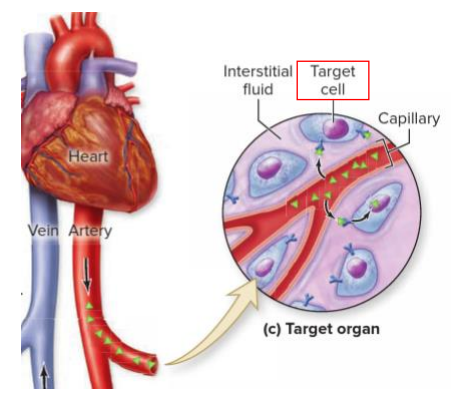
- body control system where regulation requires duration rather then speed
- glands that secrete chemical messengers (hormones) into circulatory system (blood)
- Hormone characteristics: produced in small quantities, transported some distance in circulatory system, acts on target tissues elsewhere in body
- Hormone secretion can be: acute (sudden release due to stimulus, e.g adrenaline in response to stress), chronic (small variations over long periods, e.g thyroid hormones), episodic (e.g estrogen and progesterone during menstrual cycle
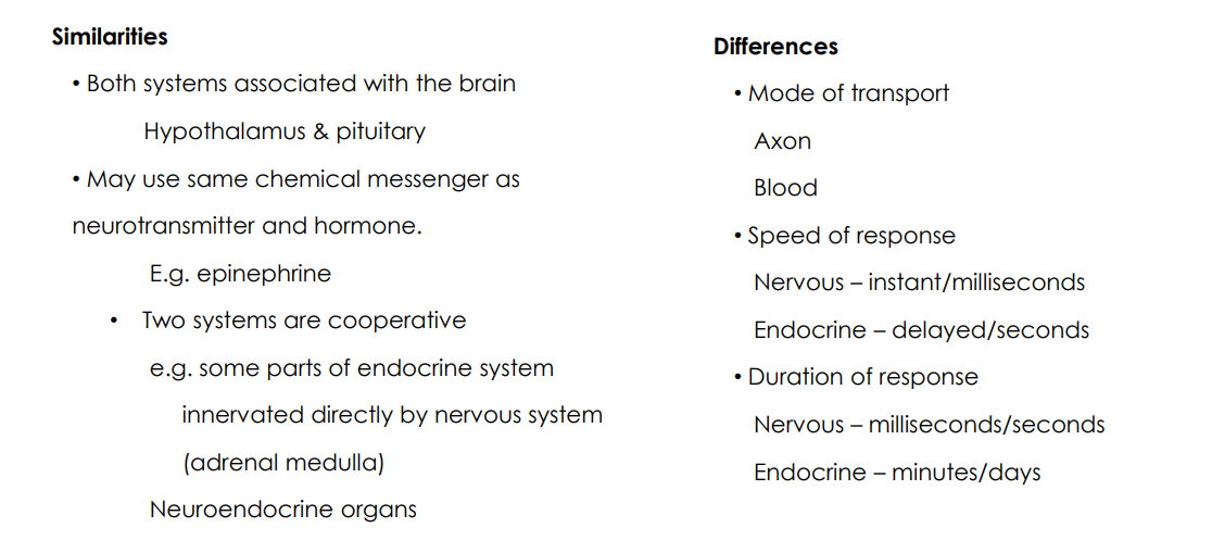
Endocrine vs Nervous Communication
SIMILARITIES:
- Both systems associated with the brain (hypothalamus and pituitary)
- may use same chemical messenger as neurotransmitter and hormone. e.g epinephrine
- two systems are cooperative, e.g some parts of endocrine system innervated directly by nervous system (adrenal medulla). Neuroendocrine organs
DIFFERENCES:
- Mode of transport (axon vs blood)
- Speed of response (nervous: instant/milliseconds, endocrine: delayed/seconds)
- Duration of response (Nervous: milliseconds/seconds, Endocrine: minutes/days)


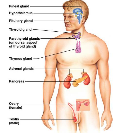
Endocrine Organs
- hypothalamus
- pituitary gland
- pineal gland
- thyroid gland
- parathyroid gland
- thymus gland
- adrenal gland (suprarenal)
- pancreas
- gonads (testes and ovaries)
- all located along midline of body so its easier to connect on aorta
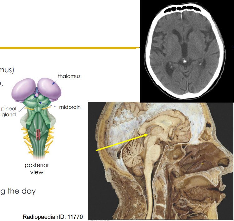
Hypothalamus
- controls centre of ANS and controls the pituitary gland
- Lies inferior and anterior to the thalamus
- Secretes hormone-releasing factors which stimulate hormone release from anterior pituitary
- Secretes release-inhibiting factors to inhibit release from pituitary
- Connected to by the infundibulum
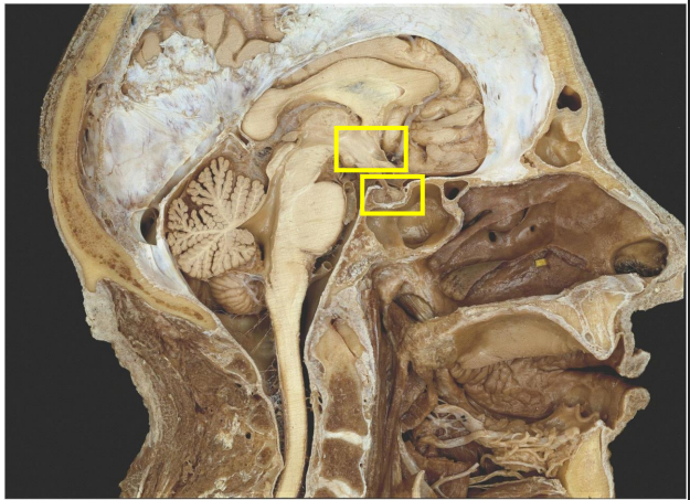
- NB: position of the pituitary gland in the pituitary fossa of the sphenoid bone

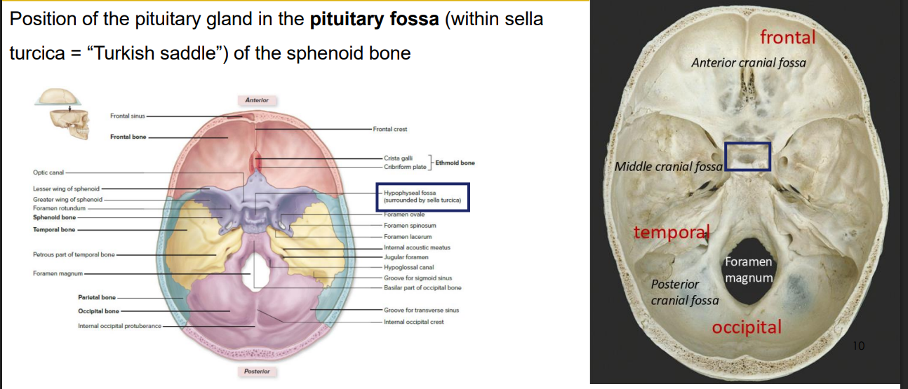
Cranial Location
- position of the pituitary gland in the pituitary fossa of the sphenoid bone


Pituitary Gland
- divided into anterior and posterior parts which have different embryological origins, functions and control mechanisms
- anterior pituitary (adenohypophysis)
- posterior pituitary (neurohypophysis)
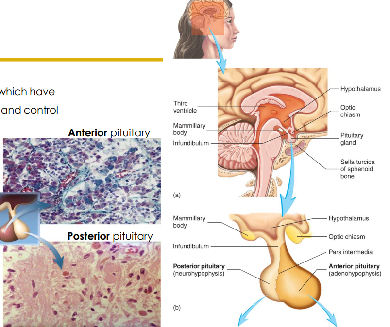

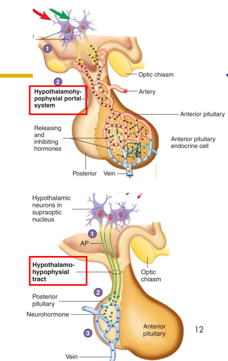
Pituitary Gland and Infundibulum
- AP and PP arise from different germ layers (embryologically), therefore formed of different tissue types.
Infundibulum contains:
- hypothalamic-hypophyseal tract and hypothalamic-hypophyseal portal system
- hypothalamic means relating to hypothalamus, hypophyseal means relating to pituitary gland and portal system means its refering to a blood network
- 70% of the pituitary mass is anterior pituitary
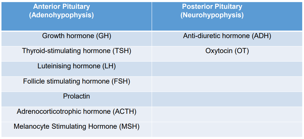
Anterior Pituitary Hormones x7 (NAMING ONLY)
1. Growth Hormone (GH)
2. Thyroid Stimulating Hormone (TSH)
3. Adrenocorticotropic Hormone (ACTH)
4. Melanocyte-stimulating hormone (MSH)
5. Lutenising Hormone (LH)
6. Follicle Stimulating Hormone
7. Prolactin
Growth Hormone
- acts on most cells of body
- stimulates uptake of amino acids; protein synthesis
- stimulates breakdown of fats to be used as an energy source
- promotes bone and cartilage growth
- regulates blood levels of nutrients after a meal
Thyroid Stimulating Hormone
stimulates thyroid to secrete T3 (Triiodothyronine) and T4 (Thyroxine)
Adrenocorticotropic Hormone
stimulates adrenal cortex to secrete cortisol and aldosterone
Melanocyte-Stimulating Hormone
causes melanocytes to produced more melanin
Lutenising Hormone and Follicle Stimulating Hormone
- both hormones regulate production of gametes and reproductive hormones
- testosterone in males
- estrogen and progesterone in females
Prolactin
role in milk production
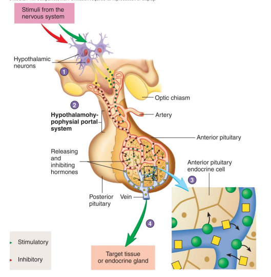
Anterior Pituitary Signalling
1. Stimuli within nervous system regulate secretion of releasing hormones from neurons in hypothalamus
2. Releasing hormones pass to anterior pituitary
3. Releasing hormones stimulate the release of hormones from anterior pituitary
4. Anterior pituitary hormones released into surrounding capillary bed and travel in bloodstream to target tissue, which may be another endocrine gland


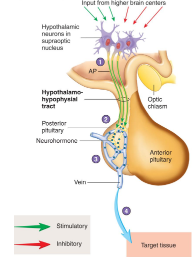
Posterior Pituitary Hormones x2 (NAMING ONLY)
1. Antidiuretic Hormone
2. Oxytocin

Antidiuretic Hormone (ADH)/Vasopressin
- stimulates increased reabsorption of water from nephrons, so less (more concentrated) urine is produced (water is conserved in the body)
- diuretic means want to urinate so anti-diuretic is opposite


Oxytocin
- uterine contractions during birth
- ejection of milk (milk let down) from lactating breast

Pituitary Hormones SUMMARY
DIAGRAM ON SLIDE 16


Pineal Gland
- Posterior part of diencephalon (epithalamus)
- located in posterior wall of 3rd ventricle, midsagittal
Pinealocytes:
- synthesises melatonin from serotonin
- secretes melatonin rhythmically on 12 hour light/dark cycle
- directed by retinal light detection
- sympathetic stim. inhibits release during the day
- begins to calcify in your 30s
- increase melatonin makes you sleep

Thyroid Gland DIAGRAM
DIAGRAM ON SLIDE 19
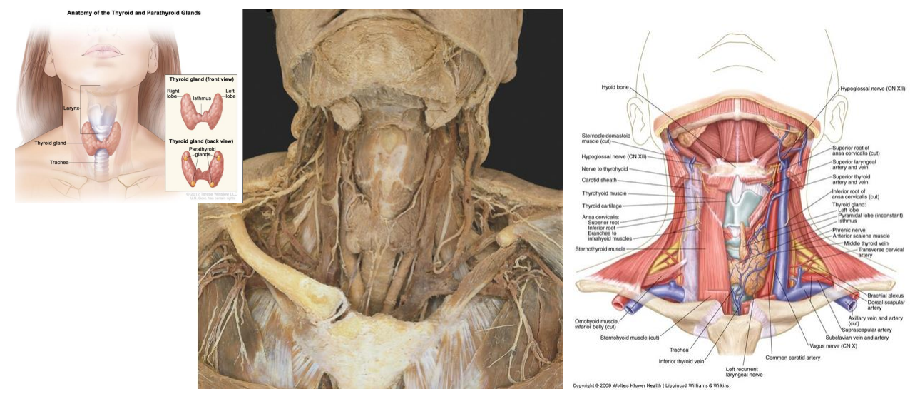

Thyroid Gland
- contains thyroid follicles and parafollicular cells
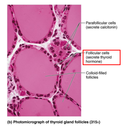
Thyroid Follicles:
- colloid storage lakes lined by follicular cells (simple cuboidal)
- produces thyroglobulin: stored inactive form in colloid, used to syntheise thyroid hormones:
- Tetraiodothyronine (T4) (aka thyroxine)
- Triiodothyronine (T3): more potent, less abundant
- TSH (from anterior pituitary) stimulates release of T3 and T4
- Effect: increase metabolic rate in almost all body tissues
- when TSH comes along, it stimulates the conversion of thyroglobulin into T3 and T4
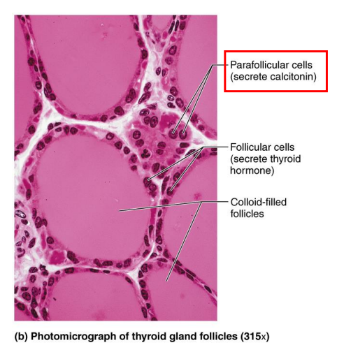
Parafollicular cells (Thyroid Gland)
- produce calcitonin which acts on bones and kidney to reduce blood calcium levels
- inhibiting osteoclasts, promotes [Ca2+] deposition in bone by osteoblasts, reduces plasma [Ca2+]
- promotes deposition of calcium into bones
![<p>- produce calcitonin which acts on bones and kidney to reduce blood calcium levels</p><p>- inhibiting osteoclasts, promotes [Ca2+] deposition in bone by osteoblasts, reduces plasma [Ca2+]</p><p></p><p>- promotes deposition of calcium into bones</p>](https://knowt-user-attachments.s3.amazonaws.com/a9edcbb5-8127-4f87-92b4-87f147c82b99.png)
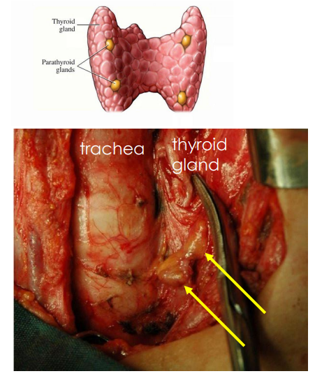
Parathyroid Gland
- location: usually two glands on posterior side of each thyroid lobe (about the size of a grain of rice)
- 4 (to 6) parathyroid glands
- secrete Parathyroid Hormone (PTH) when [Ca2+] is low
- raises blood calcium by stimulating osteoclast activity and bone resorption (opposes calcitonin)
- PTH is essential for life
- stimulates breakdown of bone
![<p>- location: usually two glands on posterior side of each thyroid lobe (about the size of a grain of rice)</p><p>- 4 (to 6) parathyroid glands</p><p>- secrete Parathyroid Hormone (PTH) when [Ca2+] is low</p><p>- raises blood calcium by stimulating osteoclast activity and bone resorption (opposes calcitonin)</p><p>- PTH is essential for life</p><p></p><p>- stimulates breakdown of bone</p>](https://knowt-user-attachments.s3.amazonaws.com/0432f446-34dc-40c8-bbdc-139394da8d6f.png)
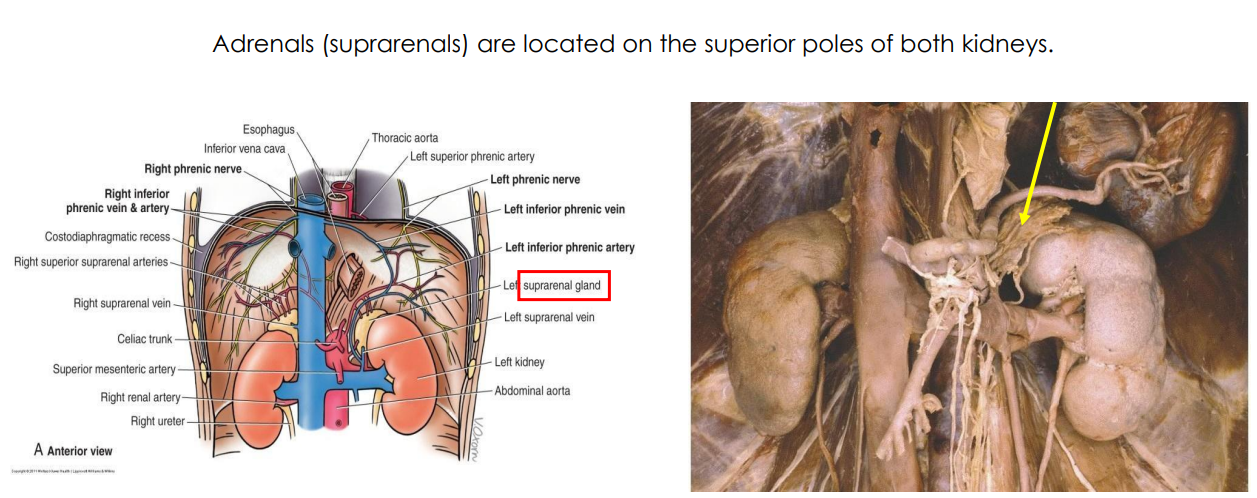
Adrenal Glands DIAGRAM
- supra means above
- adrenal: ad means adjacent, renal means to the kidneys

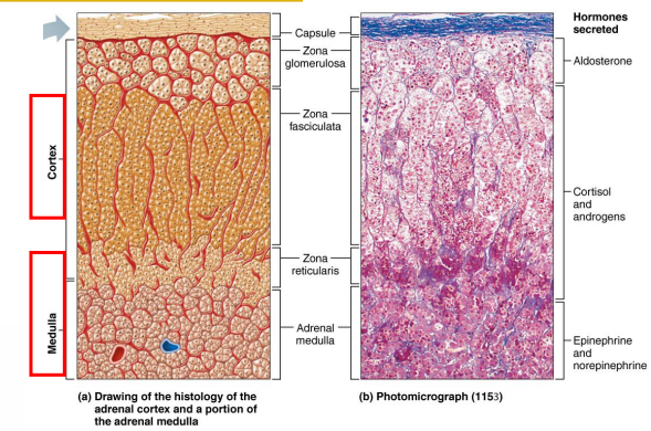
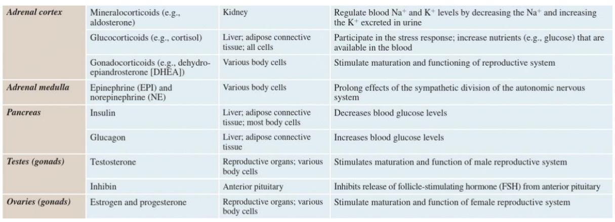
Adrenal Gland
- formed of outer cortex and inner medulla.
- cortex is regulated by ACTH from Anterior Pituitary
- Cortex produces and secretes glucocorticoids (cortisol), mineralocorticoids (aldosterone) and androgens (DHEA)
- salt, sugar and sex (order in which they are released) (aldosterone, cortisol and most inner layer androgens)
- Medulla produces and secretes adrenaline (epinephrine).
- sympathetic stimulation


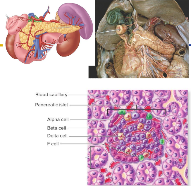
Pancreas
Recall: has both exocrine and endocrine functions.
- exocrine: digestive enzymes; delivered to GIT by ducts
- Endocrine function: produces insulin, glucagon and somatostatin which regulate blood glucose levels.
Pancreatic islets:
- alpha cell (around 20% islet cells): Glucagon increases blood glucose
- beta cells (around 70%): insulin decreases blood glucose
- Delta cells (around 5-10%): somatostatin (body static): inhibits glucagon and insulin secretion
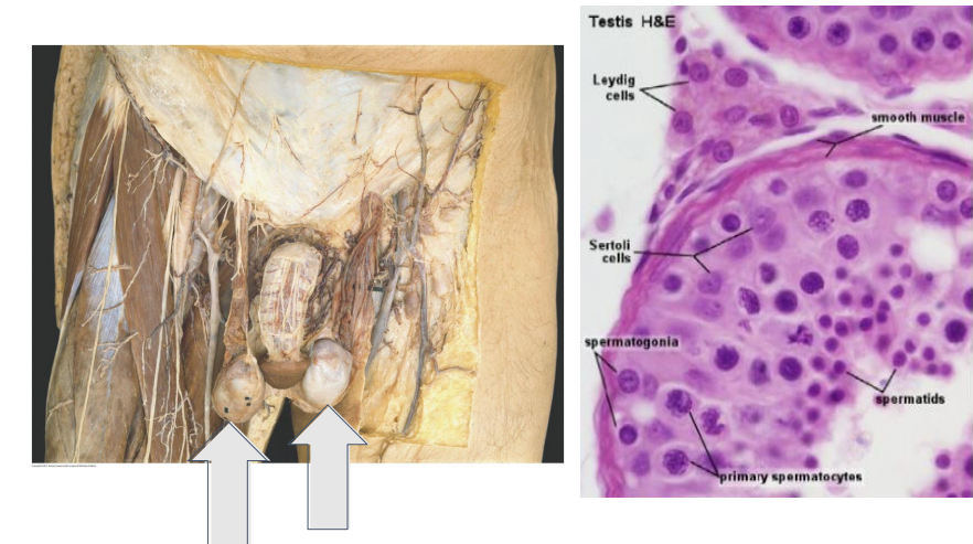
Testes
- testicular interstitial cells (Leydig) between the seminiferous tubules are stimulated by LH from Anterior Pituitary to produce testosterone required to stimulate spermatogenesis

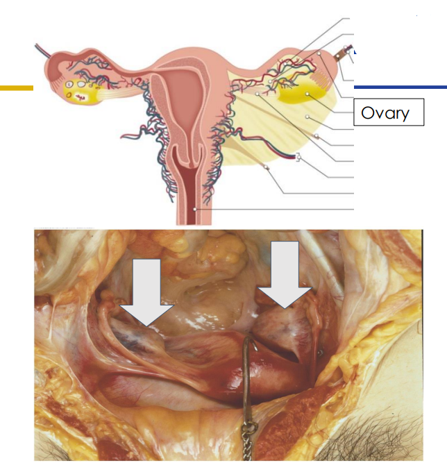
Ovaries
- stimulated by FSH and LH from the anterior pituitary
- LH and FSH required for follicle maturation and ovulation
- Produces oestrogen and progesterone which maintains the endometrium (lining of uterus)

TAKE HOME MESSAGE WORDS
DIAGRAM ON SLIDE 28

TAKE HOME MESSAGES HORMONES AND GLANDS
DIAGRAM ON SLIDE 29

TAKE HOME MESSAGES HORMONES AND GLANDS 2
DIAGRAM ON SLIDE 30