lab practical 1
1/115
There's no tags or description
Looks like no tags are added yet.
Name | Mastery | Learn | Test | Matching | Spaced | Call with Kai |
|---|
No analytics yet
Send a link to your students to track their progress
116 Terms
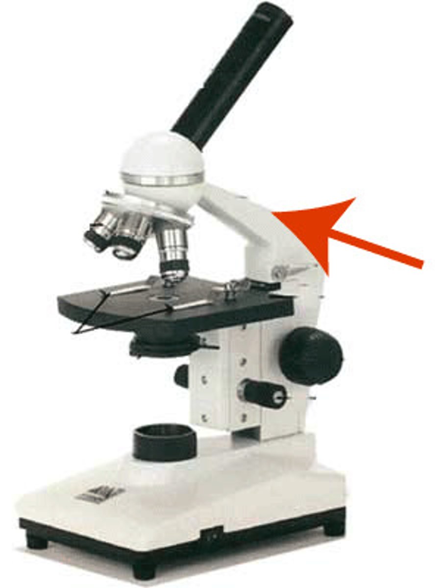
Arm
Vertical portion of the microscope that connects the base and the head
Head
Attaches to the revolving nosepiece to support the objective lens system. It also provides for attachment of the eyepieces which house the ocular lenses
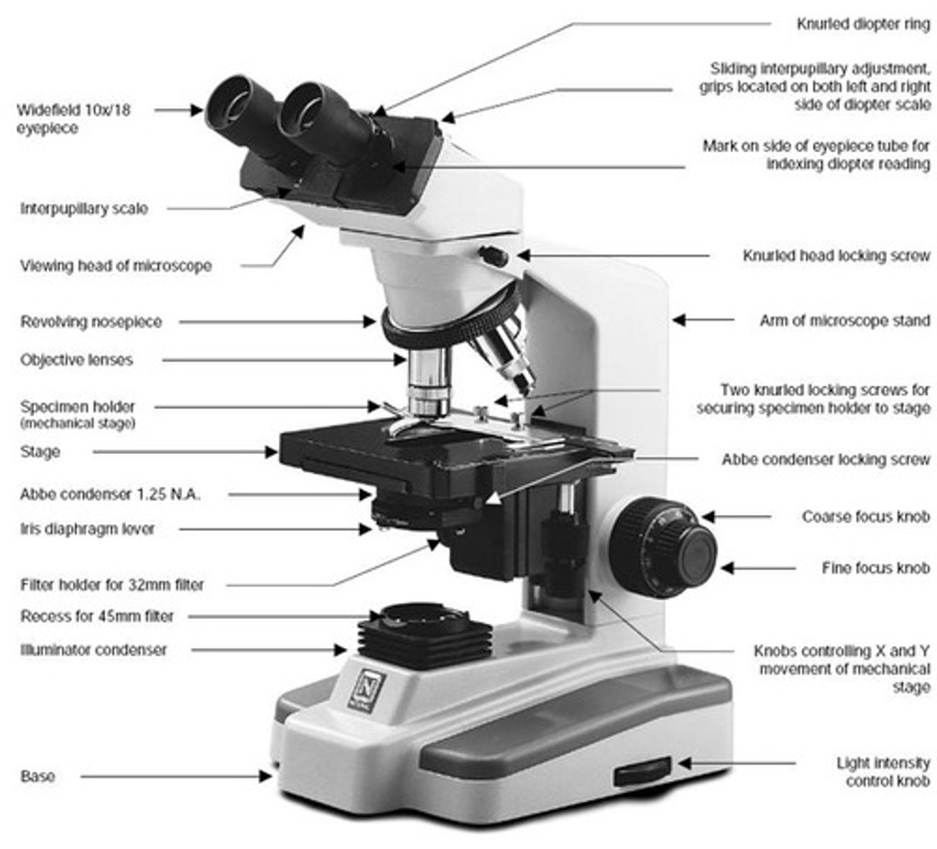
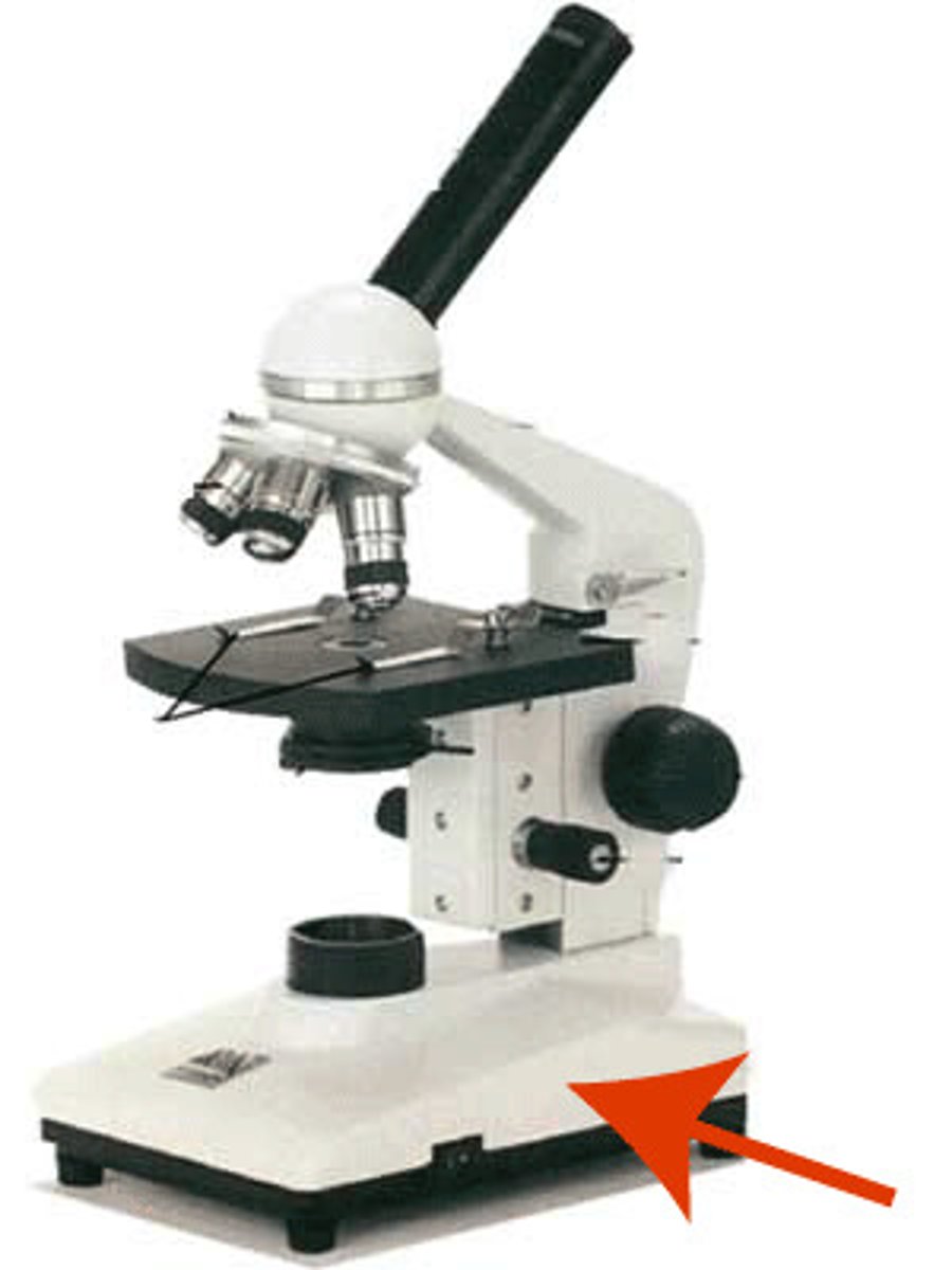
Base
The bottom of the microscope. Provides a sturdy flat surface to support and steady the microscope
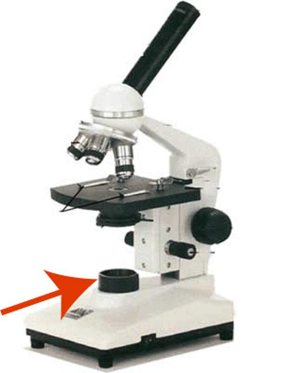
Light source or substage light
Located in the base. The light from the lamp passes directly upward through the microscope
Light control knob
Located on the base or arm. This dial allows you to adjust the intensity of the light passing upwards through the specimen
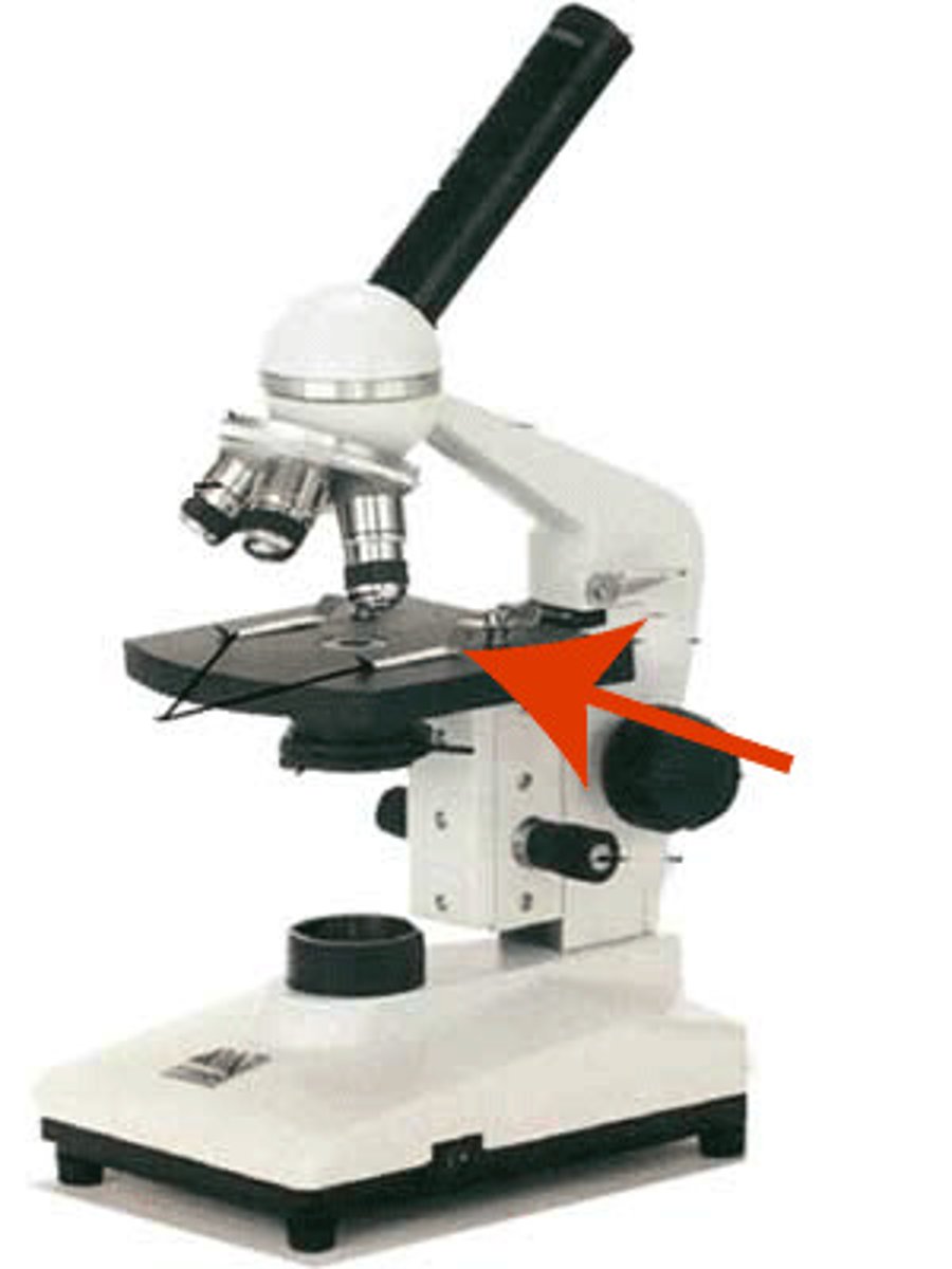
Stage
The platform that the slide rests on while being viewed. The stage has a hole in it to allow light to pass through the stage and through the specimen
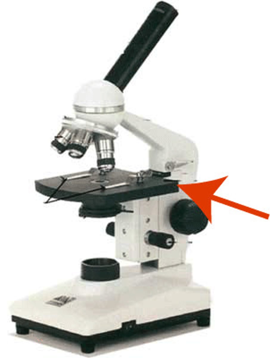
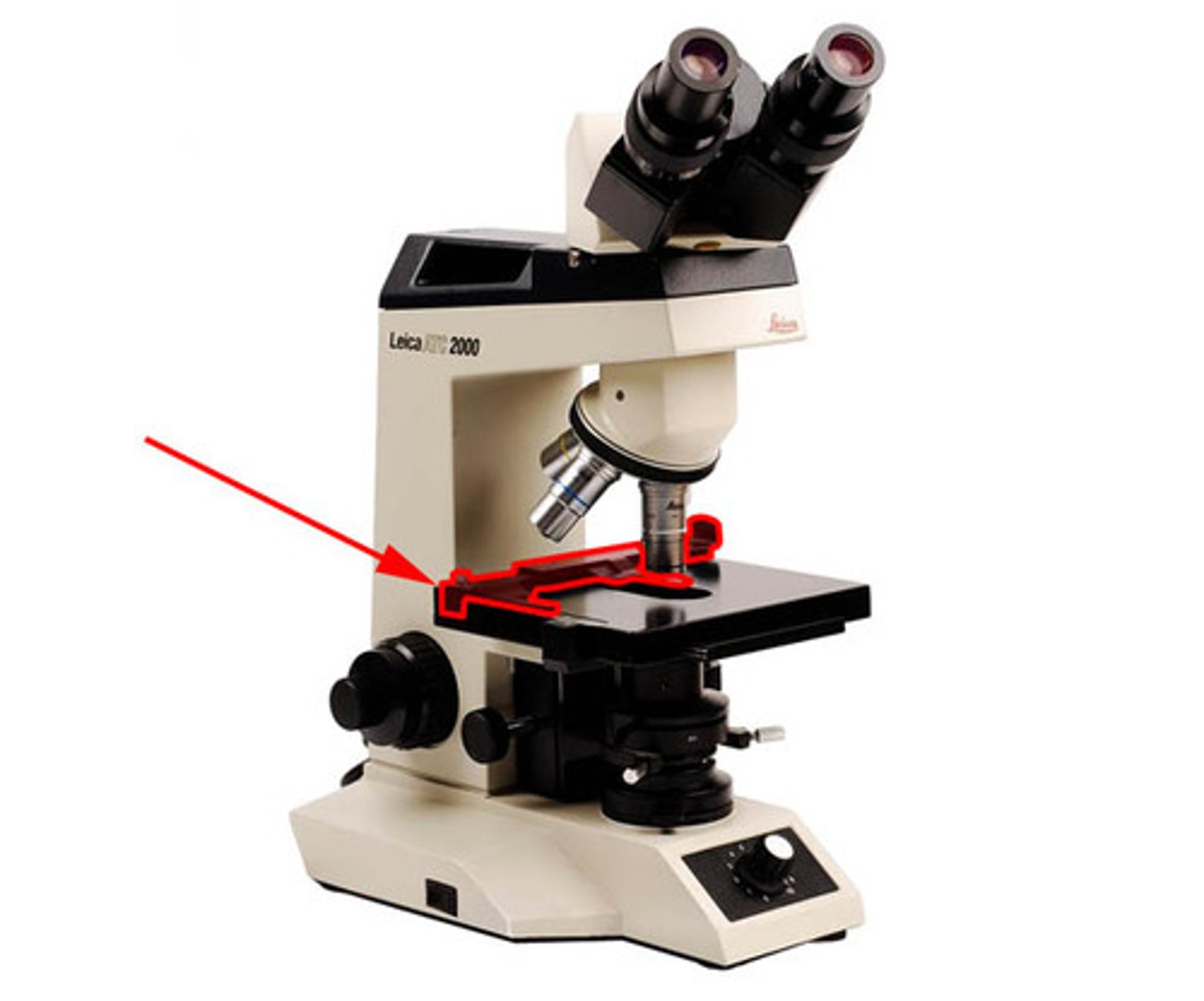
Mechanical stage
Holds the slide in position for viewing and has two adjustable (x/y adjustment or coaxial) knobs that control the precise movement of the slide
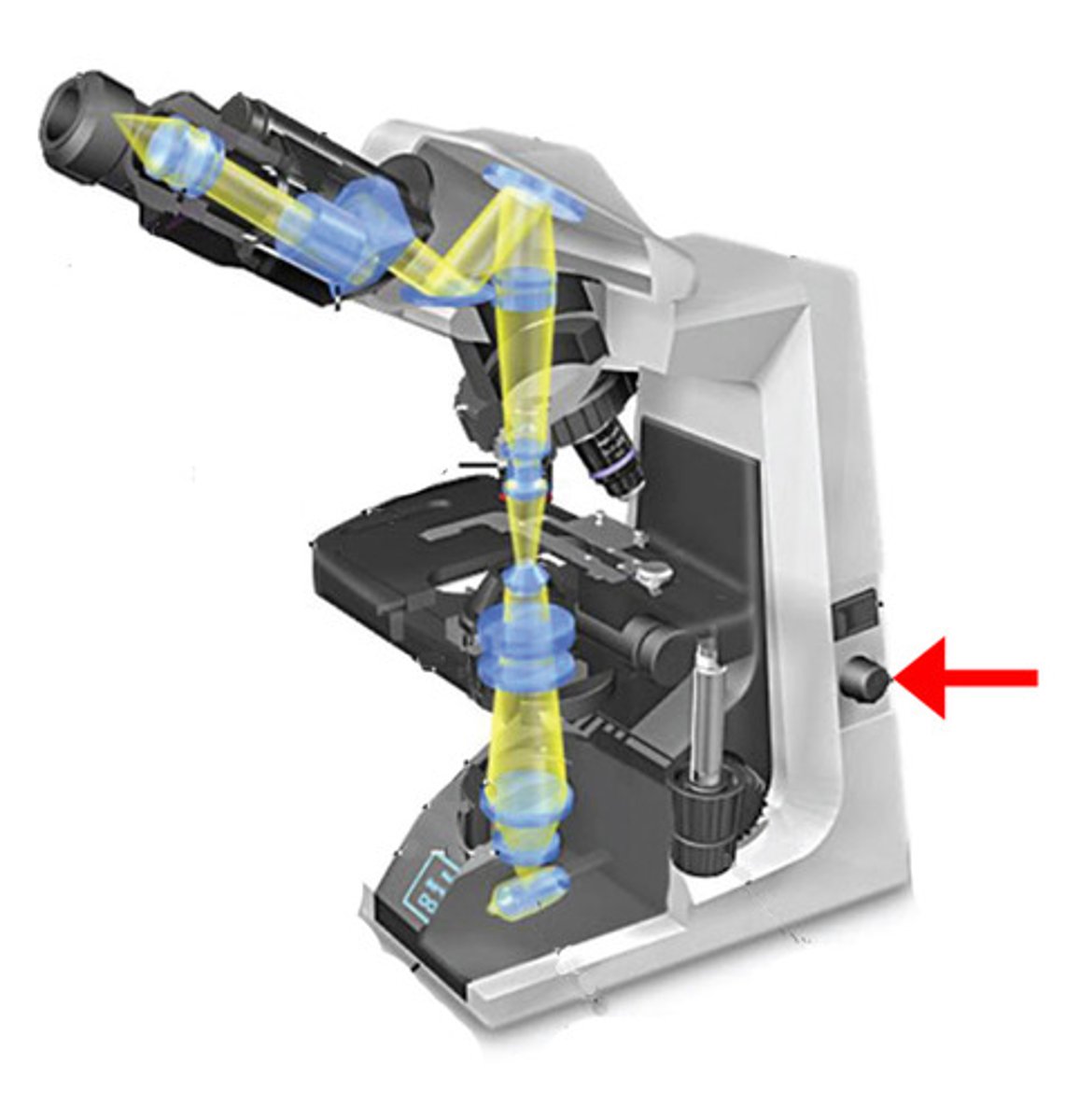
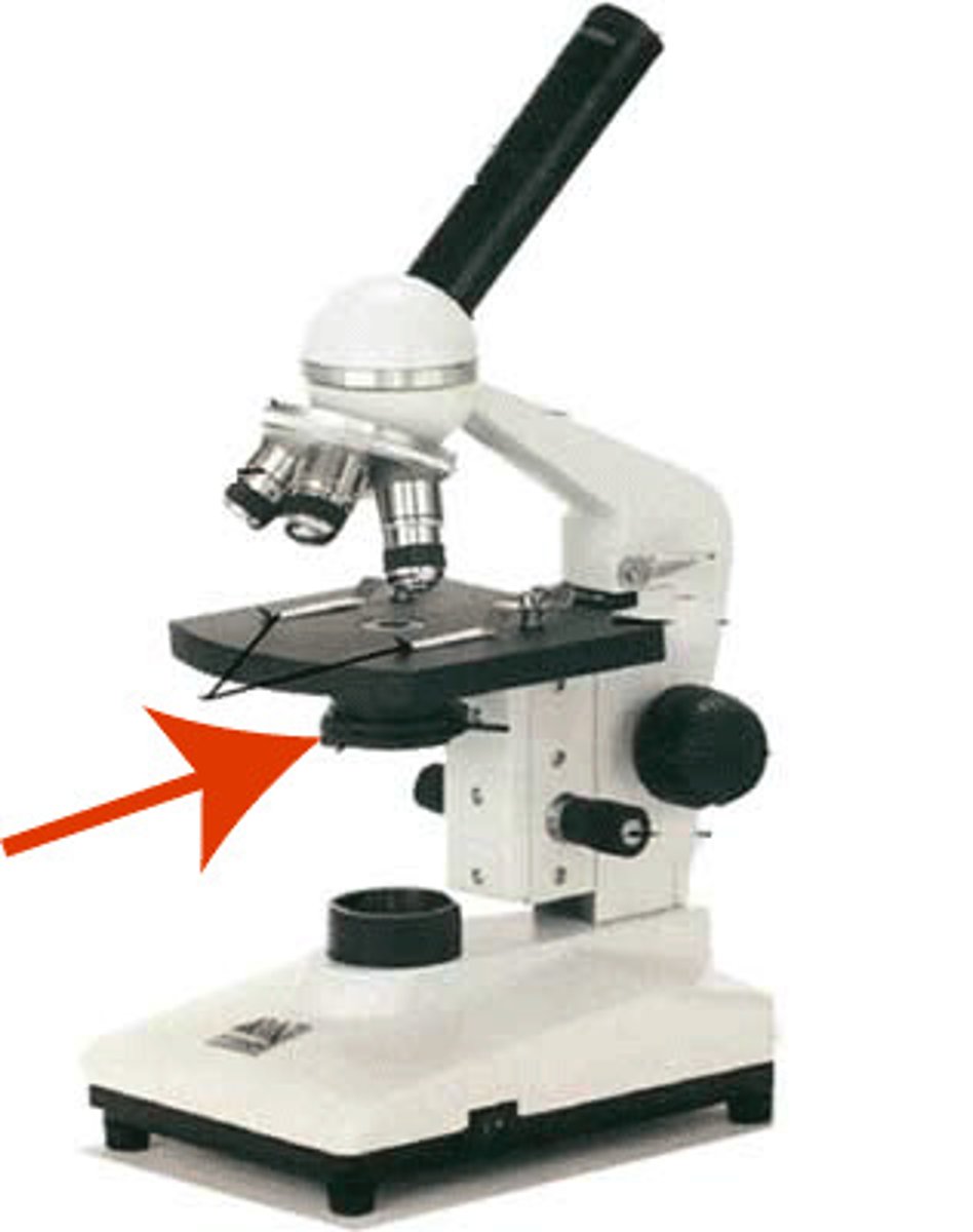
Condenser
Small non-magnifying lens located beneath the stage that concentrates the light on the specimen. The condenser may have a knob that raises and lowers the condenser to vary the light delivery. Generally, the best position is close to the inferior surface of the stage
Iris diaphragm lever
The iris diaphragm is a shutter within the condenser that can be controlled by a lever to adjust the amount of light passing through the condenser. The lever can be moved to close the diaphragm and improve contrast. If your field of view is too dark, you can open the diaphragm to let in more light
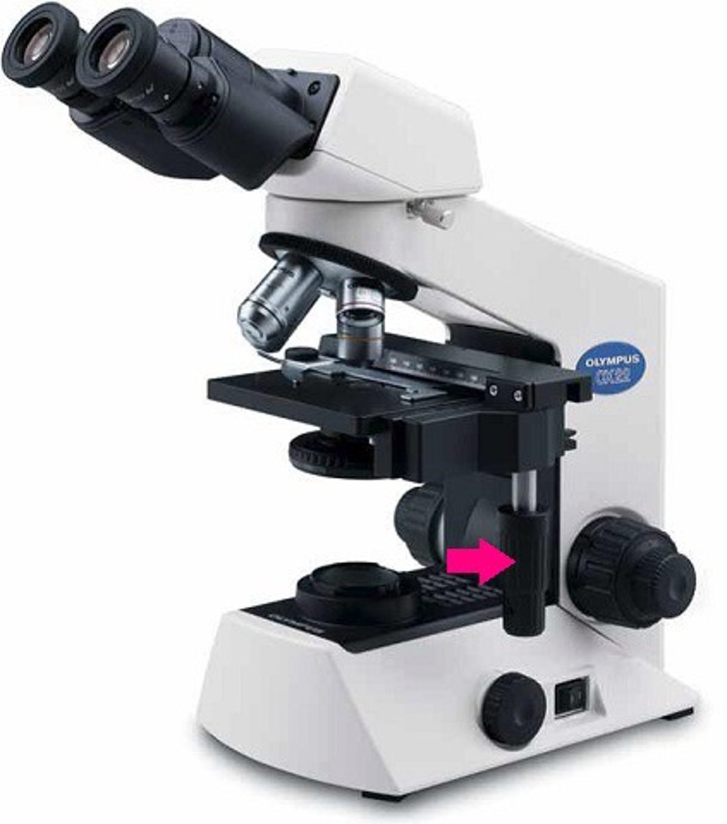
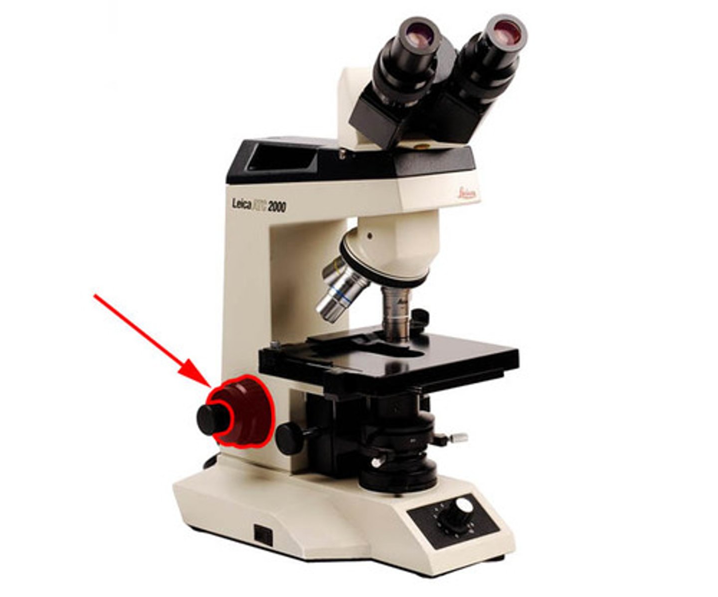
Coarse adjustment knob
This knob allows you to make large adjustments to the height of the stage to initially focus your specimen
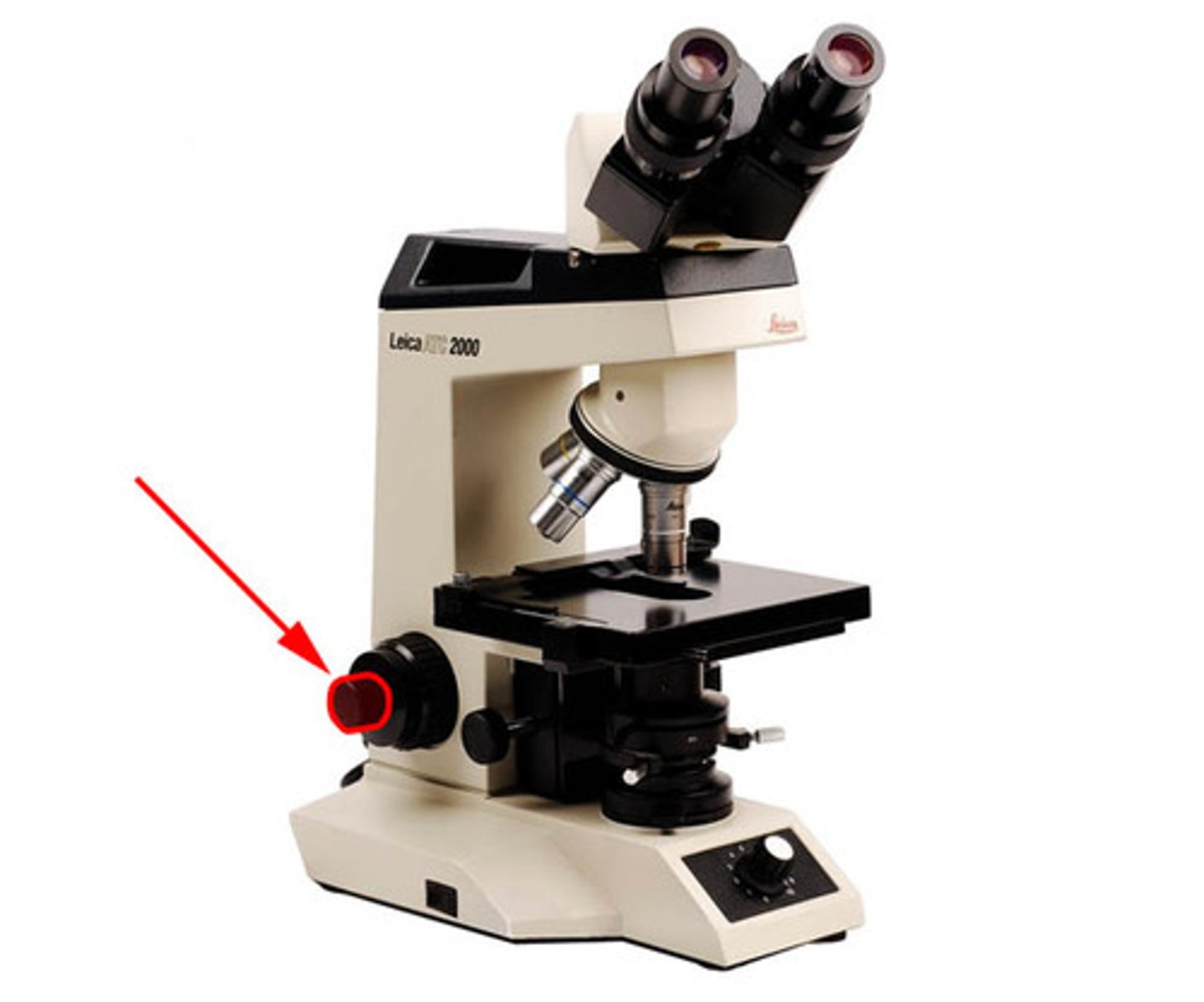
Fine adjustment knob
This knob is used for precise focusing once the initial coarse focusing has been completed
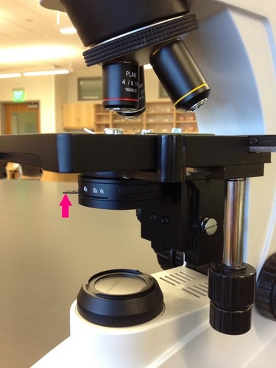
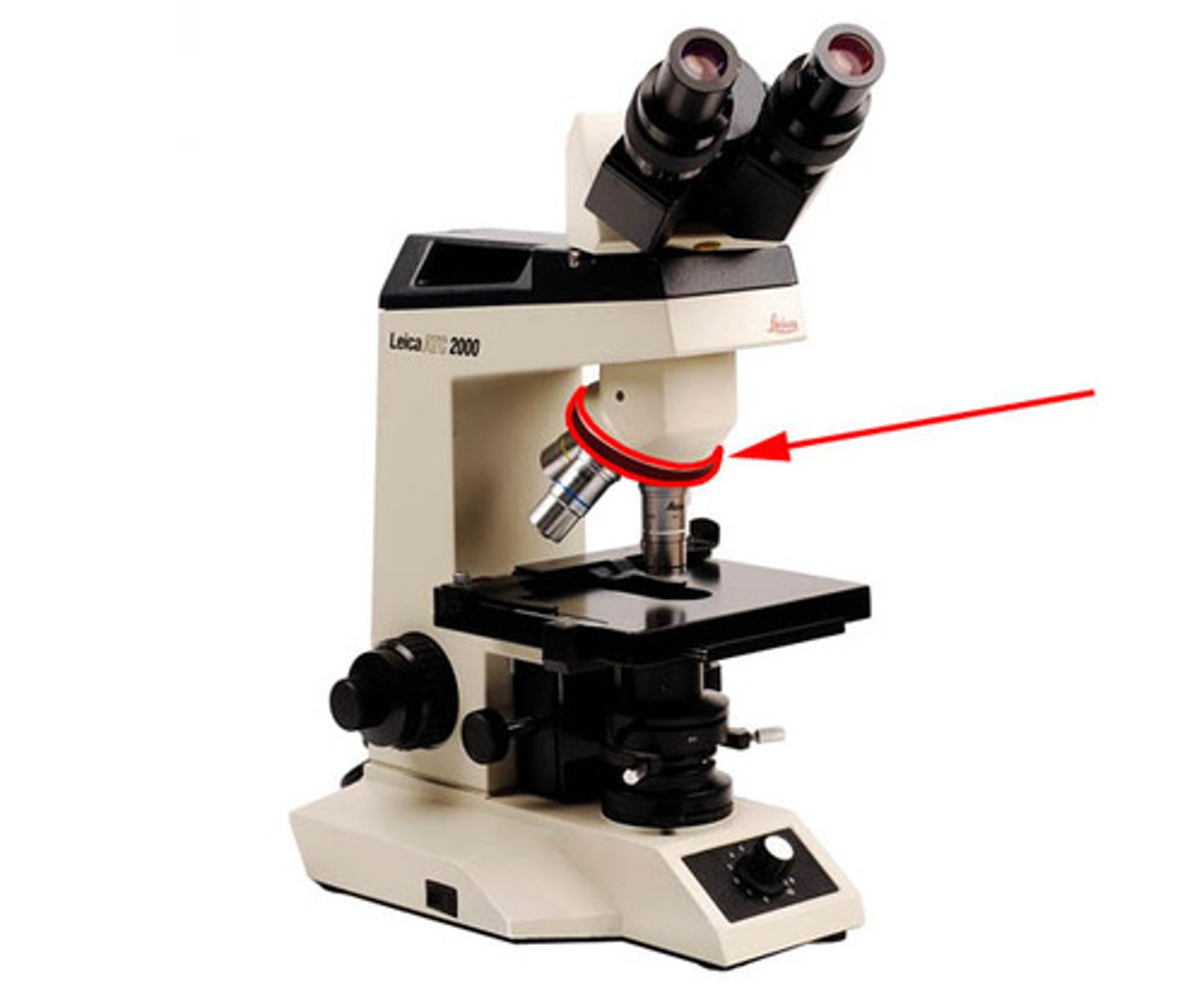
Nosepiece
Rotating mechanism connected to the head. Generally, it carries three or four objective lenses and permits positioning of these lenses over the hole in the stage
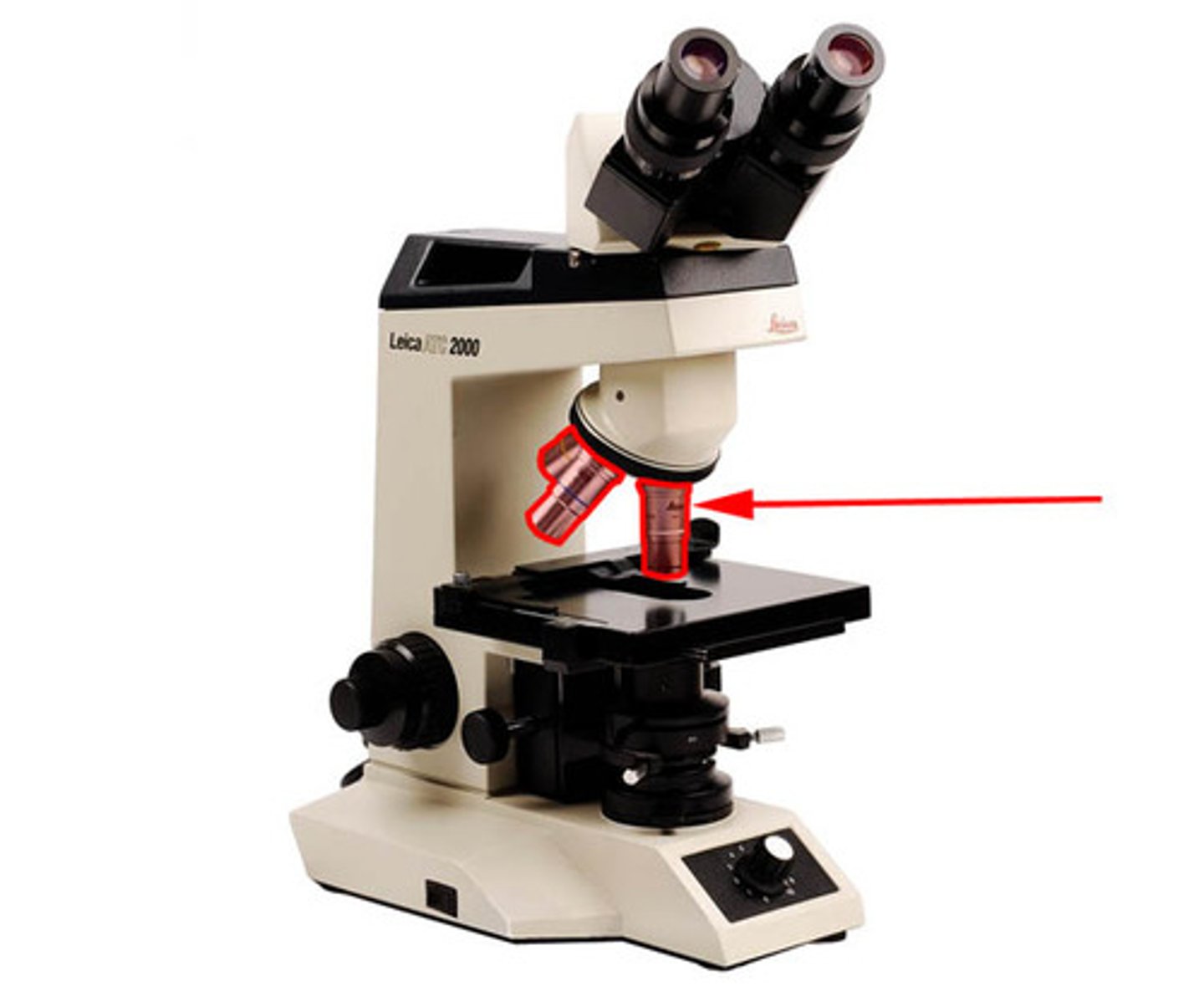
Objective lenses
These lenses are attached to the nosepiece. Usually, a compound microscope has four objective lens: scanning (4x), low-power (10x), high-power (40x), and oil immersion (100x). Typical magnifying powers for the objectives are listed in parentheses
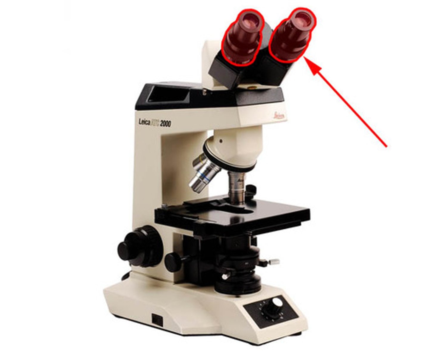
Ocular lenses
Binocular microscopes will have two lenses located in the eyepieces as the superior end of the head. Most ocular lenses have a magnification power of 10x. Some microscopes will have a pointer and / or reticle (micrometer) which can be positioned by rotating the ocular lens
Stage opening
Allows light upwards toward the specimen for better viewing via the condenser
Stage lock control
Allows stage to be locked in a single position
Slide holder
Holds the slide in place for viewing the specimen
Taenia pisiformis
is a parasitic tapeworm primarily infecting dogs and wild canids, with rabbits and other lagomorphs serving as intermediate hosts.
How is Taenia pisiformis transmitted?
what is the magnification of oil immersion?
100x
How do you calculate the total magnification?
Ocular magnification (10x) times objective magnification
What is resolution?
Is the ability of a lens to distinguish between two points in close proximity as separate and distinct.
Objective lenses with high magnification they tend to
have a decreased field of view
have a decreased depth of focus
Why do we heat fix slides? And was is heat fixing?
It’s passing the slide through the fire three times. The reason why we do it is to kill the cells, to make the cells adhere to the slide, and to facilitate staining of the cells.

What is a simple stain? Name the commonly used simple stain.
It uses one dye to visualize the organism.
Methylene blue
What is a differential stain?
uses two or more dyes to distinguish between kinds of organisms
(one category will stain one color while the other category stains another)
What is a structural stain?
It identifies parts of the cell, such as endospores, capsules, flagella, inclusions, and granules.
What is a gram stain?
It is a differential stain, differention is due to cell wall structural differences.
Gram-positive (thick cell wall)
Gram-negative (thin cell wall)
Name me the order of stains placed in a gram stain. Whicu part does the differentiation happens?
Crystal Violet - Primary stain
Iodine - mordant
Alcohol - destaining
Safranin - counterstain
Alcohol
What color do gram positive organisms stain?
Purple
What color do gram negative organisms stain?
Red
What is a acid fast stain?
Its another differential stain but its used to detect myobacterium species that have a waxy cell walls.
Like tuberculosis and leprosy
Name the order of the stains in a acid fast stain.
Carbolfuchsin (often heated)
Acid alcohol rinse
Methylene blue
Acid fast cells stain what color?
Red
Nonacid fast cells stain the color
Blue
What is a spore stain?
Its a structural and differential stain that its used to detect endospores.
What are endospores?
Are dormant resistant structures produced by some bacteria dor surviving harsh conditions
What are the order of the dyes for a spore stain?
Heated malachite green
Safranin
Vegatative cells stain ___.
Red
Endospores stain ____.
Bluish green
What is a negative stain?
It’s used for detecting capsules (slimy coverings around some bacteria)
How does the background and the capsule appear in a negative stain?
Background is dark and the capsule is light (halo effect)
What staining does the negative stain have?
Its mixed with india ink or with nigrosin, the mixture is spread over ths lide in a thin film and then you allow is to dry
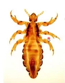
What is pediculus humanus?
It’s a parasatic insect in the kingdom animalia.
Anthropods
They cause epidemic typhus (high fever, rash, and internal bleeding)
To detect the eggs fluorescence in UV light
What is taenia pisiformis
They are intestinal parasites and they take all the hosts nutrition
They are apart of the kingdom animalia
Tapeworms is a flatworms
Name the parts of the taenia pisiformis
The head (sucker) is named the scolex
The body is called the proglottids
What does hermaphroditic
Both sexes are the same worm
What is trichinella spiralis?
They are a roundworm and they are accquired by eating undercook meat such as pork or game animals.
Kingdom animalia
They encysts (spiral) in skeletal muscle tissue
Cyst is the ovoid encastment of the worm
They can cause damage to breathing muscle (diaphragm)
What is rizopus?
Its a mold, asexual spores that are borne in a case called sporangium. They are the decomposer of bread and fruits
Kingdom fungi
Most fungu have hyphae
Some species can lead to zygomycosis with people that have supressed immune systems
What is penicillium?
It’s another type of mold, mitosporic fungus
Some species produce penicillin
Kingdom fungi
What is aspergillus?
A mold, mitosporic fungus, its has conidia
Kingdom fungi
Its important in allergies
Produces so many spores that end up in the hair
May cause diseases and its dangerous if it gets in the lungs
Some produce toxins and they are called alfatoxins
What are yeasts?
They are unicellular fungi
They divide by budding or fission
What are candida albicans?
Its a yeast thats a unicellular fungi
Causes infections in the vagina, mouth, skin, blood and organs
Has hyphal form with long threadlike extensions
What is aspergillosis?
is a respiratory infection or allergic reaction caused by Aspergillus, a common indoor/outdoor mold
What is a amoeba proteus
Its a amoeba and the shape constantly changes.
The cytoplasmic extensions are called pseudopods
Amoebas feed by phagocytosis
They feed on bacteria
Kingdom protista
What is a eugena?
They are these organisms that are green. Its a protozoan
They have one single flagellum.
They are green because they have chloroplasts.
The red eyespot is light sensing structure
Exhibit phototaxis
What is the trypanosoma gambiense?
It’s a dangerous protozoan parasite
Causes african sleeping sickness ( affects the CNS which can lead to coma and death )
Its common in africa
The Tsete flies are vector
The protozoan is also flagellated
Volvox
Its an algae that contains chlorophylls A and B
Each cell is flagellated
Kingdom protista
It contains smaller spheres within larger ones
What is spirogyra?
Its a filamentous green algae
Has a spiral chloroplast
What are diatoms?
They are unicellular algae with silica covering.
They contain A and C chlorophylls plus fucoxanthin
20% of oxygen in the atmosphere come from diatoms.
What is fucoxanthin?
s a marine carotenoid pigment found primarily in brown algae, known for its antioxidant, anti-obesity, and metabolic health properties
What mineral is stored in the cell wall of a diatom?
Silica
What is diatomaceous earth?
is fossilized layers of diatoms is can be used for toothpaste abrasion and filtration.
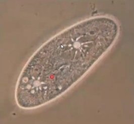
What is paramecium?
It’s a ciliate protozoan
has a macro and micro nucleus
has a gullet (stomach)
contractile vacuole
What is the difference between the macronucleus and micronucleus?
Macro - for metabolism
micro - for sexual reproduction
What is a gullet?
For ingesting food
What is the contractile vacuole?
it expels excess water
What are ciliates?
numerous hair-like structures called cilia, which they use for movement, feeding, and sensing their environment.
What is Balantidium coli?
Its another ciliated protozoan that causes human disease.
If you come into contact with a pig its a risk factor
causes diarrhea, vomiting, abdominal pain, and ulceration.
What are the symptoms of balantidiasis? And what is the risk factor for this disease?
A major risk factor to getting this disease is being in contact with pigs. Symptoms include diarrhea, vomiting, abdominal pain, and ulceration.
What is a Plasmodium?
Its an apicomplexan protozoan and it causes malaria.
transmitted by mosquitoes
Symptoms include cyclical fever, chills, RBC destruction, and chronic anemia.
What is the apical complex?
is a specialized structure found in the phylum Apicomplexa, consisting of organelles that facilitate the invasion of host cells by these parasitic organisms. (parasitic)
What is the vector of transmission to malaria? What are the symptoms of malaria?
transmitted by mosquitoes
Symptoms include cyclical fever, chills, RBC destruction, and chronic anemia.
Who is at risk for anthrax?
People who work with animals
What are the three forms are human anthrax?
Cutaneous
Intestinal
Inhalation
What is bacillus anthracis?
It’s a domain of bacteria
Kingdom Eubacteria
Chains of longer rods
Gram —positive
causative agent of anthrax
Mainly animal disease
What is Staphylococcus aureus?
Is another domain bacteria
Kingdom Eubacteria
Clusters of spherical cells
gram-positive
Important agent is nosocomial infections
Causes skin, blood, lungs, soft tissue, and organ infections
What is a healthcare-related infection of staphylococcus?
MRSA
Why have many Staphylococcus strains become dangerous?
Their ability to develop antibiotic resistance
What is Escherichia coli?
Its a domain bacterium
kingdom eubacteria
small rod shapes
gram-negative
common intestinal bacterium
Are most strains of this organism dangerous?
Most strains of E. coli are not dangerous and are part of the natural gut flora. However, a small subset, such as E. coli O157:H7, can be deadly.
What diseases can pathogenic strains cause? E.coli
Pathogenic strains of E. coli can cause a variety of diseases, including:
Gastroenteritis: Inflammation of the stomach and intestines, leading to symptoms like diarrhea, abdominal pain, and fever.
Urinary Tract Infection (UTI): Infection of the urinary tract, which can include the kidneys, ureters, bladder, or urethra.
Septic Shock: A life-threatening condition where the blood vessels are constricted, leading to reduced blood flow to organs.
Meningitis: Inflammation of the protective membranes covering the brain and spinal cord, which can cause severe headaches, fever, and sensitivity to light.
Hemolytic-uremic Syndrome (HUS): A rare but serious condition that can occur after E. coli infection, leading to kidney failure due to the destruction of red blood cells.
What is hemolytic uremic syndrome (HUS)?
A rare but serious condition that can occur after E. coli infection, leading to kidney failure due to the destruction of red blood cells.
Bloody diarrhea
what is Clostridium tetani?
They are anerobic bacteria
kingdom eubacteria
gram-positive
causative agent - tetanus
terminal endospores
What is an anaerobe?
an organism that grows without air, or requires oxygen-free conditions to live.
What is an endospore?
is a dormant, highly resistant, non-reproductive structure formed by certain bacteria to survive extreme environmental conditions.
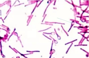
Identify the endospore.
The swollen heads
What are the symptoms of tetanus?
Its a powerful neurotoxin that gives symptoms like stiffening of muscle, lock jaw, sardonic smile, arching of back, and violet convulsive contractions
How does the toxin work? in Clostridium tetani
tetanospasmin, works by disrupting the release of neurotransmitters in the nervous system.
What is Proteus vulgaris?
• Domain Bacteria
– Kingdom Eubacteria
• Rod-shaped cells
• Gram-negative
• Wispy filaments surrounding cells are bacterial flagella
• Flagella for motility
• Frequent cause of urinary tract, skin abscesses, and nosocomial infections
What infection is Proteus vulgaris most associated with?
Complicated urinary tract infections (UTIs)
What is Klebsiella pneumoniae?
• Domain Bacteria
– Kingdom Eubacteria
• Rod-shaped cells
• Bacteria stained with the negative stain
• Halo-like structures
• around cells are capsules.
• Capsule helps bacterium evade phagocytes
• Causes pneumonia, nosocomial infections, and CRE
What disease does Klebsiella pneumoniae cause?
Pneumonia: Klebsiella pneumoniae is a leading cause of hospital-acquired pneumonia. It can cause severe lung infections, particularly in individuals with weakened immune systems or those on ventilators.
Who was Robert Whittaker?
He developed the 5-Kingdom classification scheme
It was based on cell type and nutritional mode.
Distinguish between a prokaryotic cell and eukaryotic cell.
A prokaryotic cell does not have a nucleus.
Simple structure
DNA not enclosed by
special membrane
Eukaryotic has a nucleus.
Complex structure
DNA enclosed by the nuclear envelope.
Membrane-bound organelles
What is the difference among autotrophic, heterotrophic by ingestion, and heterotrophic
by absorption? Which is the feeding mode of decomposers and pathogens?
Autotrophs make their own food (most of these are photosynthetic + Plants and algae) meanwhile Heterotrophs consume other organisms for their nutrition.
There are two types of heterotrophs
ingestion- Food is ingested. Food broken down in digestive system
Absorption -organism secretes exoenzymes onto food source. Exoenzymes break down the complex food molecules
The feeding mode of decomposers and pathogens is heterotrophs absorption.
What were Whittaker’s 5 kingdoms?
Monera (prokaryotic organisms)
Protista (simple eukaryotic organisms)
Fungi (yeasts, molds, mushrooms)]
Plantae (plants)
Animalia (animals)
Who was Karl Woese? How did he approach classification?
He sequenced rRNA of organisms. He found that life clustered in three domains
Woese’s work suggested a modification of the 5-
Kingdom system.
Archaea
Bacteria
Eukarya
What are the three domains?
Archaea
Bacteria
Eukarya - contains Protista, fungi, Plantae, and Animalia
What are the general characteristics of the 6 kingdoms?
a. Eubacteria
b. Archaebacteria
c. Protista
d. Fungi
e. Plantae
f. Animalia
a. Eubacteria- Unicellular and prokaryotic, Cell walls made of peptidoglycan, Important human pathogens
b. Archaebacteria - Unicellular and prokaryotic, Some live in extreme
environments like boiling water and extreme saltiness!, Cell membrane composition
different from all other organisms
c. Protista - Eukaryotic and multicellular and unicellular, Common examples include
protozoa and algae, Some have cell walls while
others don’t. Amoeba and diatoms
d. Fungi - Multicellular and Eukaryotic, In most fungi, the basic unit of
growth is the hypha, Cell wall composed of chitin
e. Plantae - Multicellular and Eukaryotic, Autotrophic by
photosynthesis, Cells walls composed of
cellulose
f. Animalia - Multicellular and Eukaryotic, no cell walls ,quite motile, arthropods