LO's for Final
1/377
There's no tags or description
Looks like no tags are added yet.
Name | Mastery | Learn | Test | Matching | Spaced | Call with Kai |
|---|
No analytics yet
Send a link to your students to track their progress
378 Terms
Four parts of medical terms
prefixes: beginning of the word
suffixes: end of the word
word root: give meaning to the word
combining vowels: connect word roots to other word roots and suffixes
Four steps to dissect a medical term
First: break word into parts: prefixes, suffixes, word roots, combining vowels with slashes
Second: label what word part they are at the top
Third: give the meaning of the word parts at the bottom
Fourth: write the whole meaning of the word
Correct order to read word parts
start with suffix, followed by prefix, and read across until the suffix
Combining Vowel and Suffixes
when suffix starts with a consonant the CV is kept
when suffix starts with a vowel the CV is dropped
Combining Vowel and another WR
if other WR starts with vowel or consonant then the CV is kept
What is a macron?
a long dash above a vowel, long vowel sound for the vowel
What is a breve?
a “u” shaped symbol above a vowel, short vowel sound for the vowel
Phonetic spelling has Capital letters
Capital letters signify that those letters have more stress (drawn out pronunciation)
Singular ends in A
for plural keep A and add E
Singular ends in AX
for plural drop X and add CES
Singular ends in EN
for plural drop EN and add INA
Singular ends in IS
for plural drop IS and add ES
Singular ends in IX
for plural drop IX and add ICES
Singular ends in EX
for plural drop EX and add ICES
Singular ends in MA
for plural keep MA and add TA
Singular ends in ON
for plural drop ON and add A
Singular ends in UM
for plural drop UM and add A
Singular ends in US
for plural drop US and add I
Singular ends in Y
for plural drop Y and add IES
Different levels of organization and definitions
cell: most basic
tissue: similar cells work together for a function
organ: which are made up of tissues to achieve a specific function
organ system: organs that work together for a common function
organism: made of complex organ systems
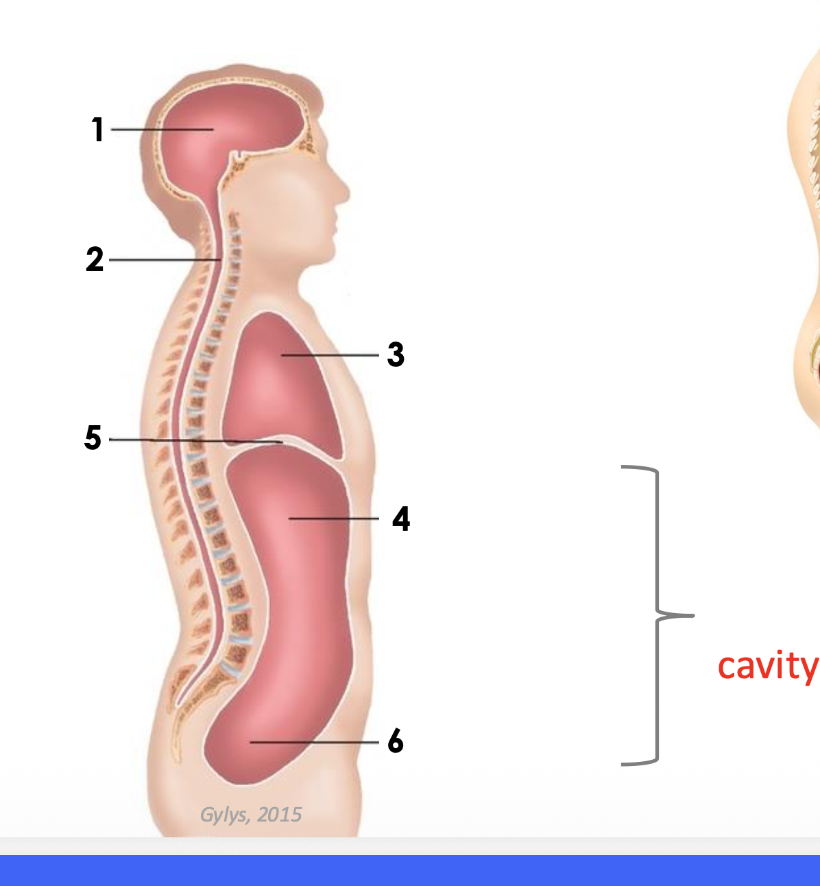
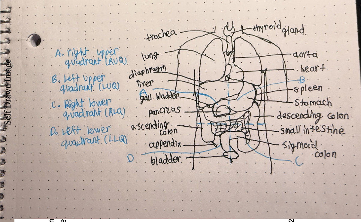
Different body cavities
cranial
spinal
thoracic
diaphragm
abdominal
pelvic
abdominopelvic
how does a pathogen travel between cavities?
through the blood and lymphatic vessels or through connected cavities like abdominopelvic cavity
What is anatomical position?
where the person is standing straight, with palms facing up and feet straight
midline
line in the exact middle of the body
medial
closer to midline
lateral
to the side or farther from midline
proximal
closer to limb root
distal
farther from limb root
superior and other words for it
above or closer to the head
cranial or cephalic
inferior and other word for it
below; farther from head; closer to the tail (feet)
caudal
posterior and other word for it
back of the body
dorsal
anterior and other word for it
front of the body
ventral
superficial
closer to the surface of the body
deep
farther from the surface of the body
prone
lying on the belly and facing down
supine
looking up and lying flat on back
sagittal
a plane of the body separating the body into right and left parts
frontal and another word for it
a plane of the body separating the body into anterior and posterior parts
coronal
transverse and another word for it
a plane of the body separating the body into superior and inferior portions
axial or horizontal
midsagittal
a plane of the body that divides the body into equal right and left parts
How to determine the right and left of a image in transverse plane?
my right is patients left and my left is patients right and we are looking at image from feet up
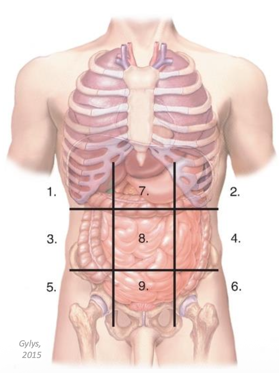
nine abdominopelvic regions
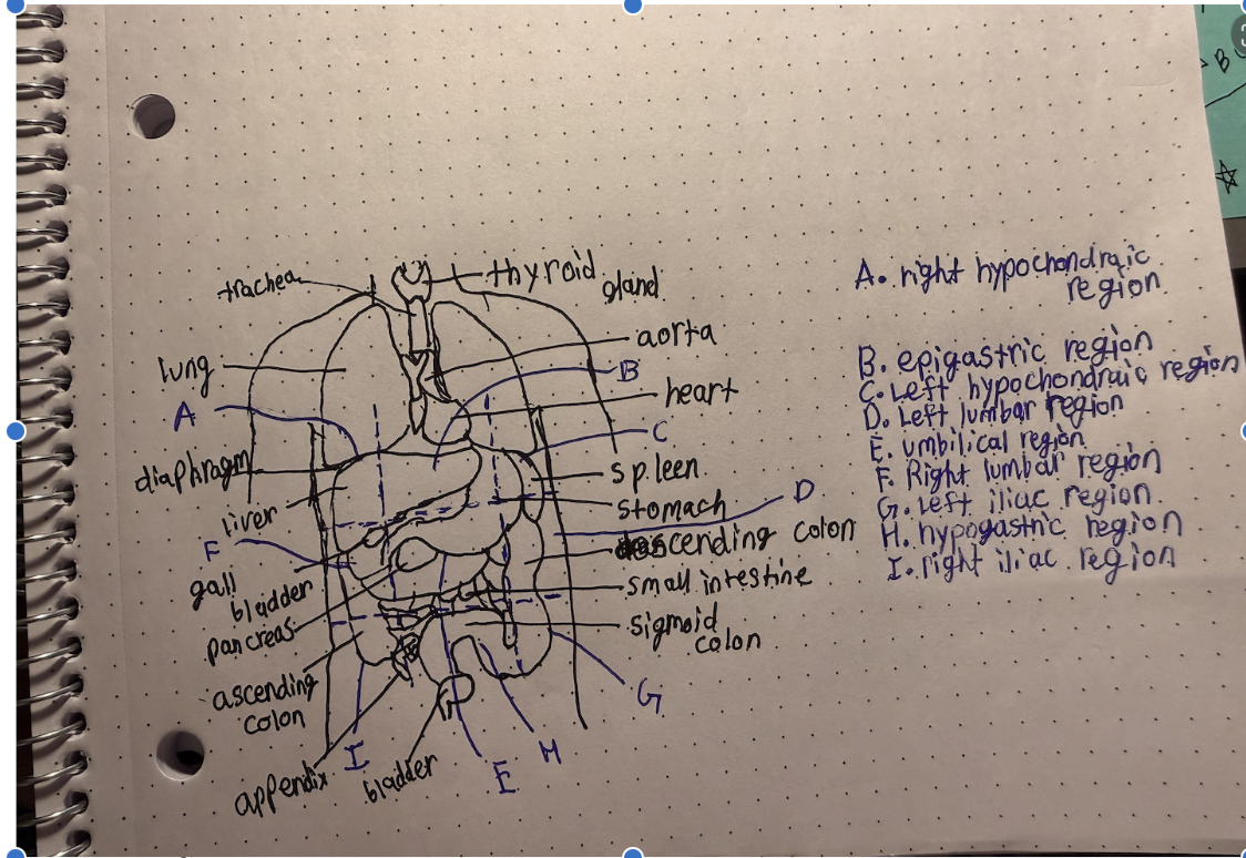
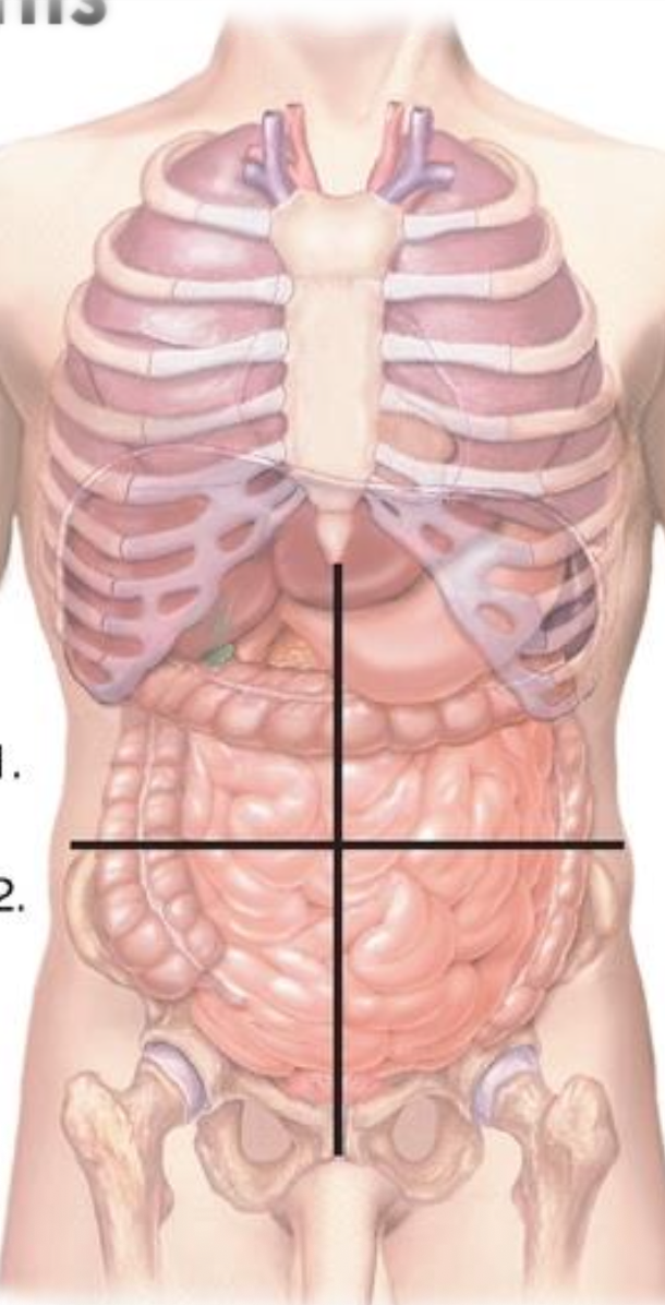
four abdominopelvic quadrants
What is integumantary system composed of?
skin and accessory organs such as hair, nails, sweat glands and sebaceous (oil) glands
three layers of skin
epidermis, dermis, and hypodermis
epidermis
the top layer of skin and doesn’t have any blood vessels or nerves
dermis
below the epidermis and has blood vessels, nerves, hair follicles, and glands
hypodermis
below the dermis and has blood vessels and subcutaneous tissue
Accessory organs (of integumentary system)
sudoriferous glands, sebaceous glands, hair, nails
sudoriferous glands
sweat glands that produce sweat
sebaceous glands
oil glands that secrete oil through hair follicles
hair
important for temperature regulation and sensation
nails
important for protection
functions of the integumentary system
protection, waste removal, temperature regulation, sensation
what structures contribute to the protection (integumentary system)
epidermis, hair, nails, sebaceous glands
what structures contribute to the waste removal (integumentary system)
epidermis, suderiferours glands
what structures contribute to the temperature regulation (integumentary system)
dermis, hypodermis, hair and suderiferours glands
what structures contribute to the sensation (integumentary system)
dermis, hypodermis, and hair
what are keratinocytes and where are they found
cells that produce horny (hard) tissue called keratin
found in epidermis
what are basal cells
deepest cells and above the dermis
what are squamous cells
flat cells and at the top layer of epidermis
what are melanocytes and where are they found
cells that produce melanin
found at border of epidermis and dermis
what is melanin, where is it produced, and what is its function
is a skin pigment ranging from orange-red to brown-black
produced in melanosomes vesicles inside the melanocytes
function is to protect fro UV radiation
How are individuals with darker and lighter skin different in terms of the number of
melanocytes and the amount/type of melanin?
everyone has the same amount of melanocytes
the different amount of melanin gives different skin colors
darker skin is due more melanin that’s darker
lighter skin is due to less melanin that’s lighter
non-melanoma
a type of skin cancer that’s more common but less aggressive
melanoma
a type of skin cancer developed in melanocytes that’s less common but more aggressive, and is a change in an existing mole or new abnormal growth
basal cell and squamous cell carcinoma
are a non-melanoma cancer meaning that it isn’t developed in melanocytes
how to check for melanoma
ABCDE test
ABCDE test
Asymmetry
Border irregularity
Color changes
Diameter increases
Evolution in size, color, or shape
erythema
skin turns red, due to increase in blood in dermis because of heat release, trauma, infection
pallor
paleness due to decrease in blood in dermis because of heat conservation, low blood pressure, and anemia
Cyanosis
blue skin color due to low oxygen, respiratory or heart problems
contusion
black and blue skin due to a rupture of blood vessels causing blood to pool in tissues
brusie
jaundice
yellow color of skin due to increased bilirubin in blood, liver disease, or blockage of bile ducts, or in babies as liver isn’t developed yet
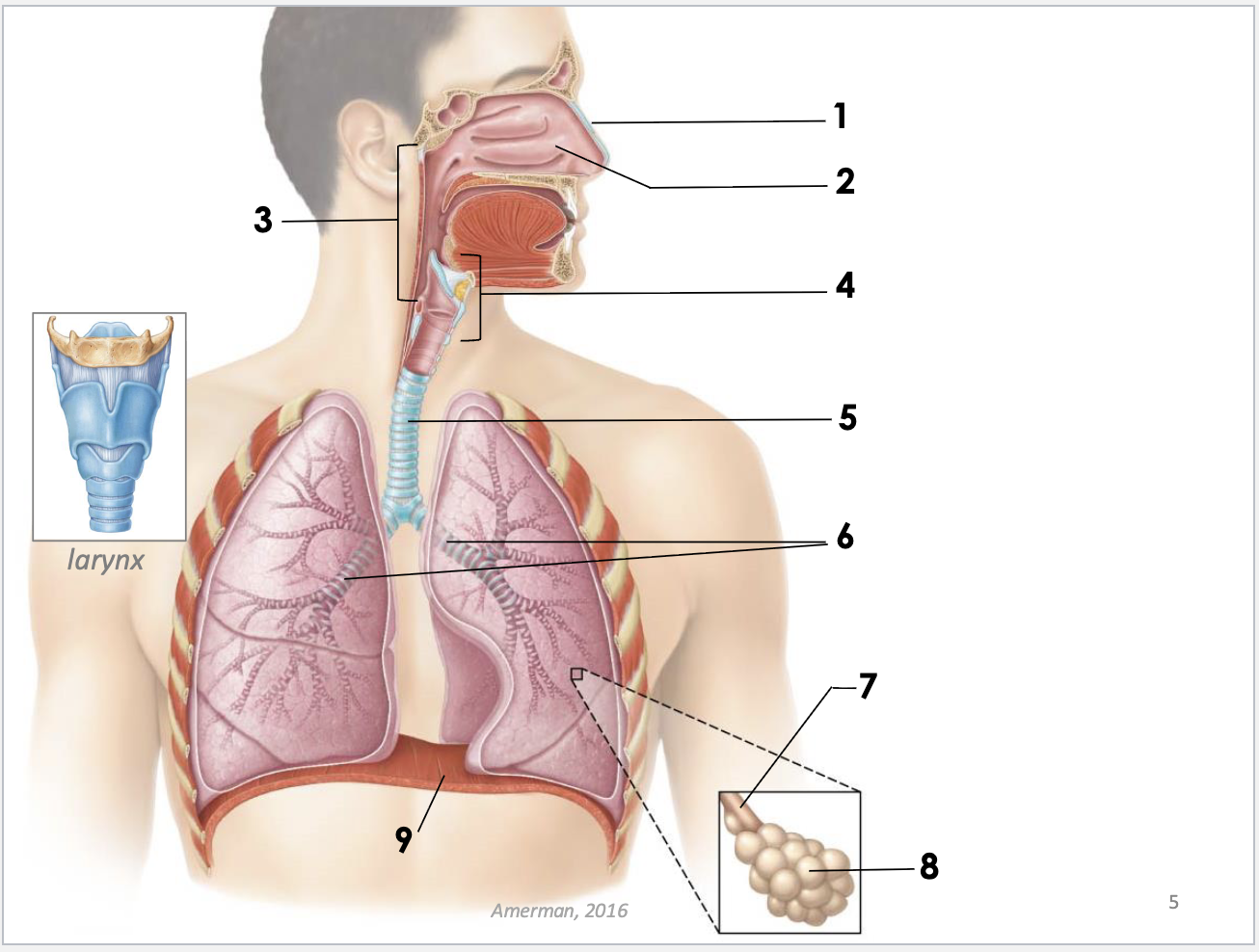
structures of respiratory system and function
nose: for breathing and smell
nasal cavity: through which air travels to lungs
pharynx: a tube that connects to larynx
larynx: for voice production
trachea: carries air to lungs
bronchi: branches that branch off trachea and carry air to alveoli
bronchioles: smaller branches connect bronchi alveoli, bring air to alveoli
alveoli: grape-like air sacs structures where gas exchange happens
diaphragm: breathing muscle
gas exchange
happens in alveoli capillaries
oxygen deficient blood from heart to lungs
oxygen rich blood from lungs to heart
O2 from alveoli to capillaries
CO2 from capillaries to alveoli
pleura
membrane that attaches either to body wall or lungs
parietal pleura
attaches to body wall
visceral pleura
attaches to internal organs (lungs for pleura)
pleura cavity
space between two pleurae (for the lungs in this case) has some fluid for suction with diaphragm to create a low pressure environment so sir can be drawn in
hilum of lung
is the place where blood vessels, bronchi, and nerves enter and exit lungs
tonsils
collections of lymphatic tissue made from infection fighting cells, located major orifices like nasal and oral cavities
adenoids
lymphatic tissue located superior to pharynx
atelectasis
incomplete dilation or a collapsed lung
general causes of atelectasis
obstruction from blockage of airway due to foreign body, tumor, mucus
fluid accumulation from blood, infection, congestive heart failure
air accumulation from pentrating chest wound
treatment for atelectasis
thoracocentesis, surgical puncture of chest that draws fluid or air from thoracic cavity
cystic fibrosis
genetic disorder that affects mucus producing cells
affects respiratory, digestive and reproductive systems
caused due to gene mutation that is inherited from parents
produces thick and sticky mucus that blocks up tracts causing body’s process to be affected
COPD and two conditions that contribute to it
chronic obstructive pulmonary disease
two conditions that contribute are emphysema and chronic bronchitis
emphysema
caused due to smoking and damages alveoli causing a loss of lung tissue elasticity so gas exchange doesn’t work as effectively
chronic bronchitis
inflammation of bronchi and increases mucus production, causes inflammation and swelling of airways making them narrow and increased mucus production leads to blocked airways
can also be caused due to smoking
function of cardiovascular system
transport oxygen and nutrients through blood to tissues and CO2 and waste from tissues
two components of cardiovascular system
heart: pumps blood
blood vessels through which blood travels
systemic circulation
takes oxygenated blood from heart to tissues and de-oxygenated blood from tissues to heart
systemic arteries
blood vessels that go from heart to tissues with oxygen rich blood
systemic veins
blood vessels that go from tissues to heart with de-oxygenated blood
pulmonary circulation
where de-oxygenated blood is taken to lungs from heart
pulmonary artery
carries de-oxygenated blood from heart to lungs
pulmonary veins
carries oxygenated blood from lungs to heart
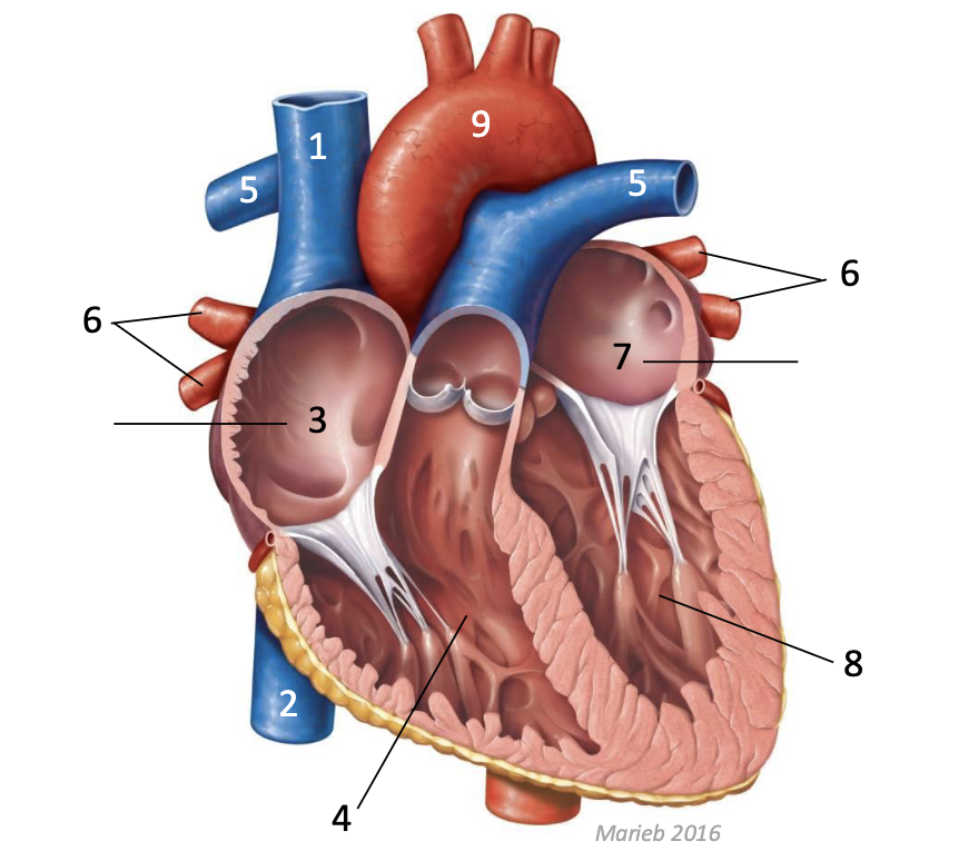
structures of heart
superior vena cava: de-oxygenated blood from organs superior to heart comes through here
inferior vena cava: de-oxygenated blood from organs inferior to heart comes through here
right atrium: upper right side of heart (left if looking at picture) de-oxygenated blood present
right ventricle: lower right side of heart (left if looking at picture) de-oxygenated blood present
pulmonary artery: takes blood to lungs to become oxygenated
pulmonary veins: blood from lungs to heart that is oxygenated
left atrium: upper left side of heart (right if looking at picture) oxygenated blood present
left ventricle: lower left side of heart (right if looking at picture) oxygenated blood present
aorta: major blood vessel that that takes the oxygenated blood to tissues
arteries
have thick muscular layer, decreases in size as move away from heart, don’t have valves
transport oxygenated blood to tissues, but pulmonary artery which takes de-oxygenated blood away from the heart to the lungs