Looks like no one added any tags here yet for you.
What are Eukaryotes?
- All living organisms are made of cells, which have the same basic features in common, suggesting all living things evolved from the same common ancestor.
- There are two main types of organism: Eukaryotes and Prokaryotes; both contain organelles.
- Eukaryotic organisms are made of eukaryotic cells; they are complex.
What are Prokaryotes?
- All living organisms are made of cells, which have the same basic features in common, suggesting all living things evolved from the same common ancestor.
- There are two main types of organism: Eukaryotes and Prokaryotes; both contain organelles.
- Prokaryotic organisms are prokaryotic cells; they are single-celled organims; they are smaller and simpler.
What are organelles?
- Organelles are parts of cells.
- Each one has a specific function.
- If you examine a cell through an electron microscope, you can see it's organelles and the internal structure of most of them.
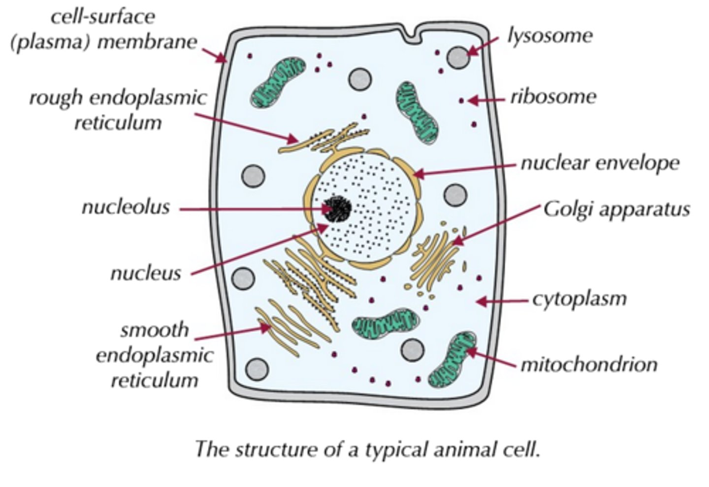
What are Eukaryotic cells?
- Eukaryotic cells are generally a bit more complicated than prokaryotic cells and have more organelles.
- Animal, plant, algal and fungal cells are all eukaryotic.
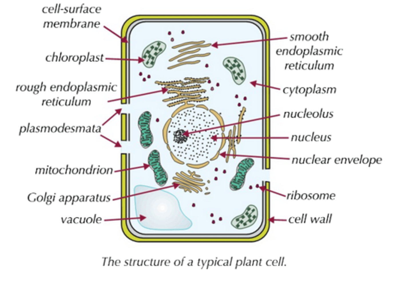
What are plant cells?
Plant cells have the same organelles as animal cells, but with a few added extras such as:
- a cellulose cell wall with plasmodesmata ('channels' for exchanging substances between adjacent cells).
- a vacuole (fluid-filled compartment).
- chloroplasts (organelles involved in photosynthesis).
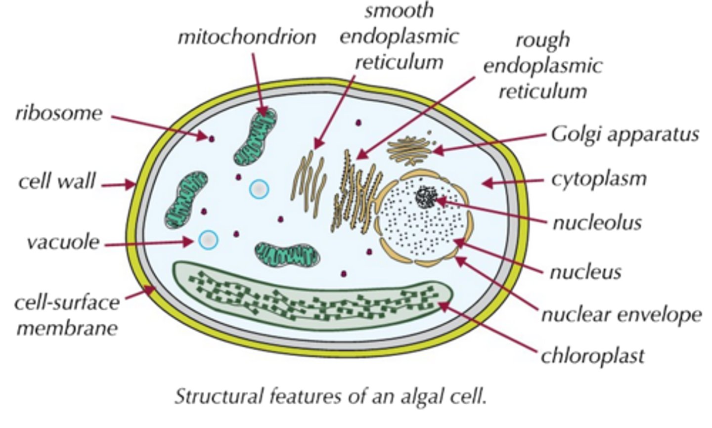
What are algal cells?
- Algae carry out photosynthesis, like plants, but unlike plants they can be unicellular (e.g. Chlorella) or multicellular (e.g. seaweed).
- Algal cells are a lot like plant cells; they have all the same organelles, including a cellulose cell wall and chloroplasts; however, the chloroplasts in many algal cells are a different shape and size to plant chloroplasts; for example, some algae have one large chlorplast rather than several smaller chloroplasts.
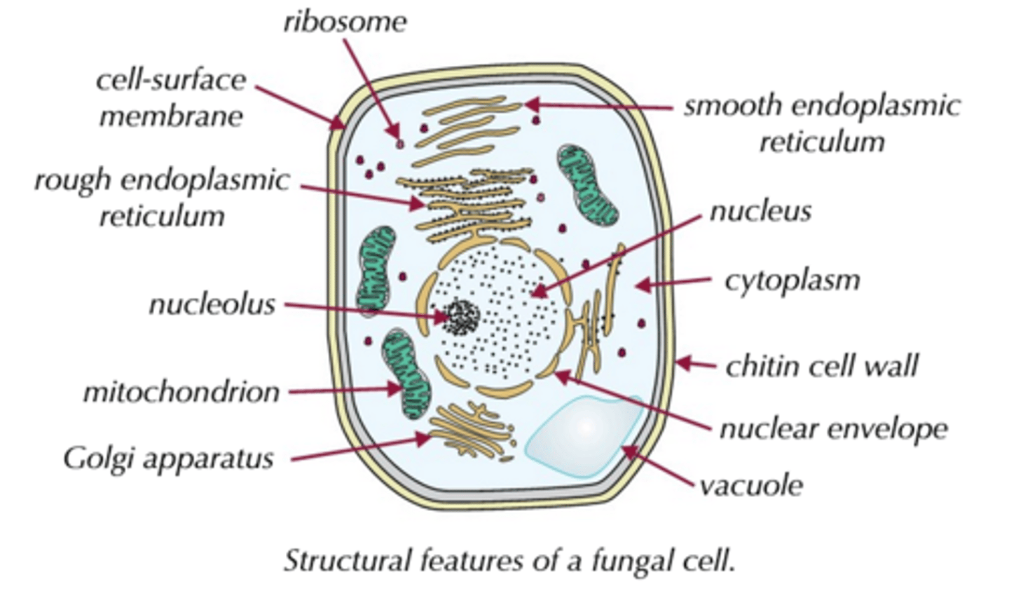
What are fungal cells?
- Fungi can also be multicellular (e.g. mushrooms) or unicellular (e.g. yeast).
- Fungal cells are also a lot like plant cells, but with two key differences:
1) Their cell walls are made of chitin, not cellulose.
2) They don't have chloroplasts (because they don't photosynthesise).
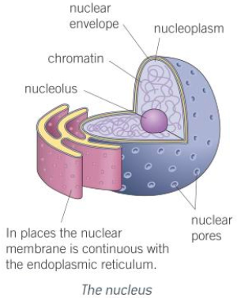
What is the Nucleus?
Description:
Most prominent feature of eukaryotic cells, such as an epithelial cell; spherical; 10-20 μm in diamter; contains organism's hereditary material; controls cell's activities; has a number of parts:
1) Nuclear envelope:
- Double membrane that surrounds the nucleus.
- Outer membrane is continous with the ER of the cell and often has ribosomes on its surface.
- Controls entry/exit of materials in/out of nucleus, and contains reactions taking place within it.
2) Nuclear pores:
- Allow passage of large molecules (e.g. mRNA) out of the nucleus.
- Typically around 3000 pores in each nucleus, each 40-100nm in diameter.
3) Nucleoplasm:
- Granular, jelly-like material.
- Makes up the bulk of the nucleus.
4) Nucleolus:
- Small, spehrical region within nucleoplasm.
- Manufactures ribosomal RNA and assembles the ribosomes.
- There can be more than one nucleolus in a nucleus.
Function:
- Acts as the control centre of the cell through production of mRNA and tRNA, and hence protein synthesis.
- Retains genetic material of the cell in the form of DNA and chromosomes.
- Manufacture ribosomal RNA and ribosomes.
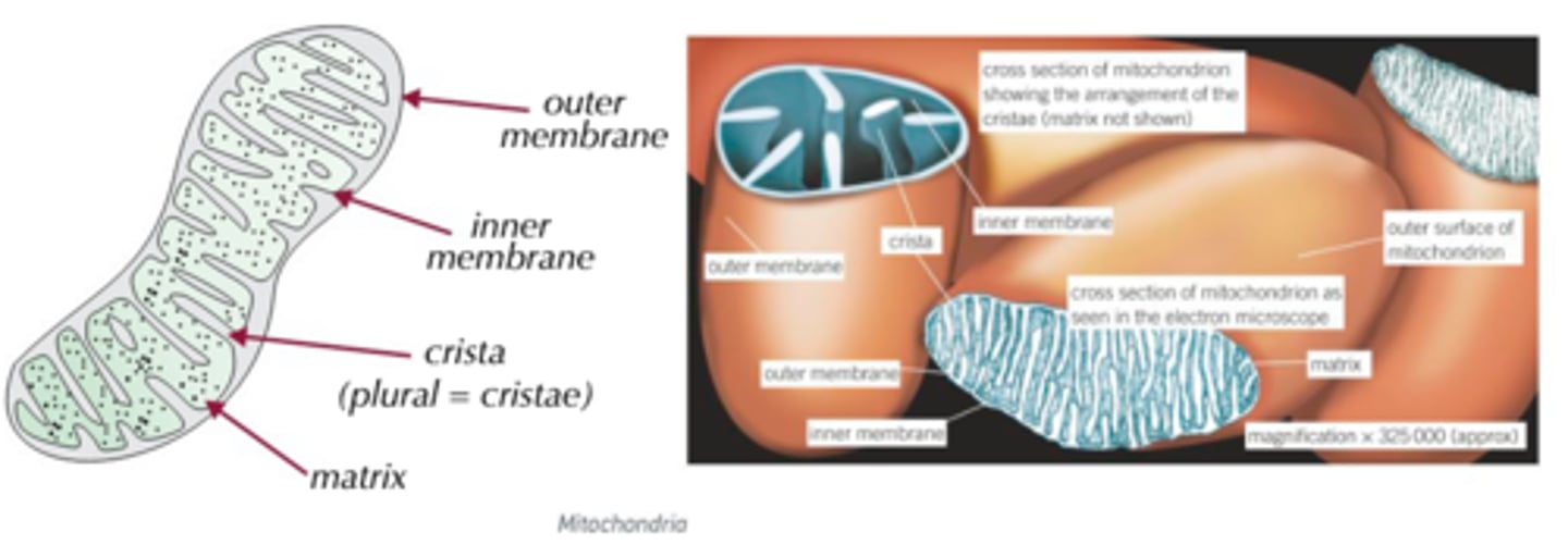
What are Mitochondria?
Description:
Rod-shaped; 1-10 μm in length, made up of:
1) Double membrane:
- Controls entry/exit of material.
- Iner membrane folded to form extensions known as cristae.
2) Cristae:
- Extensions of inner membrane.
- Extend across the whole width of the mitochondrion in some species.
- Provide large surface area for attachment of enzymes and other proteins involved in respiration.
3) Matrix:
- Makes up remainder of mitochondrion.
- Contains protein, lipids, ribosomes and DNA that sllows mitochondria to control production their own proteins.
- Location of many respiratory enzymes.
Function:
- Sites of aerobic stages of respiration (the Krebs cycle and the oxidative phosphorylation pathway).
- Responsible for production of ATP (the energy-carrier molecule), from respiratory substrates such as glucose.
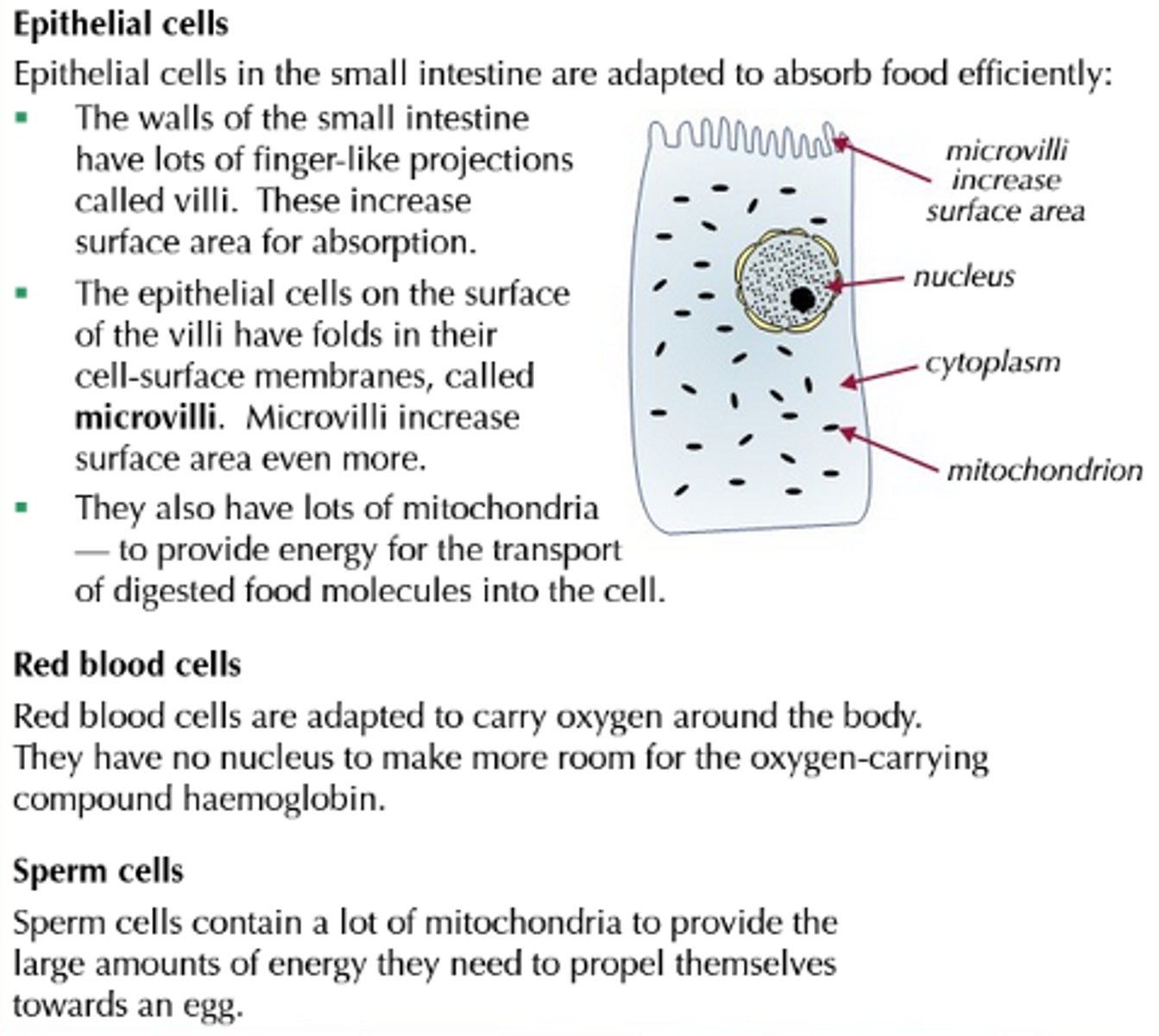
- Number/size of mitochondria/cristae are high in cells with high levels of metabolic activity, and require plentiful supply of ATP (these include muscle and epithelial cells; epithelial cells in the intestine require a lot of ATP for absorption of substances from the intestines via active transport).
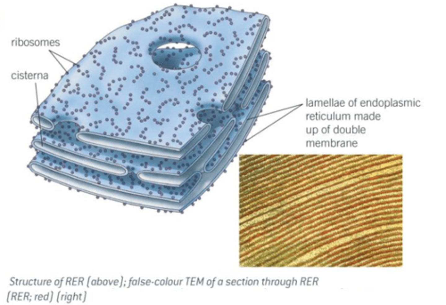
What is the Endoplasmic Retiuclum (ER)?
Description:
Elaborate, three-dimensional system of sheet-like membranes; spread throughout cytoplasm of cells; continuous with outer nuclear membrane; membranes enclose network of tubules and flattened sacs called cristernae; cells that manufacture/store large quantities of carbohydrates/proteins/lipids have a very extensive ER (e.g. liver and secretory cells such as epithelial cells which line the intestine); there are two types of ER:
1) Rough endoplasmic retiuclum (RER):
- Has ribosomes present on outer surface of membranes.
- Provides large surface area for synthesis of proteins & glycoproteins.
- Provides pathway for transport of materials (especially proteins) throughout cell.
2) Smooth endoplasmic reticulum (SER):
- Lacks ribosomes on surface.
- Often more tubular in apperance.
- Synthesises/stores/transports lipids and carbohydrates.
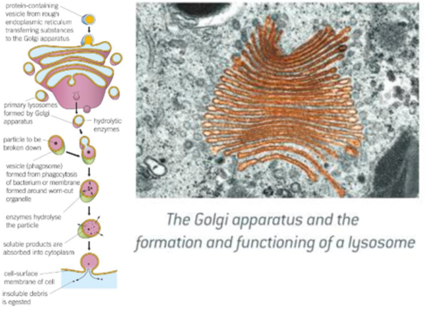
What is the Golgi Apparatus?
Description:
Occurs in almost all eukaryotic cells; similar to SER in structure, but is more compact; it is especially well developed in secretory cells, such as the epithelial cells that line the intestines; the Golgi is made up of:
1) Cisternae:
- Stack of membranes that make up flattened sacs.
2) Golgi Vesicles:
- Small, rounded, hollow structures.
- Once accurately sorted and sent to correct destinations, after being labelled and modified (often with non-protein components such as carbohydrates) whilst being passed through the Golgi in a strict sequence, after being produced by the ER; lipids/proteins are transported in Golgi vesicles (which are regularly pinched off from the ends of the Golgi cisternae).
- Vesicles may move to the cell surface, where they fuse with the membrane and release their contents to the outside.
Function:
- Adds carbohydrates to proteins to form glycoproteins.
- Produces secretory enzymes, such as those secreted by panceas.
- Secreted carbohydrates, such as those used in making cell walls in plants.
- Transport, modify, and store lipids.
- Form lysosomes.
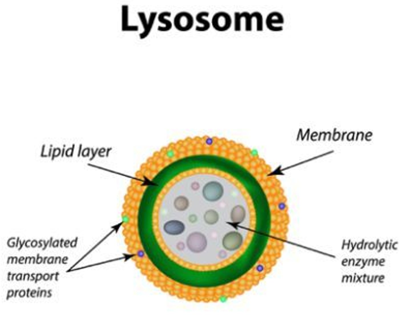
What are Lysosomes?
Description:
Formed when the vesicles produced by Golgi contain enzymes such as proteases and lipases; up to 1.0 μm in diameter; they're abundant in secretory cells (e.g epithelial cells) and phagocytic cells; they contain:
1) Lysozymes:
- Enzymes which hydrolyse the cells walls of certain bacteria.
- There can be up to 50 lysozymes in a single lysosome.
- Lysosomes isolate lysozymes from the rest of the cell before releasing them to either the outside or into a phagocytic vesicle within the cell.
Function:
- Hydrolyses material ingested by phagocytic cells (e.g. white blood cells and bacteria).
- Release enzymes to the outside of the cell (exocytosis) in order to destroy material around the cell.
- Digest worn out organelles so that the useful chemicals they're made of can be re-used.
- Completely break down cells affter they have died (autolysis).
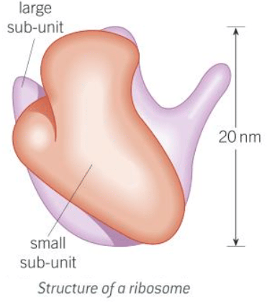
What are Ribosomes?
Description:
Small cytoplasmic granules found in all cells; they have two subunits - one large and one small, each of which contains ribosomal RNA and protein; may occur in cytoplasm or be associated with the RER; despite small size, they occur in vast numbers and can account for up to 25% of the dry mass of a cell; there are two types, depending on the cells in which they are found:
1) 80S Ribosomes:
- Found in eukaryotic cells.
- Around 25 nm in diameter.
2) 70S Ribosomes:
- Found in prokaryotic cells, mitochondria and chloroplasts.
- Slightly smalled than 80S ribosomes.
Function:
- Site of protein synthesis.
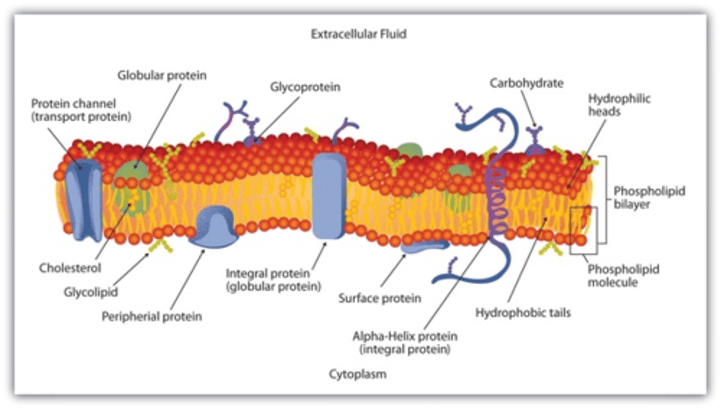
What is the Cell-surface Membrane/Plasma Membrane?
Description:
- The membrane found on the surface of animal cells and just inside the cell wall of other cells.
- Mainly made of lipids and protein.
Function:
- Regulates the movement of substances into and out of the cell.
- Has receptor molecules on it, which allow it to respond to chemicals like hormones.
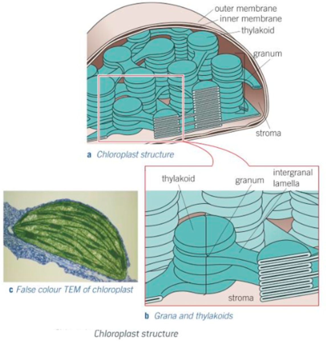
What is a Chloroplast?
Description:
Organelles which carry out photosynthesis; vary in shape and size but are: typically disc-shaped, typically 2-10 μm long, typically 1 μm in diameter; their main features include:
1) Chloroplast envelope:
- Double plasma membrane that surrounds the organelle.
- Highly selective in what it allows to enter/leave the chloroplast.
2) Grana:
- Stacks of up to 100 disc-like structures called thylakoids, where first stage of photosynthesis (light absorption) takes place.
- Thylakoids contain photosynthetic pigment called chlorophyll.
- Thylakoids have tubular extensions which join up with thylakoids in the adjacent grana.
3) Stroma:
- Fluid-filled matrix where the second stage of photosynthesis (synthesis of sugars) takes place.
- Within stroma are numerous other structures (e.g. starch grains).
Function:
Chloroplasts are adapted to their function of harvesting sunlight and carrying out photosynthesis in the following ways:
- Granal membranes provide a large surface area for attachment of chlorophyll, electron carries and enzymes that carry out light absorption; these chemicals are attached to the membrane in a highly ordered fashion.
- Fluid of stroma possesses all enzymes needed to make sugars in sugar synthesis.
- Chloroplasts contain both DNA and ribosomes, and can quickly and easily manufacture proteins required for photosynthesis.
What is the Cell Wall?
Description:
Consists of microfibrils of the polysaccharide cellulose, embedded in a matrix; cell walls of algae are made up of either cellulose or glycoproteins (or a mixture of both); cell walls of fungi don't contain cellulose, but comprise a mixture of a nitrogen-containing polysaccharide called chitin, a polysaccharide called glycan and glycoproteins; cell walls have the following features:
1) Numerous Polysaccharides:
- Microfibrils of cellulose have considerable strength and so contribute to the overall strength of the cell wall.
2) Middle lamella:
- Thin layer which marks the boudary between adjacent cell walls and cements adjacent cells together.
Function:
- Provides mechanical strength, preventing the cell from bursting under the pressure created by the osmotic entry of water (osmotic lysis).
- Provides mechanical strength to the plant as a whole.
- Allows water to pass along it, contibuting to the movement of water through the plant.
What are Cell Vacuoles?
Description:
1) Fluid-filled sac bounded by a single membrane.
2) It contains a solution of mineral salts, sugars, amino acids, wastes and sometimes pigments such as anthocyanins.
3) The single membrane around it is called the tonoplast.
4) Within mature plant cells, there is usually one large central vacuole.
Function:
- Support herbaceous plants, and herbaceous parts of woody plants, by making cells turgid.
- The sugars and amino acids may act as a temporary food store.
- The pigments may colour petals to attract pollinating insects.
How can you relate a cell's ultrastructure to it's function?
- As each organelle has it's own fucnction, it's possible to deduce, with reasonable accuracy, the role of a cell by looking at the number and size of the organelles it contains.
- E.g. mitchondria produce ATP (mainly used as a temporary energy store), meaning cells with many mitochondria are likely to require a lot of ATP and therefore high rates of metabolism; even within each mitochondrion, the more dense and numerous the cristae, the greater the metabolic rate of the cell possessing these mitochondria.
How, and why do cells specialise?
- To stay alive, all cells of a multicellular organism perform certain, basic functions.
- However, no one cell can provide the best conditions for all functions.
- Therefore, cells of multicellular organisms are each specialised in different ways to perform particular roles.
- Each specialised cell has evolved more or fewer of certain organelles and structures to suit the role it carries out, and each cell becomes specialised in structure to suit the role it will carry out (as an embryo matures, the first group of initially identical cells, take on their own individual charecteristics to suit to the function they will perform when they mature).
- All cells in an organism, such as a human, are produced by mitotic divisions from the fertilised egg.
- All cells in an organism contain the same genes, and every cell contains the genes needed for it to decelop into any one of many different cells in an organism; however, only some of these genes are expressed in any one cell, at any one time.
- Different genes are expressed in each type of specialised cell, whilst the rest are not.
- Alongside shape, numbers of each organelle may vary (e.g. a muscle or sperm cell has many mitochondria, whilst a bone cell has very few); these cells are adapted to their own particular function and perform it more effectively, and the organism functions efficiently as a result.
What are tissues?
- For working efficiency, cells are normally aggregated together.
- A collecution of similar cells that perform a specific function is known as a tissue.
- E.g. Epithelial tissues (found in animals) consist of sheets of cells; they line the surfaces of organs and often have a protective or secretory function; there are many similar types, including those made up of thin, flat cells that line organs where diffusion takes place, for example the alveoli of the lungs, and ciliated epithelium that lines a duct such as the trachea; the cilia are used to move mucus over the epithelial surface.
- E.g. Xylem, which occurs in plants, is made up of numerous similar cell types, and is used to transport water and mineral ions throughout the plant and provides mechanical support.
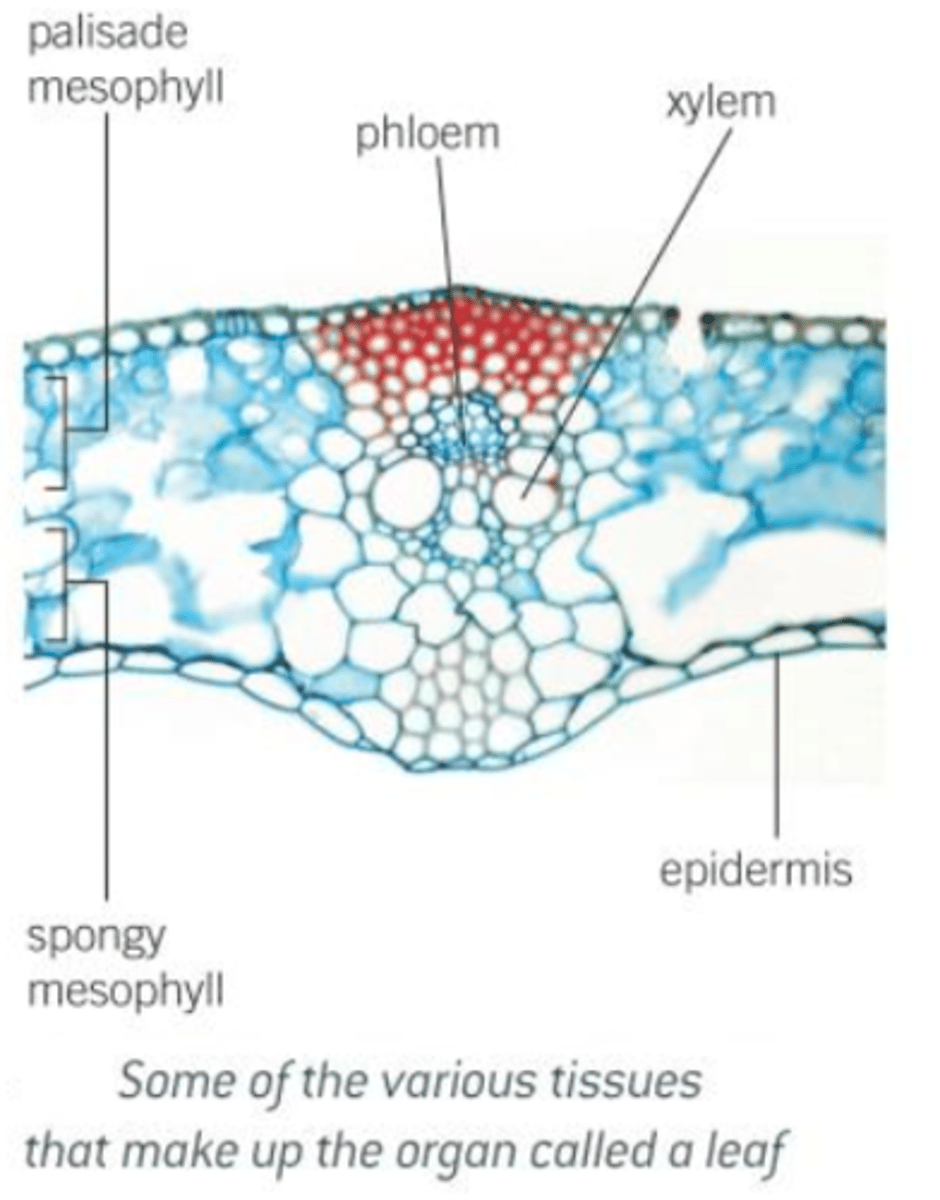
What are organs?
- Just as cells are aggregated into tissues, tissues are aggregated into organs.
- An organ is a combination of tissues that are coordinated to perform a variety of functions, although they often have one predominant major function.
- In animals, for example, the stomach is an organ involved in the digestion of certain types of food, and is made up of tissues such as: muscle (to churn and mix the stomach contents), epithelium (to protect the stomach wall and produce secretions), connective tissue (to hold together the other tissues).
- In plants, a leaf is an organ made up of: palisade mesophyll (made up of leaf palisade cells that carry out photosynthesis), spongy mesophyll (adapted for gaseous diffusion), phloem (to transport organic materials away from the leaf), xylem (to transport water and ions into the leaf).
- It's not always easy to determine which structures are organs, however: blood capillaries aren't organs whereas arteries and veins are; all three structures have the same major function, but the capillaries are made up of just one tissue (epithelium), whereas arteries and veins consist of numerous tissues (muscle, epithelial, etc).
What are organ systems?
- Organs work together as a single unit known as an organ system.
- These systems may be grouped together to perform particular functions more efficiently.
- There are a number of organ systems in humans: the digestive system (digests and processes food; made up of organs including: the salivary glands, oesophagus, stomach, duodenum, ileum, pancreas and liver), respiratory system (used for breathing and gas exchange; made up of: trachea, bronchi and lungs) and the circulartory system (pumps and circulates blood; made up of organs that include: the heart, arteries and veins).
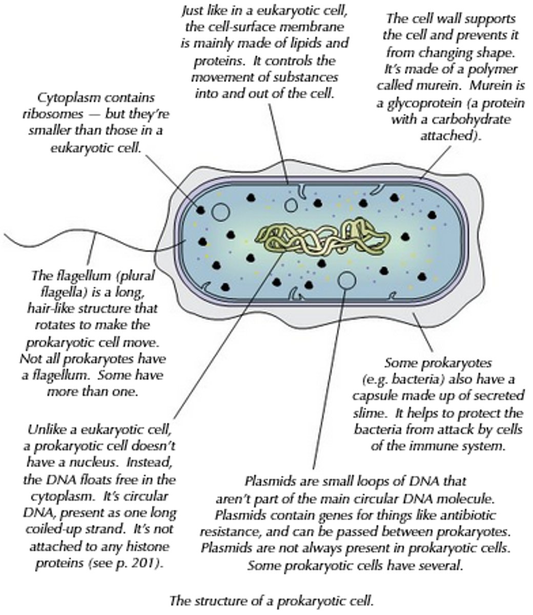
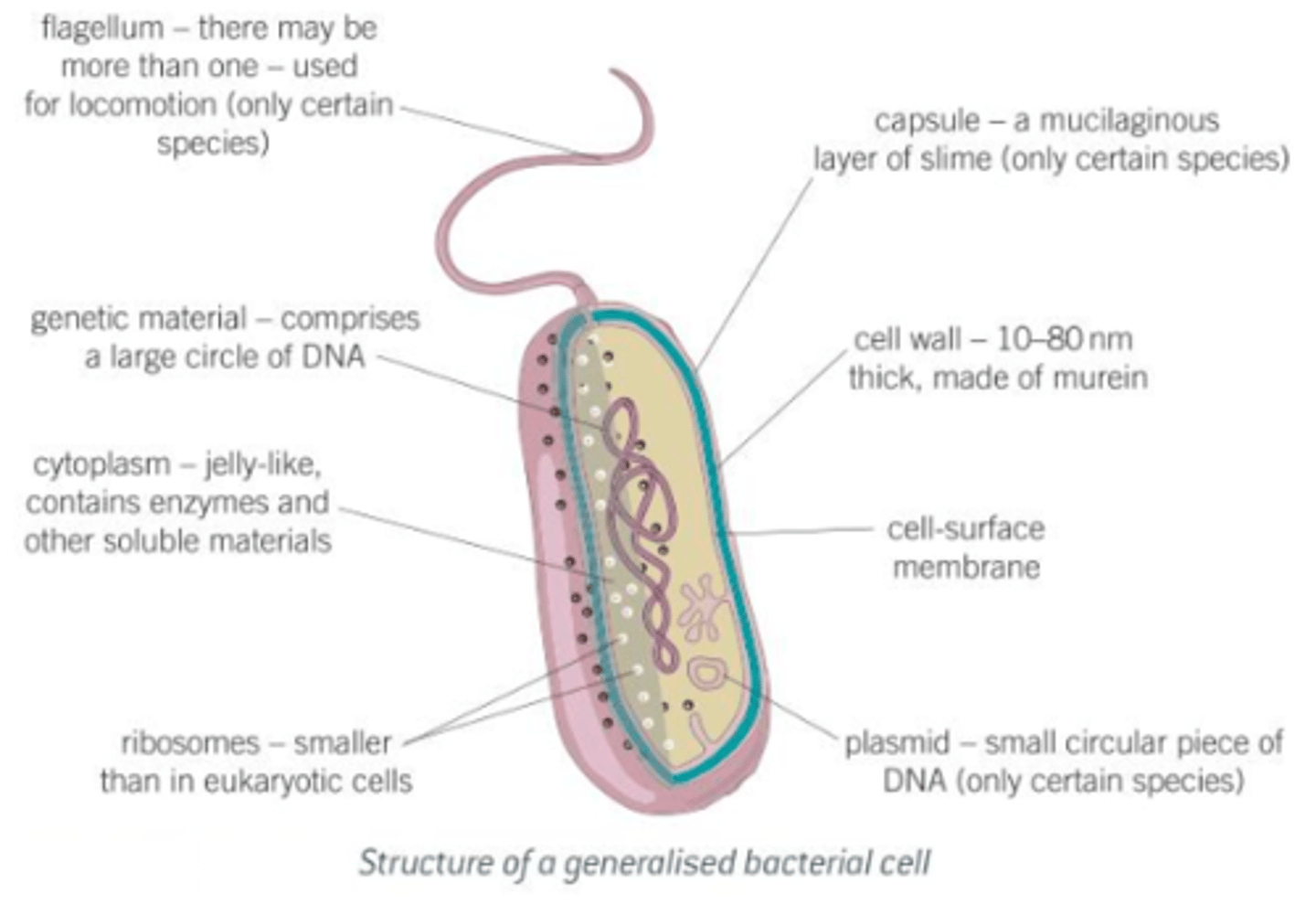
What is the structure of a bacterial cell?
- Bacteria normally range from 0.1-10 μm in length.
- All bacteria posses a cell wall made up of murein (a polymer of polysaccharides and peptides).
- Many bacteria further protect themselves by secreting a capsule of mcilaginous slime around this wall.
- Inside the cell wall is the cell-surface membrane, within which the cytoplasm contains 70S ribosomes.
- Bacteria store food reserves as glycogen granules and oil droplets.
- The genetic material in bacteria is in the form of a circular strand of DNA; seperate from this are smaller circular pieces of DNA, called plasmids (which can reproduce themselves independently and may give the bacterium resistance to harmful chemicals such as antibiotics).
- Plasmids are used extensively as vectors (carriers of genetic information) in genetic engineering.
What are the roles of the structures found in a bacterial cell?
1) Cell wall: Physical barrier that excludes certain substances and protects against mechanical damage and osmotic lysis.
2) Capsule: Protects bacterium from other cells and helps groups of bacteria to stick together for further protection.
3) Cell-surface membrane: Acts as a differentially permeable layer, which controls the entry and exit of chemicals.
4) Circular DNA: Possesses genetic information for the replication of bacterial cells.
5) Plasmid: Possesses genes that may aid the survival of bacteria in adverse contions, e.g. produces enzymes that break down antibiotics.
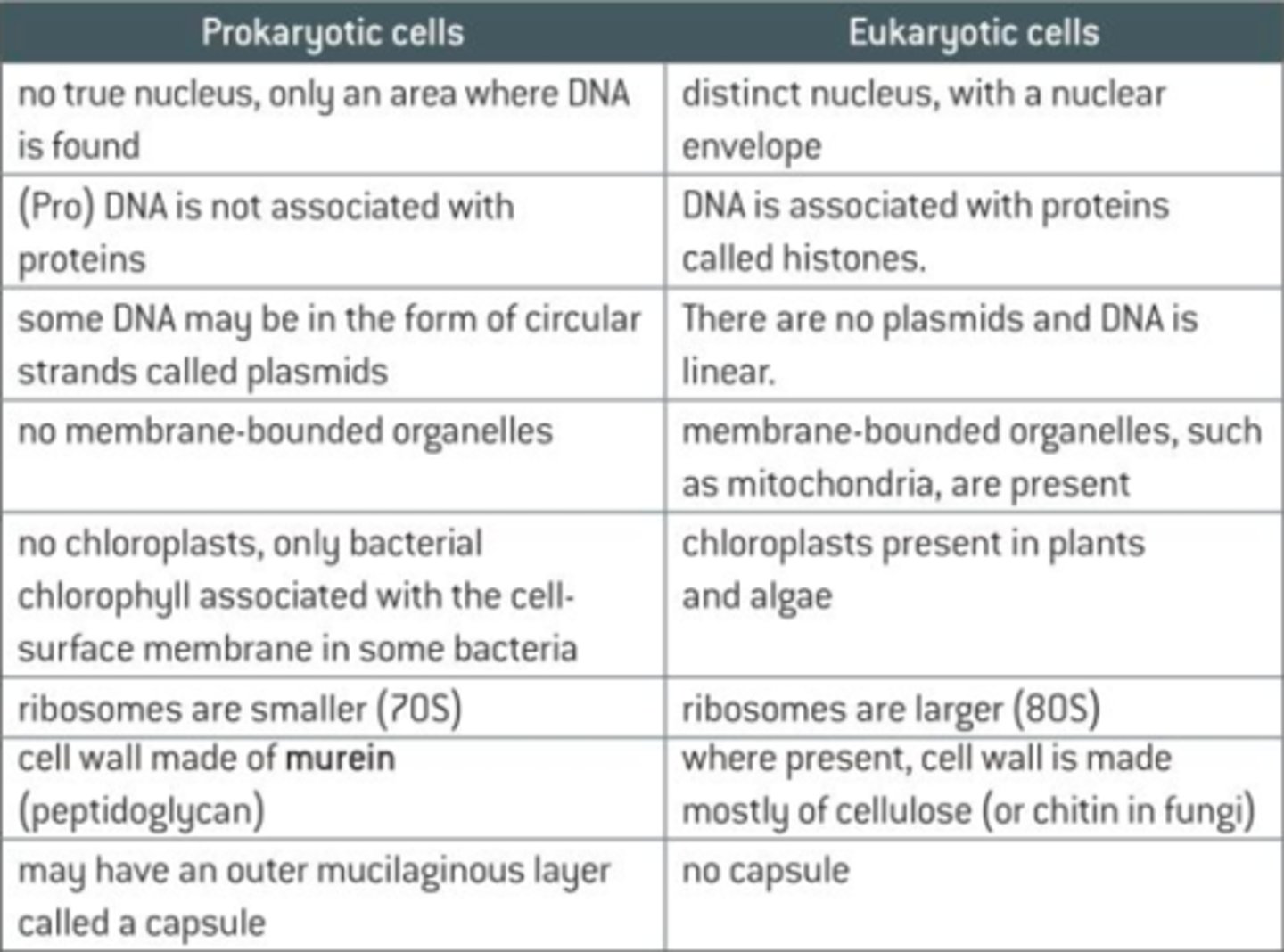
What are the differences between prokaryotic and eukaryotic cells?
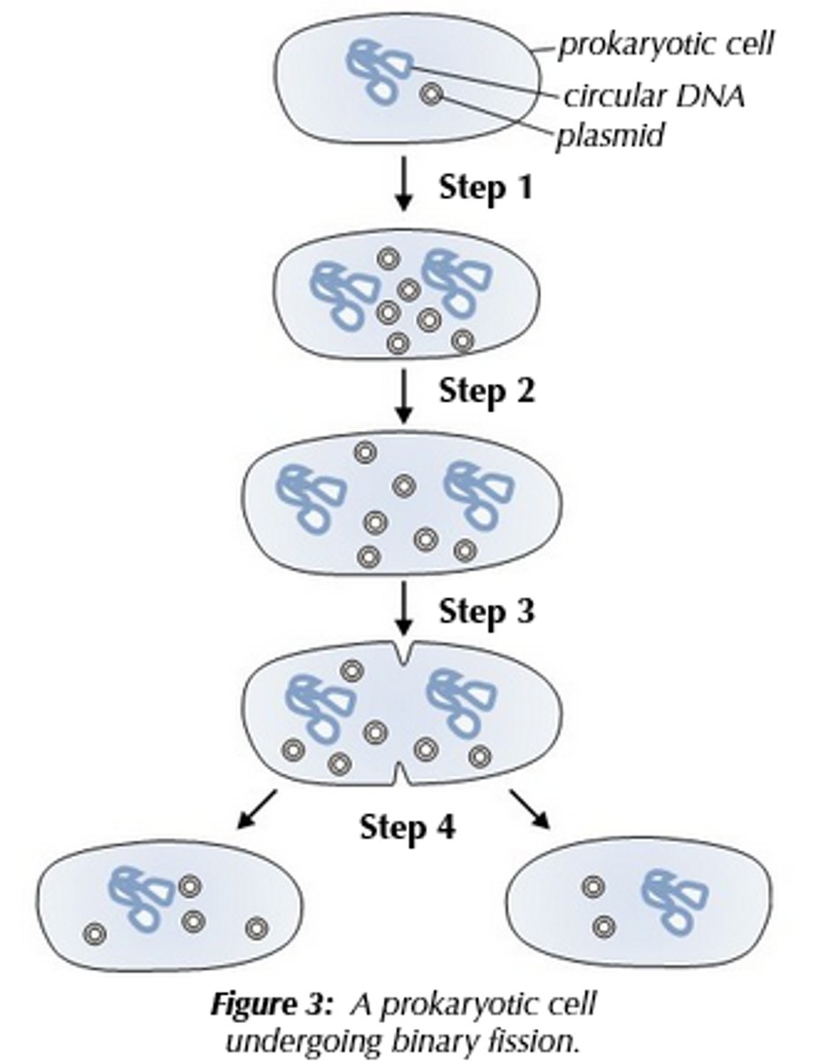
How do prokaryotic cells replicate?
Prokaryotic cells replicate by a process known as binary fission, in which the cell replicates its genetic material, before physically splitting into two daughter cells.
What is the process of binary fission?
1) The circular DNA and plasmid(s) replicate; the main DNA loop is only replicated once, but plasmids can be replicated many times.
2) The cells get bigger, and the DNA loops move to opposite 'poles' (ends) of the cell.
3) The cell membrane begins to grow between the two DNA molecules and begins to pinch inward, and the cytoplasm begins to divide (and new cell walls begins to form).
4) The cytoplasm divides and two daughter cells are produced; each daughter cells has one copy of the cicular DNA, but can have a variable number of copies of the plasmid(s).
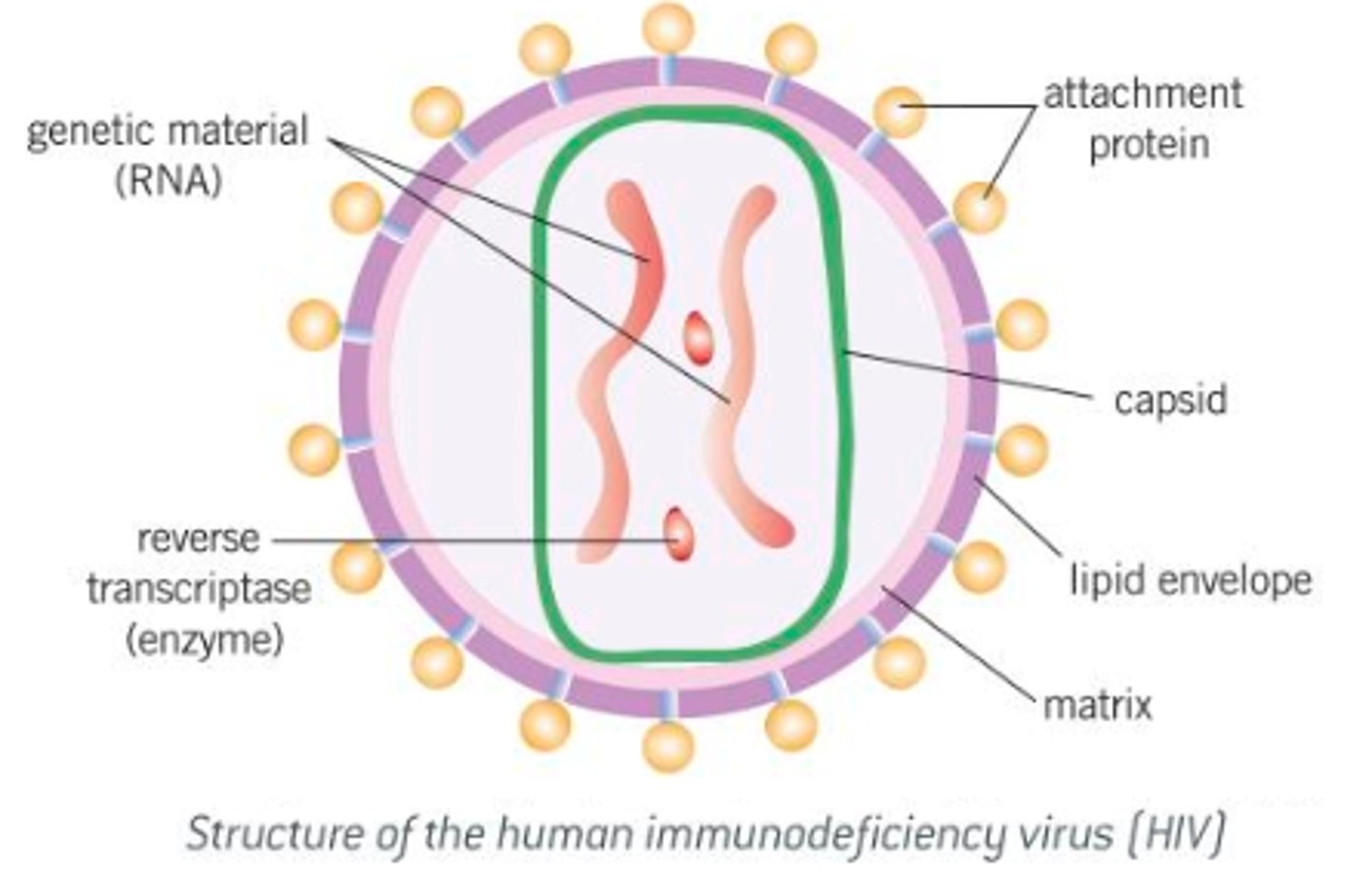
What are viruses?
- Acellular, non-living particles.
- Smaller than bacteria, ranging in size from 20-300 nm.
- Contain nucleic acids such as DNA or RNA as genetic material, but can only multiply inside living host cells.
- Nucleic acid is enclosed within a protein coat known as a capsid.
- Some viruses, like the human immunodeficiency virus, are further surrounded by a lipid envelope.
- The lipid envelope (or the capsid, if the lipid envelope isn't present) have attachment proteins, essential to allow the virus to identify and attach to host cells.
- Viruses don't have a cell-surface membrane, cytoplasm or ribosomes.
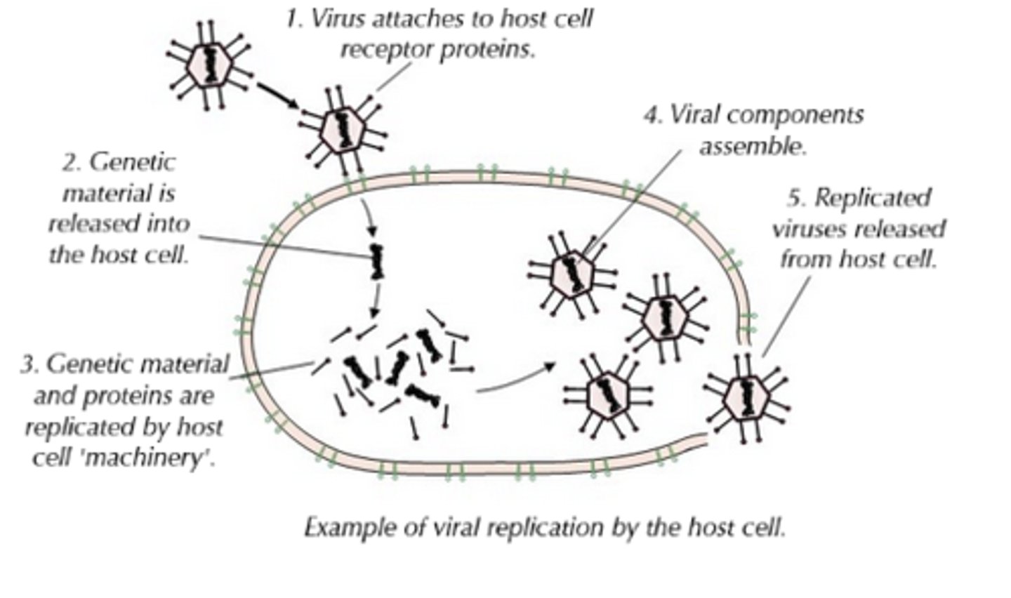
How do viruses replicate?
- Because they're not alive, viruses don't undergo cell division.
- They inject their DNA/RNA into the host cell, and this hijacked cell then uses its own 'machinery' (e.g. enzymes, ribosomes) to replicate the viral particles.
- In order to inject their DNA/RNA, viruses much first attach to the host cell surface, and they need their attachment proteins to achieve this, and bind to the complementary receptor proteins on the cell-surface membrane of the host cells.
- Different viruses have different attachment proteins and, therefore, require different receptor proteins on host cells; as a result some viruses can only infect one type of cell (e.g. some viruses can only infect one species of bacteria), whilst others can infect lots of different cells (e.g. influenza).
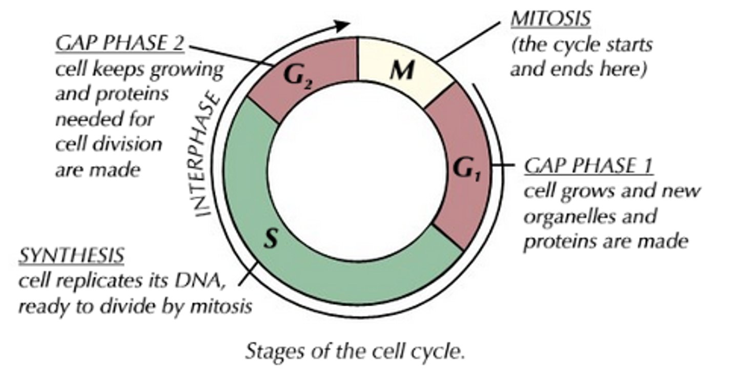
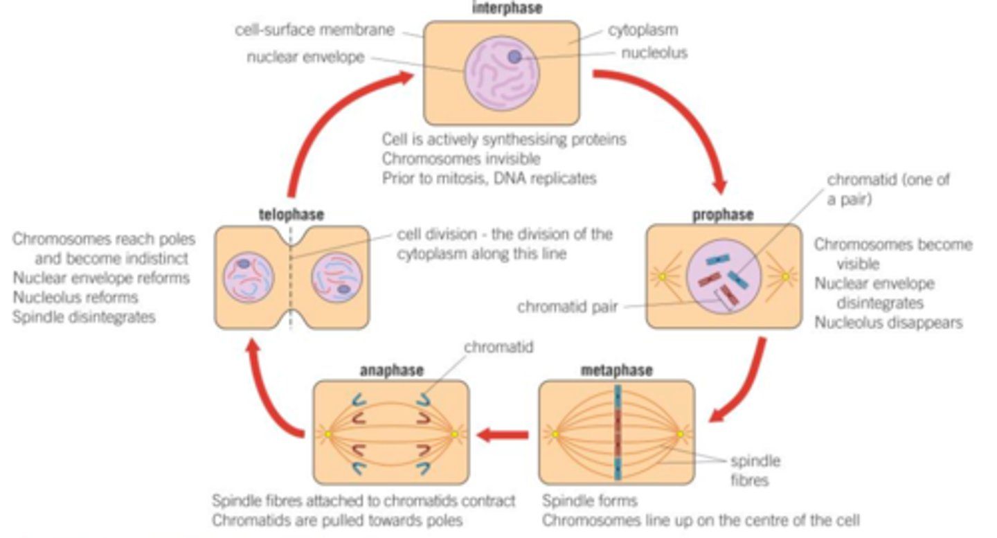
What is the cell cycle?
- In multicellular organisms, not all cells keep their ability to divide, but ones that do, follow a process called the cell cycle.
-The cell cycle starts when a cell has been produced by cell division and ends with the cell dividing to produce two identical cells.
- The cell cycle consists of a period of cell growth and DNA replication, called interphase, and a period of cell division, called mitosis.
What is Interphase?
- Interphase occupies most of the cell cycle, and is sometimes known is the resting phase because no division takes place.
- Interphase is subdivided into three seperate growth stages called G₁, S and G₂.
- During interphase, the cell carries out normal functions, but also prepares to divide.
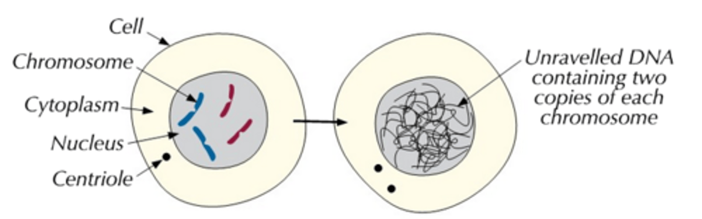
- The cell's DNA is unravelled and replicated, to double its genetic content.
- The organelles are also replicated so it has spare ones, and its ATP content is increased (ATP provides the energy needed for cell division).
How does cell division take place?
Cell division can take place either by mitosis or meiosis:
- Mitosis produces two daughter cells that have the same number of chromosomes as the parent cell and each other.
- Meiosis produces four daughter cells, each with half the number of chromosomes of the parent cell.
What is mitosis?
- The division of a cell, resulting in each of the daughter cells having an exact copy of the DNA of the parent cell (unless the rare event of a mutation occurs).
- Mitosis is always preceded by a period during which the cell is not dividing, called interphase.
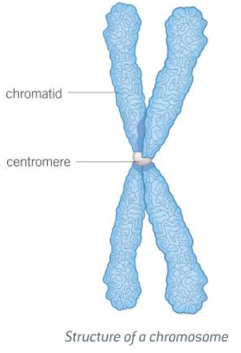
- Interphase is a period of considerable cellular activity including the replication of DNA; the two copies of DNA after replication remain joined at a place called the centromere.
What are the stages of mitosis, and what happens in each one?
1) Prophase:
- Chromosomes first become visible, initally as long thin threads, which later shorten and thicken.
- Animal cells contain two cylindrical organelles called centrioles, each of which move to opposite poles of the cell.
- From each centriole, spindle fibres develop, which span the cell from pole to pole.
- Spindle fibres, collectively, form the spindle appartus.
- As plant cells lack centrioles, but do develop a spindle appartus, centrioles are not essential to spindle fibre formation.
- Nucleolus disappears, and the nuclear envelope breaks down, leaving chromosomes free in the cytoplasm of the cell.
- These chromosomes are drawn towards the equator of the cell by the spindle fibres attached to the centromere.
2) Metaphase:
- Chromosomes are seen to be made up of two chromatids.
- Each chromatid is an identical copy of DNA from the parent cell.
- The chromatids are joined by the centromere.
- The centromere is what some microtubules from the poles are also attached, and the chromosomes are pulled along the spindle apparatus, arranging themeselves across the equator of the cell.
3) Anaphase:
- The centromeres divide into two and spindle fibres pull the individual chromatids, making up the chromosome, apart.
- The chromatids move rapidly to their respective, opposite poles of the cell and we now refer to them as chromosomes.
- The energy for anaphase is provided by mitochondria, which gather around the spindle fibres.
- If cells are treated with chemicals that destroy the spindle, the chromosomes remain at the equator, unable to reach the poles.
4) Telophase:
- Chromosomes reach their respective poles and become longer and thinner, finally disappearing altogether, leaving only widespread chromatin.
- Spindle fibres disintegrate and the nuclear envelope and nucleolus re-form.
- Finally, the cytoplasm divides in a process called cytokinesis.
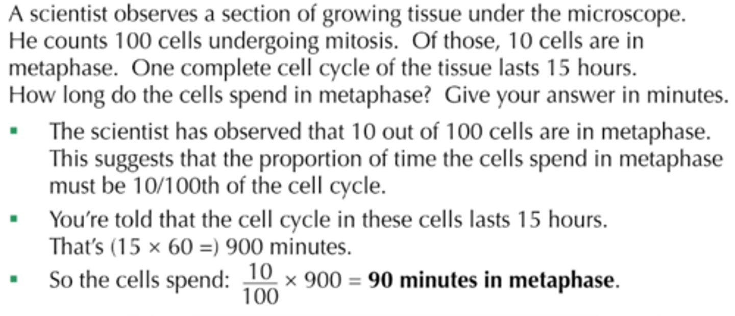
How can you calculate the time taken for each stage of mitosis?
- The time taken for each stage of mitosis varies depending on the cell type and the environmental coditions.
- You can calculate how long each stage of mitosis lasts with the right information (how many of the cells are undergoing mitosis, how many of the cells are going undergoing each phase/the phase in question, how long one complete cell cycle of the tissue you're investigating lasts).
What is cancer?
- Cancer is a group of diseases (around 200 in total) caused by a growth disorder of cells, resulting from damage to the genes that regulate mitosis and the cell cycle, leading to uncontrolled growth and division of cells.
- As a consequence, a group of abnormal cells, called a tumour, develops and constantly expands in size.
- Tumours can develop in any organ of the body, but are most commonly found in the lungs, prostate gland (male), breast and ovaries (female), large intestine, stomach, oesophagus and pancreas.
- A tumour becomes cancerous if it changes from benign to malignant.
How does mitosis sometimes result in tumours?
- Most cells divide by mitosis, either to increase the size of a tissue during development (growth), or to replace dead/worn out cells (repair).
- The rate of mitosis can be affected by the environment of the cell and by growth factors.
- It is also controlled by two types of gene, and a mutation in either of these genes results in uncontrolled mitosis.
- The mutant cells formed as a result of uncontrolled mitosis are usually structurally and functionally different from normal cells.
- Most mutated cells die, however, any that survive are capable of replicating and forming tumours.
- Malignant tumours grow rapidly, are less compact and are more likely to be life-threatening, while benign ones grow more slwoly, are more compact, and are less likely to be life-threatening.
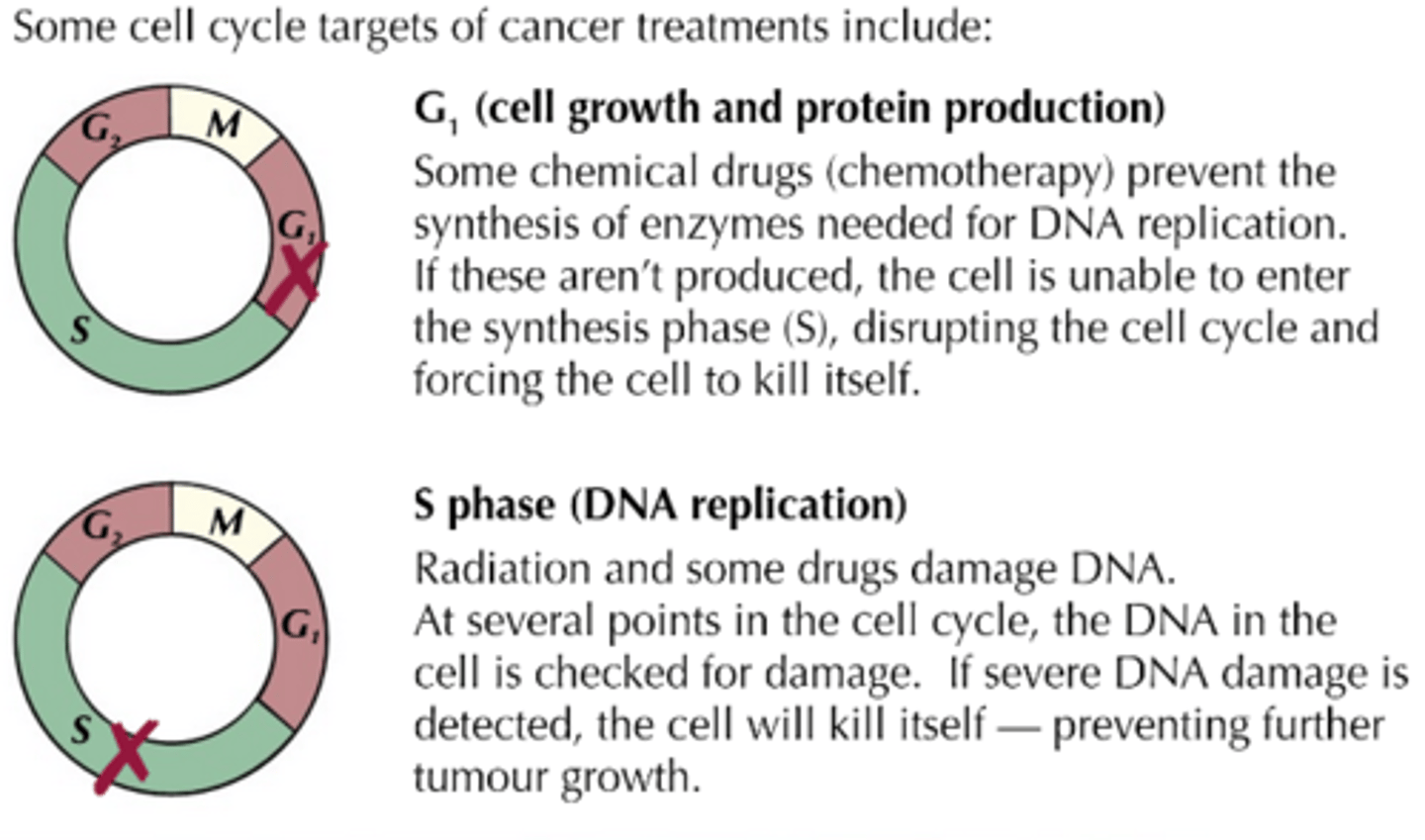
How can cancer be treated?
- The treatment of cancer often involves killing dividing cells by blocking a part of the cell cycle.
- In this way, the cell cycle is disrupted and cell division, and hence cancer growth, ceases.
- Drugs used to treat cancer (chemotherapy) usually disrupt the cell cycle by preventing DNA from replicating or inhibiting the metaphase stage of mitosis by interfering with spindle formation.
- The problem with such drugs, however, is that they also dirsrupt the cell cycle of normal cells; but, the drugs are more effective against rapidly dividing cells, and as cancer cells have a particularly fast rate of division, they are damaged to a greater degree than normal cells.
- Those normal body cells, such as hair-producing cells, that divide rapidly are also vulnerable to damage, explaing the hair loss frequently seen in patients undergoing cancer treatment.
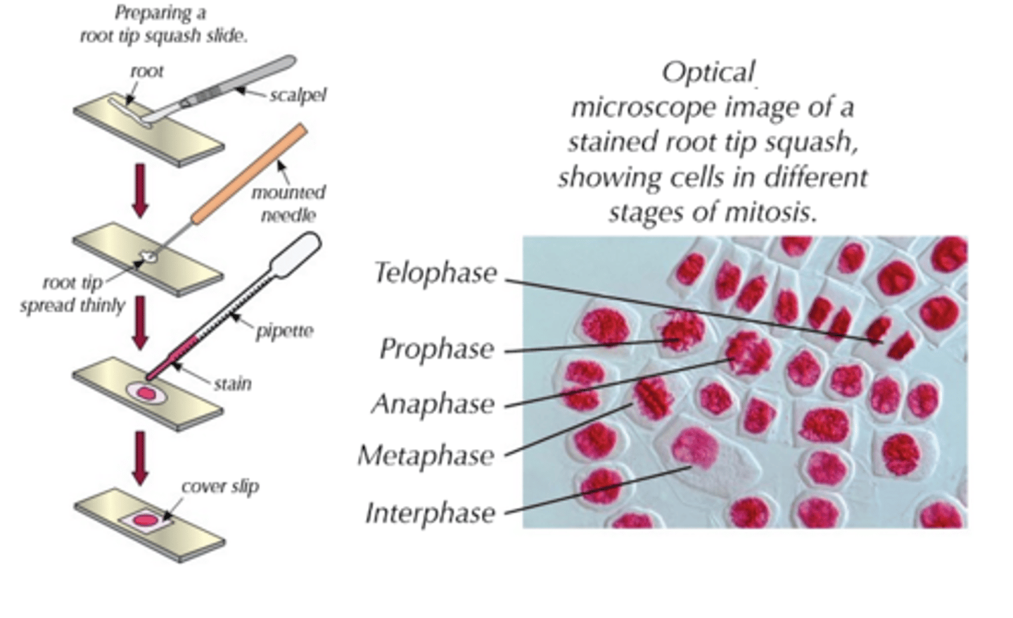
How do you prepare a root tip cell squash?
(REQUIRED PRACTICAL 2)
- You need to know how to prepare and stain a root tip in order to observe the stages of mitosis.
- Make sure you're wearing safety goggles and a lab coat before starting; you should also wear gloves when handling stains.
1) Add some 1 M HCL to a boiling tube - just enough to cover the root tip (the acid should therefore be a few millimetres deep); put the tube in a water bath that has equilibrated at 60℃.
2) Use a scalpel to cut 1 cm from the tip from a growing root (e.g. of an onion) - this needs to be the tip, as that's where growth occurs, and hence where mitosis takes place.
3) Carfully transfer root tip into boiling tube containing acid; incubate for approximately 5 minutes.
4) Use tweezers to remove tip from the tube and use a pipette to rinse it well with cold water; leave the tip to dry on a paper towel.
5) Place the root tip on a microscope slide and cut 2 mm from the very tip of it, getting rid of the rest.
6) Use a mounted needle to break the tip open and spread out cells thinly.
7) Add a few drops of stain and leave it for a few minutes - the stain will make the chromosomes easier to see under a microscope (toluidine blue O, ethano-orcein and Feulgen stain are a few examples of the stains you can use).
8) Place a cover slip over the cells and put a piece of folded filter paper on top, pushing it down firmly to squash the tissue; squashing makes the tissue thinner, and allows light to pass through it; don't smear the cover slip sideways, or you'll damage the chromosomes.
9) Now, all the stages of mitosis can be observed under an optical microscope.
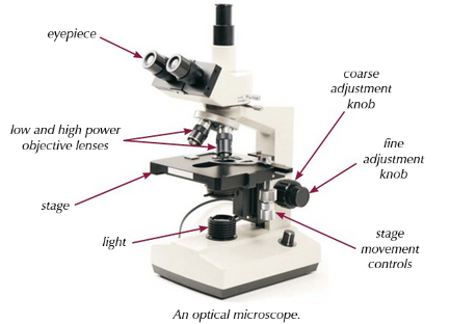
How do you use an optical microscope?
(REQUIRED PRACTICAL 2)
You need to know how to set up and use an optical microscope to observe your prepared root tip cells:
1) Start by clipping the slide you've prepared onto the stage.
2) Select the lowest-powered objective lens (i.e. the one that produces the lowest magnification).
3) Use the coarse adjustment knob to bring the stage up to just below the objective lens.
4) Look down the eyepiece (which contains the ocular lens); use the coarse adjustment knob to move the stage downwards, away from the objective lens, until the image is roughly in focus.
5) Adjust the focus with fine adjustment knob, until you get a clear image of what's on the slide.
6) If you need to see the slide with a greater magnification, swap to a higher-powered objective lens and refocus.
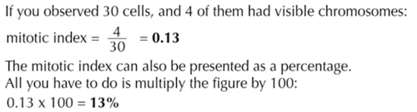
How do you calculate mitotic index?
(REQUIRED PRACTICAL 2)
- The mitotic index is the proportion of cells in a tissue sample, that are undergoing mitosis.
- It lets you work out how quickly a tissue is growing, and if there's anything abnormal going on.
- Mitotic Index = Number of cells with visible chromosomes ÷ Total number of cells observed
- A plant root tip is constantly growing, so you'd expect a high mitotic index (i.e. lots of cells in mitosis); in other tissue samples, a high mitotic index could mean that tissue repair is taking place or that there is cancerous growth in the tissue.
How can you calculate the actual size of cells?
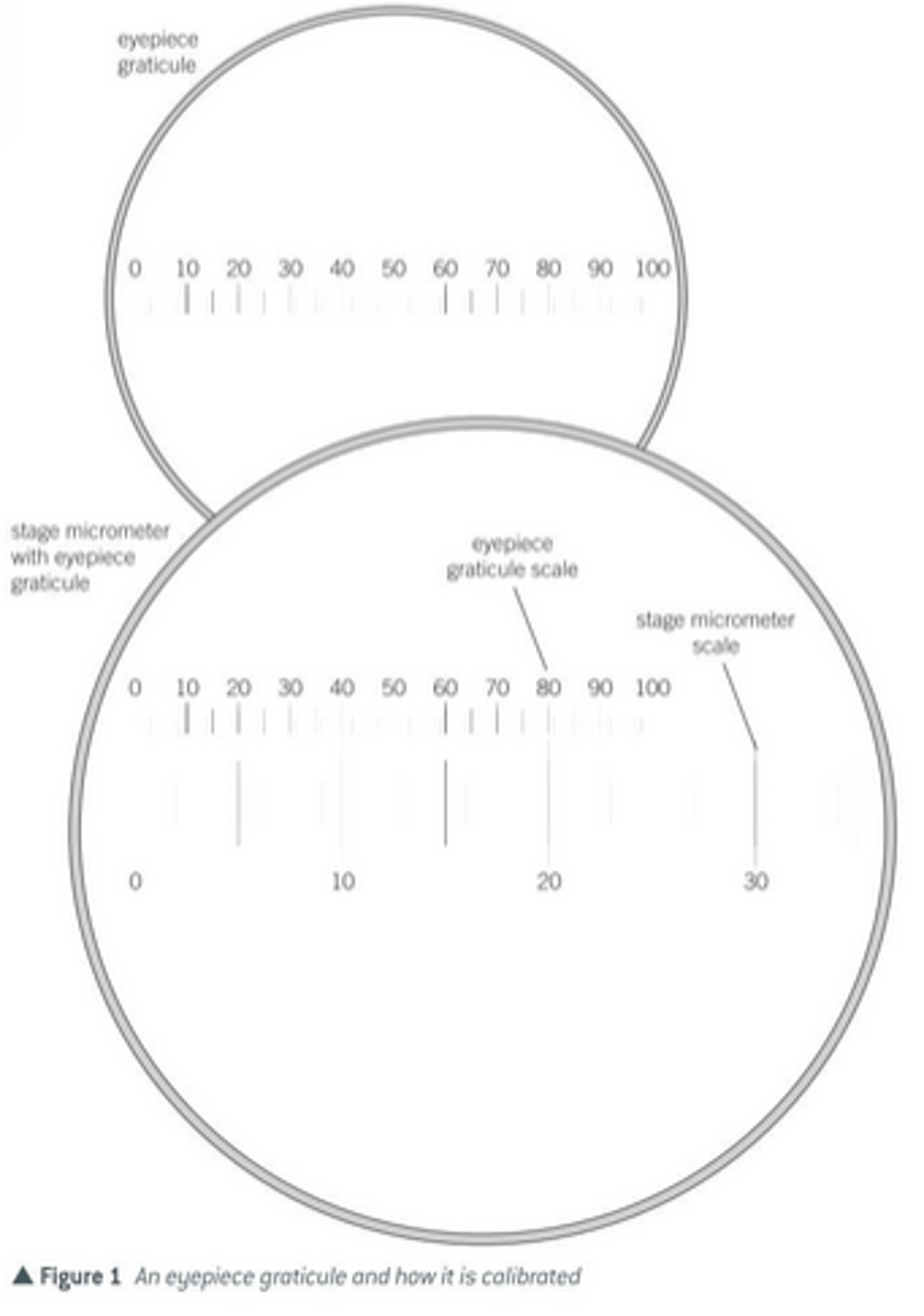
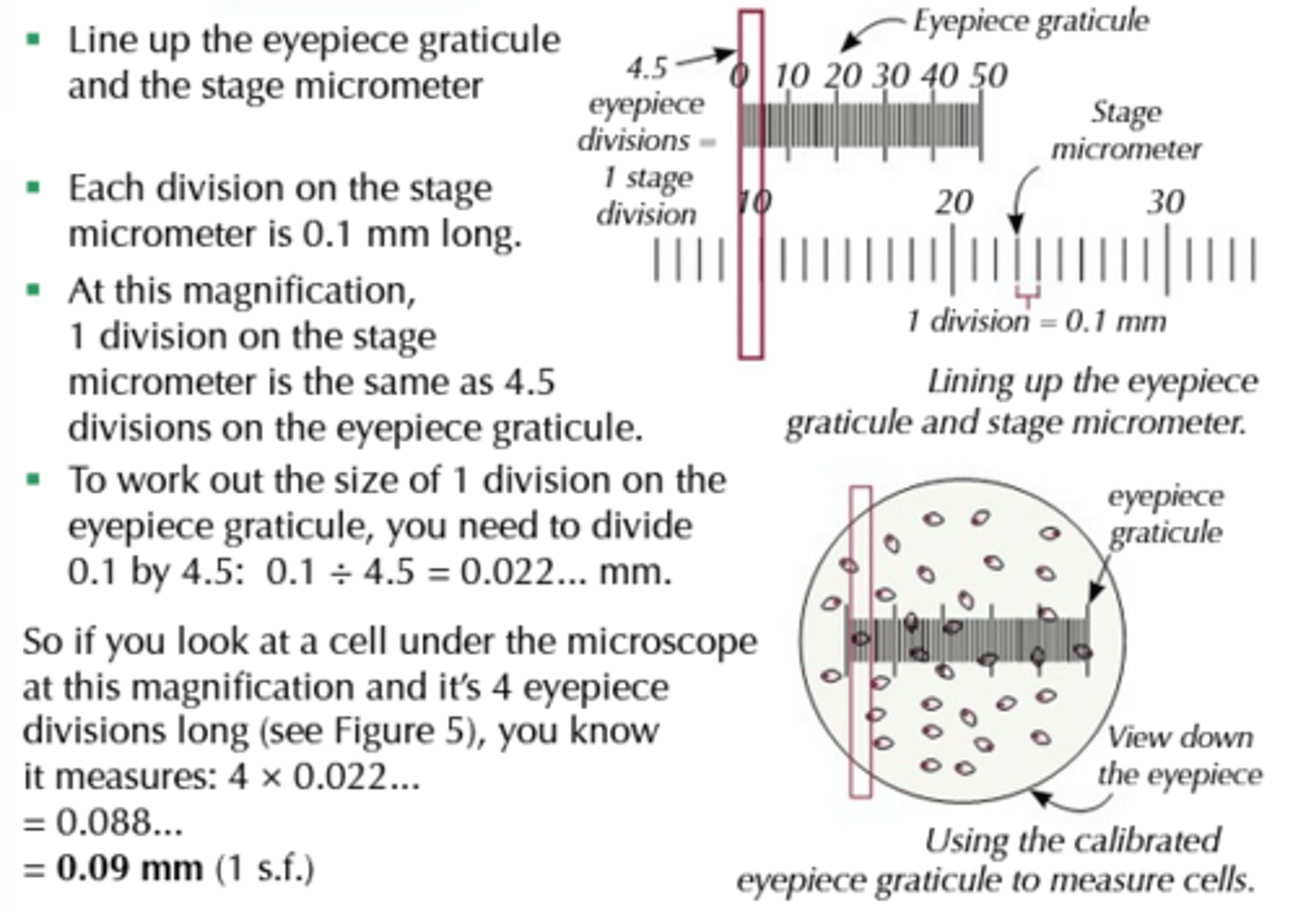
- You need to be able to calculate the size of the cells you're looking at, which is where the eyepiece graticule and stage micrometer come in.
- An eyepiece graticule is a glass disc that is fitted onto the eyepiece; a scale is etched on the glass disc, and is typically 10 mm long and is divided into 100 sub-divisions.
- The scale on the eyepiece graticule cannot be used directly to measure the size of objects under a microscope's objective lens, because each objective lens will magnify to a different degree.
- The graticule must first be calibrated for a particular objective lens; once calibrated in this way, the graticule can remain in position for future use, provided the same objective lens is used; hence why it's a good idea to record the results of the calibration for a particular objective lens, and to leave this attached to the microscope, so you don't have to recalibrate each time you want to measrue the size of an object being viewed under the microscope.
How is the eyepiece graticule calibrated?
- The stage micrometer is placed on the stage; it's a microscope slide with an accurate scale (it has units) etched onto it, and is used to work out the value of the divisions on the eyepiece graticule at a particular magnification.
- The scale is typically 2 mm long and its smallest sub-divisions are 0.01 mm (10 μm).
- When the eyepiece graticule scale and the stage micrometer are lined up, it's possible to calculate the length of the divisions on the eyepiece graticule.
- It's easy to calculate the scale for different objective lenses by dividing the differences in magnification; e.g. if an objective lens magnifying x40 gives a calibration of 25 μm per graticule unit, then an objective lens magnifying x 400 (10 times greater) will mean the graticule unit is equivalent to 25 μm ÷ 10 = 2.5 μm.
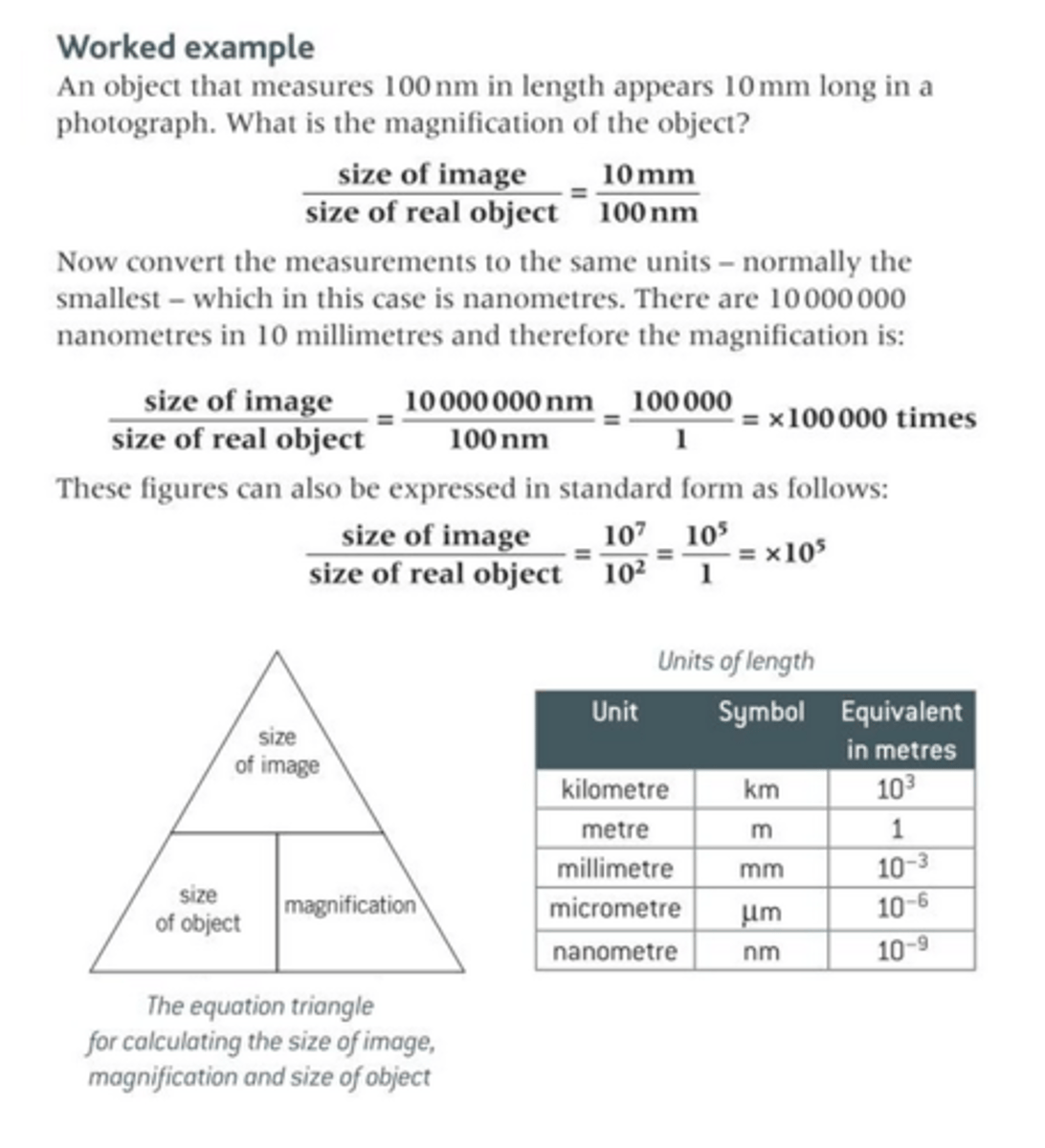
What is magnification?
- The material that is put under a microscope is referred to as the object.
- The appearance of this material when viewed under the microscope is referred to as the image.
- The magnicfication of an object is how many times bigger the image is when compared to the object.
- The important thing to remember when calculating the magnification is to ensure that the units of length are the same for both the object and the image.
- Magnification = Size of Image ÷ Size of Real Object
What is resolution?
- The resolution/resolving power of a microscope is the minimum distance apart that two objects can be in order for them to appear as two separate items.
- Whatever type of microscope, the resolving power depends on the wavelength or form of radiation used; in a light microscope, this typically 0.2 μm, meaning any two objects 0.2 μm or more apart will be seen separately, but any objects closer than 0.2 μm will appear as a single item.
- Greater resolution = Greater clarity (clearer and more precise image).
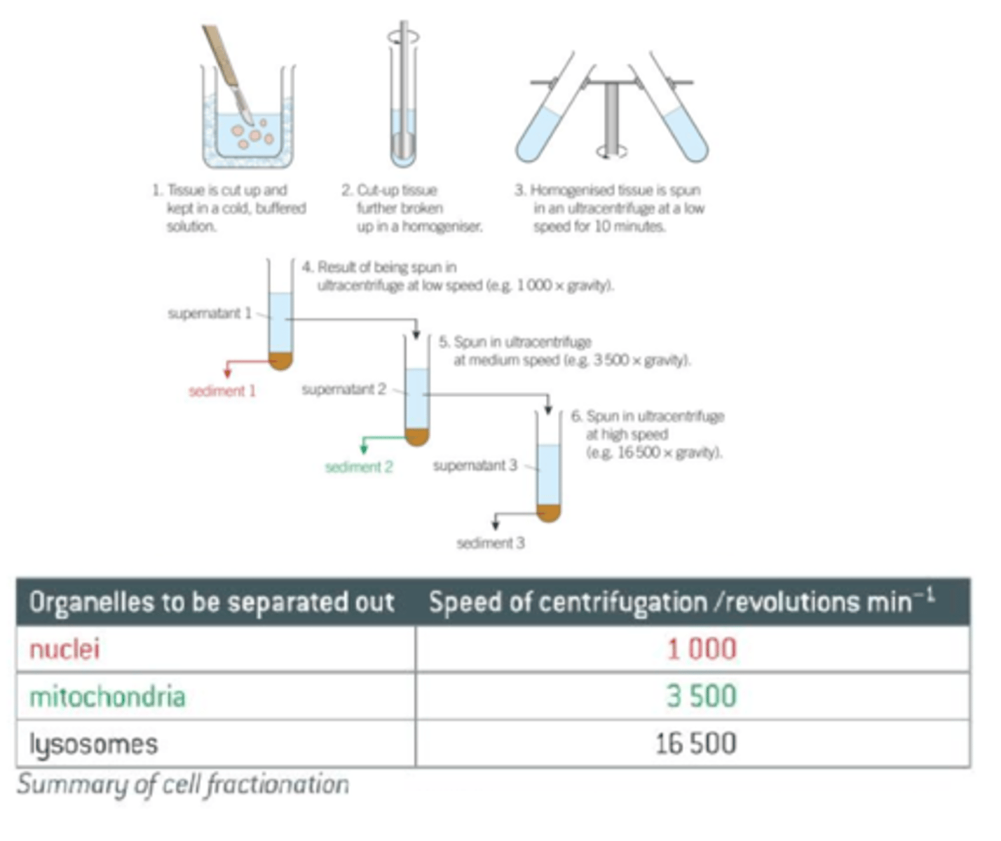
What is cell fractionation?
- In order to study the structure/function of various organelles that make up cells, it's neccessary to obtain large numbers of isolated organelles.
- Cell fractionation is the process where cells are
broken up and the different organelles they contain are separated out.
- Before cell fractionation can begin, the tissue is placed in a cold, buffered solution of the same water potential as the tissue.
- The solution is:
1) Cold: to reduce enzyme activity that might break down the organelles.
2) Isotonic to the tissue: to prevent organelles bursting or shrinking via osmotic gain or loss of water.
3) Buffered: So that the pH doesn't fluctuate - any change in pH could alter the structure of the organelles or affect the functioning of enzymes.
- There are two stages to cell fractionation: Homogenation and Ultracentrifugation.
What is Homogenation?
- The cells are broken up by a homogeniser.
- This releases the organelles from the cell.
- The resultant fluid known as the homogenate, is then filtered to remove any complete cells and large pieces of debris.
What is Ultracentrifugation?
- The process by which the fragments in the filtered homogenate are separated in a machine called a centrifuge.
- This spinds tubes of homegenate at very high speed in order to create a centrifugal force.
- For animal cells, the process is as follows:
1) The tube of filtrate is placed in the centrifuge and spun at a slow speed.
2) The heaviest organelles, the nuclei, are forced to the bottom of the tube, where they form a thin sediment or pellet.
3) The fluid at the top of the tube (supernatant) is removed, leaving just the sediment of nuclei.
4) The supernatent is transferred to another tube and spun in the centrifuge at a faster speed than before.
5) The next heavier organelles, the mitochondria, are forced to the bottom of the tube.
6) The process is continued in this way so that, at each increase in speed, the next heaviest organelle is sedimented and separated out.
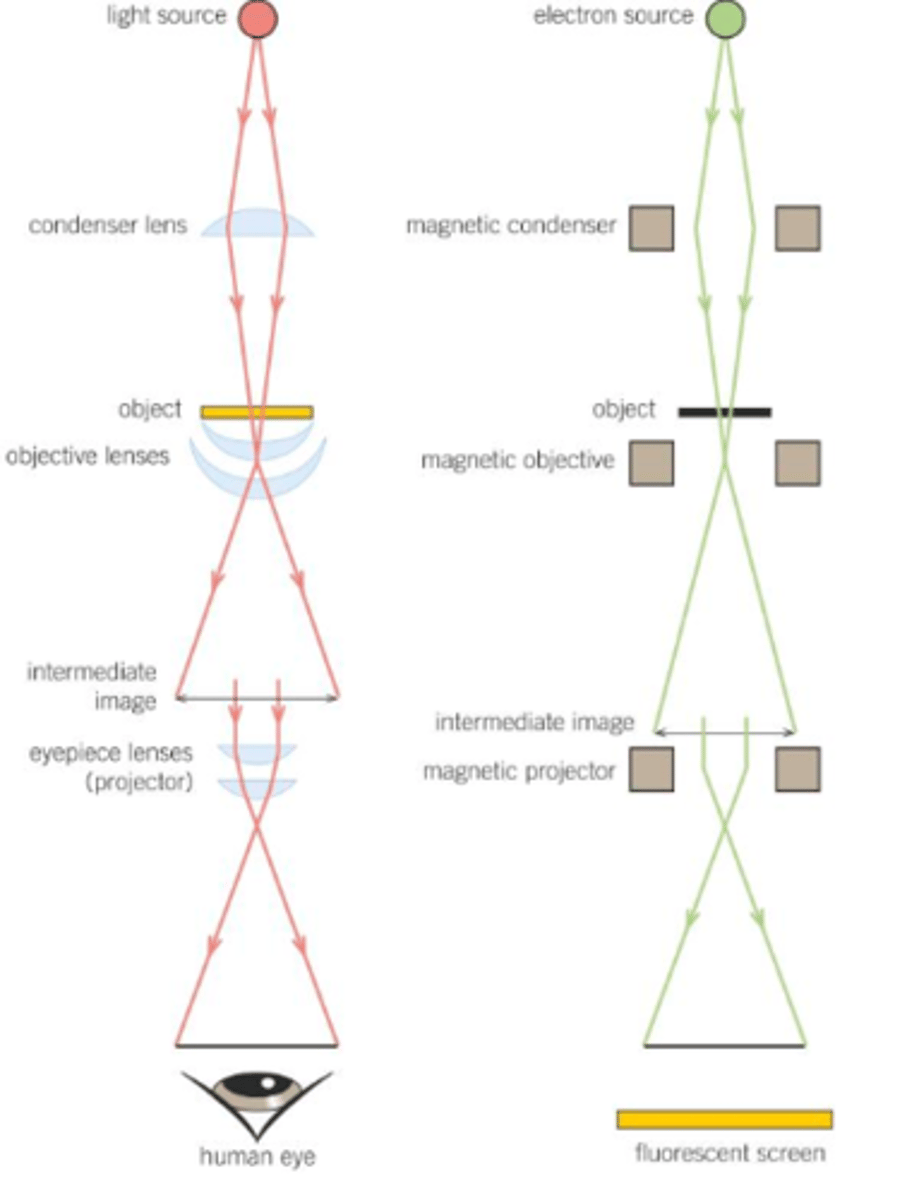
What are optical microscopes?
- They use a relatively long wavelength of light to form an image.
- They have a maximum resolution of 0.2 μm and they can't be used to view organelles smaller than 0.2 μm.
- This means they cannot view ribosomes, ER or lysosomes.
- Mitochondria might be viewable, but not in perfect detail.
- You can also see the nucleus.
- The maximum useful magnification of an optical microscope is about x 1500.
What are electron microscopes?
- They use electrons to form an image.
- They have a higher magnification & resolution than optical microscopes, so give a more detailed image (and can be used to look at more organelles).
- They have a maximum resolution of about 0.0002 μm (about 1000 times higher than optical microscopes); the best modern electron microscopes can resolve objects 0.1 nm apart (2000 times better than a light microscope); the electron beam has a very short wavelength and the microscope can therefore resolve objects well (it has a high resolving power); as electrons are negatively charged the beam can be focused using electromagnets.
- The maximum useful magnification of an electron microscope is about x 1,500,00.
- Electron microscopes produce black and white images, but these are often coloured by a computer.
- Because electrons are absorbed or deflected by the molecules in air, a near-vaccum has to be created within the chamber of an electron microscope for it to work effectively.
- There are two types of electron microscopes: Transmission Electron Microscopes (TEMs) and Scanning Electron Microscopes (SEMs).
What are TEMs?
DESCRIPTION:
- The TEM consists of an electron gun that produces a beam of electrons that is focused onto the specimen by a condenser electromagnet.
- In a TEM, the beam passes through a thin section of the specimen; parts of this specimen absorb electrons and therefore appear dark, whilst other parts of the specimen allow the electrons to pass through and so appear bright.
- An image is produced on a screen and this can be photographed to give a photomicrograph.
- The resolving power of the TEM is 0.1 nm, although this cannot always be achieved in practice due to: difficulties preparing the specimen limit the resolution that can be achieved, and a hgiher energy electron beam is required which may destroy the specimen.
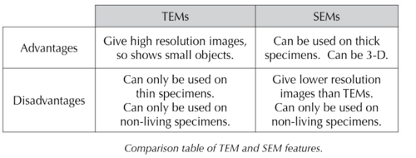
LIMITATIONS:
1) The whole system must be in a vaccum, and therefore living specimens cannot be observed.
2) A complex 'staining' process is required and even then the image is not in colour.
3) The specimen must be extremely thin.
4) The image may contain artefacts (things that result from the way the specimen is prepared); artefacts may appear on the finished photomicrograph, but are not part of the natural specimen, and it's therefore not always easy to be sure that what we see on a photomicrograph really exists in that form.
5) Specimens must be extremely thin to allow electrons to penetrate; the result is therefore a flat, 2D image; we can partially get over this by taking a series of sections through a specimen and build up a 3D image of the specimen by looking at a series of photomicrographs produced, but this is a slow and complicated process; the SEM can overcome this problem.
What are SEMs?
- All the limitations of the TEM also apply to the SEM, except that specimens don't need to be extremely thin as electrons do not penetrate.
- Basically similar to a TEM, the SEM directs a beam of electrons onto the surface of the specimen from above, rather than penetrating it from below.
- The beam is then passed back and forth across a portion of the specimen in a regular pattern.
- The electrons are scattered by the specimen and the pattern of this scattering depends on the contours of the specimen surface.
- A 3D image can be built up by computer analysis of the pattern of scattered electrons and secondary electrons produced.
- The bassic SEM has a lower resolving power than a TEM, around 20 nm, but is still ten times better than a light microscope.
BIOLOGY TOPIC 2A: CELL STRUCTURE AND DIVISION TOPIC SUMMARY
(MAKE SURE YOU KNOW THE FOLLOWING)
- Specialised cells are organised into tissues, tissues into organs and organs into systems.
- Prokaryotic cells are smaller and less complex than eukaryotic cells.
- How the structure of prokaryotic cells is different to eukaryotic cells (no membrane bound organelles in the cytoplasm, circular DNA and a muein cell wall).
- Some prokaryotic cells also have a capsule, flagella and plasmids.
- How a prokaryotic cell replicates by binary fission (the circular DNA and plasmids are replicated and then the cell divides to produce two daughter cells, each with a single copy of the circular DNA and a variable number of copies of plasmids.
- Viruses are non-living and acellular, so they don't undergo cell division, and instead invade host cells and use the host cell 'machinery' to replicate themselves.
- The structure of a typical virus, including the genetic material, capsid and attachment proteins.
- How to calculate magnification (how much bigger the image is than the sample) and resolution (how detailed the image is, based on the microscope's ability to distinguish between two points that are close together).
- The principles and limitations of optical miscroscopes, transmission electron microscopes (TEMs) and scanning electron microscopes (SEMs).
- How to prepare temporary mout microscope slides for viewing specimens with optical microscopes.
- That it wasn't easy for early scientists using microscopes to distinguish between artefacts and cell organelles - and how they eventually did this.
- How cell fractionation separates out organelles (homogenisation, filteration and ultracentrifugation).
- Not all cells are able to keep dividing in multicellular organisms.
- The eukaryotic cell cycle, including how DNA replication takes place in interphase and that the division of cells occurs during mitosis.
- Mitosis produces two daughter cells that are genetically identical to each other and to the parent cell.
- How the chromosomes behave during interphase, prophase, metaphase, anaphase and telophase of mitosis and the role of spindle fibres during mitosis.
- Mitosis ends with cytokinesis (division of the cytoplasm).
- How to recognise the stages of the cell cycle, including being able to explain the appearance of cells at each stage of mitosis.
- Tumours and cancers form when mitosis becomes uncontrolled.
- Cancer treatments are often aimed at controlling the rate of cell division.
- How to prepare stained squashes of root tip cells, use an potical microscope to observe the stages of mitosis in those cells, and calculate mitotic index (the proportion of cells undergoing mitosis) of the cells (REQUIRED PRACTICAL 2).
- How to measure the size of an object viewed with an optical microscope, including how to use an eyepiece graticule and stage micrometer.
- How to use the formula: Actual size = size of image ÷ magnification, to calculate the actual size of an object viewed through a microscope.