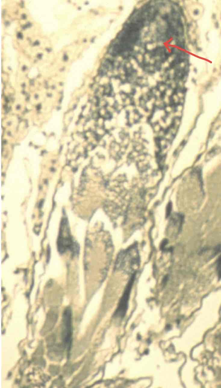
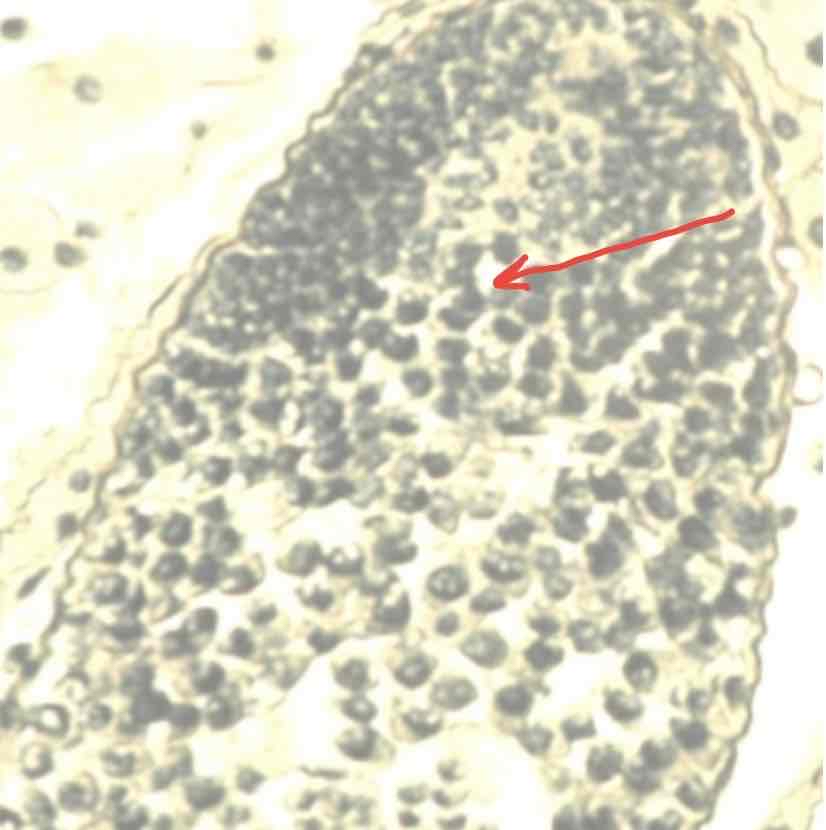
What does the following image depict? Provide name, ploidy number and location
Spermatogonia which are diploid and found in grasshopper testes
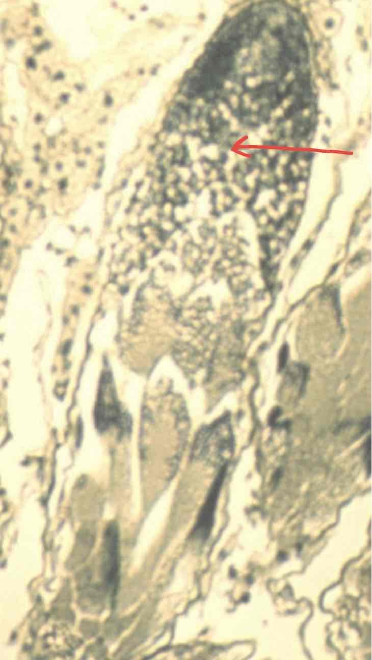
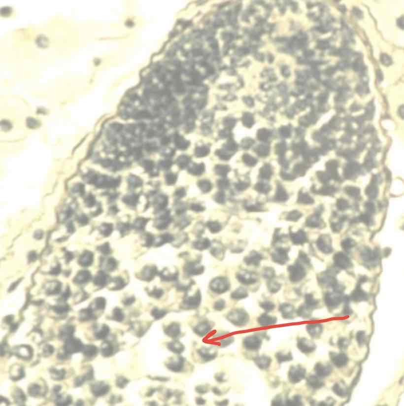
What does the following image depict? Provide name, ploidy number and location
Primary spermatocyte which are diploid and found in grasshopper testes. Product of spermatogonia going through mitosis
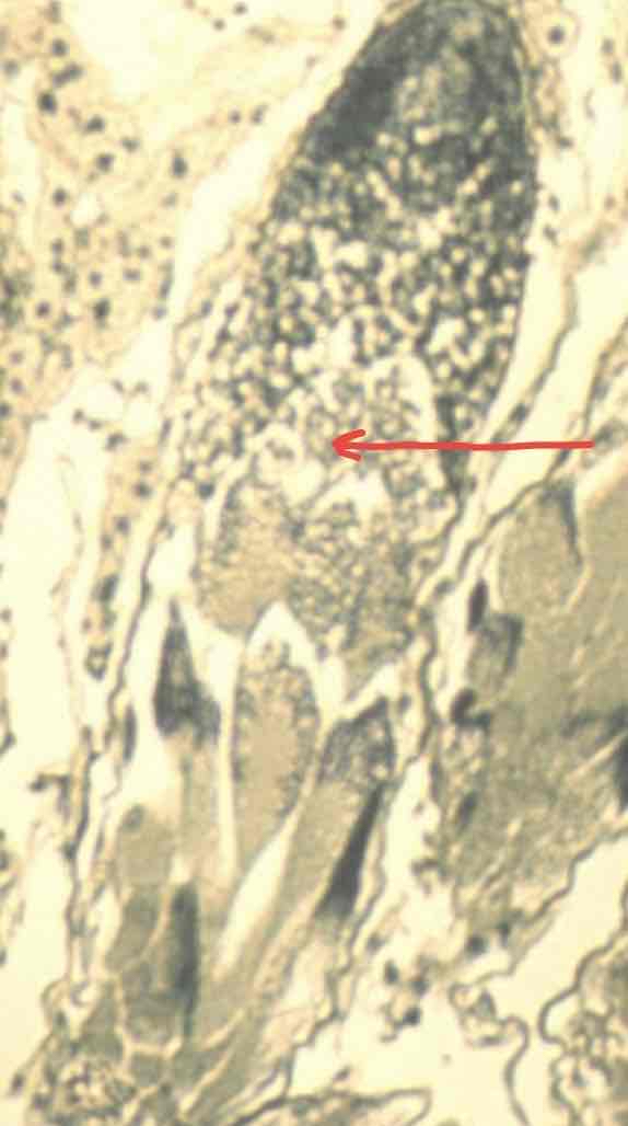
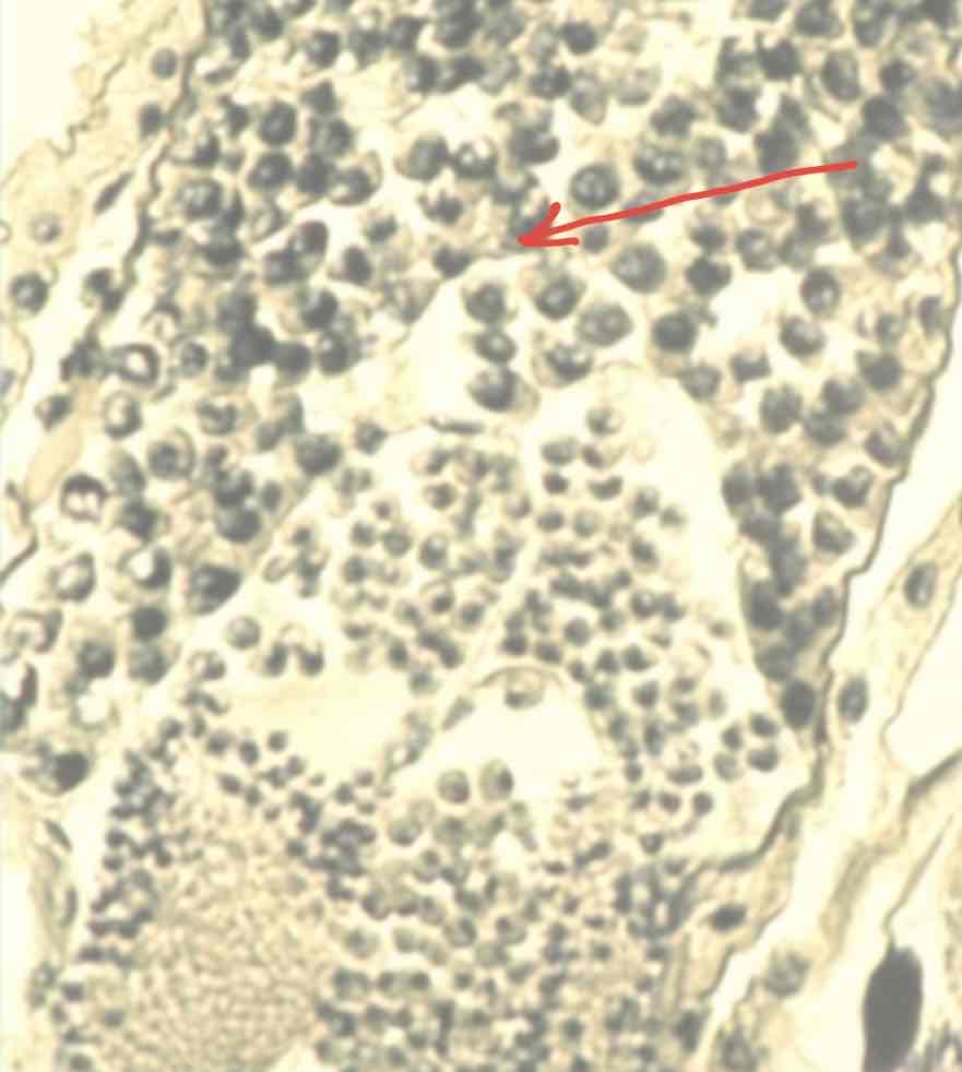
What does the following image depict? Provide name, ploidy number and location
Secondary Spermatocyte which are haploid and found in grasshopper testes. Product of primary spermatocytes going through meiosis.
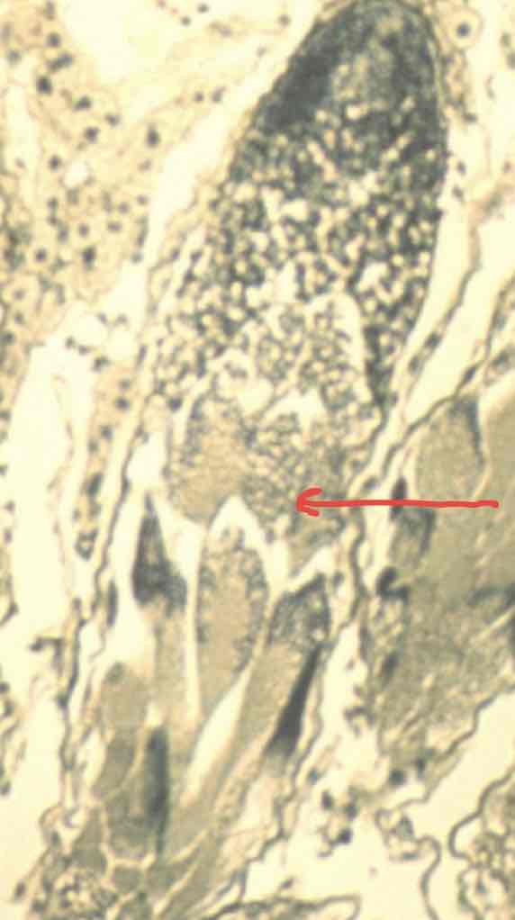
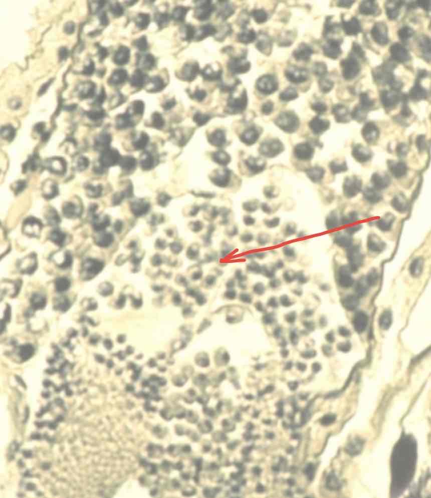
What does the following image depict? Provide name, ploidy number and location
Spermatids which are haploid and found in grasshopper testes. Product of secondary spermatocytes going through mitosis
What does the following image depict? Provide name, ploidy number and location
Spermatogonia which are diploid and found in grasshopper testes
What does the following image depict? Provide name, ploidy number and location
Primary Spermatocyte which are diploid and found in grasshopper testes
What does the following image depict? Provide name, ploidy number and location
Secondary Spermatocyte which are haploid and found in grasshopper testes
What does the following image depict? Provide name, ploidy number and location
Spermatids which are haploid and found in grasshopper testes. Product of mitosis from secondary spermatocytes
What does the following image depict? Provide name, ploidy number and location
Spermatids which are haploid and found in grasshopper testes
What does the following image depict? Provide name, ploidy number and location
Spermatozoa which are haploid and found in grasshopper testes
What does the following image depict? Provide name, ploidy number and location
Spermatozoa which are haploid and found in grasshopper testes
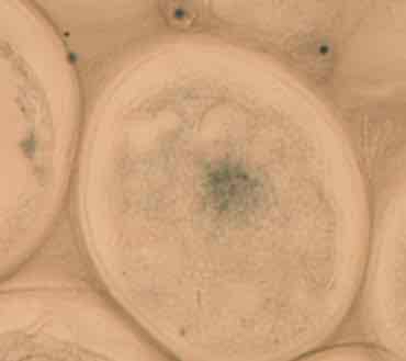
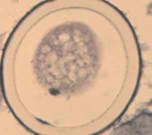
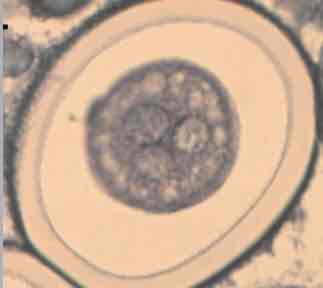
What does the following image depict? Provide name, ploidy number and location
Unfertilized egg (oogonia) which is diploid and found in Ascaris ovary (nematode)
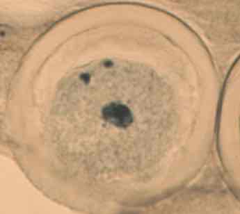
What does the following image depict
Sperm hit egg and fertilized the egg in the ascaris ovary
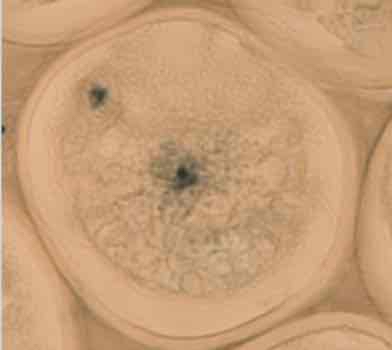
What does the following image depict?
Sperm penetration, the male pronucleus (haploid) moves towards the center of the primary oocyte (diploid), while the female nucleus moves towards the edge of the egg within the ascaris ovary
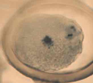
What does the following image depict?
Prophase 1: a fertilization membrane has formed around the primary oocyte. Chromosomes become visible along the egg of the ascaris ovary
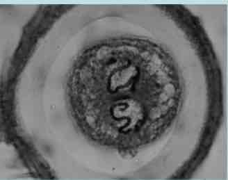
What does the following image depict?
Metaphase 1: tetrads are aligned in a linear arrangement within the ascaris ovary
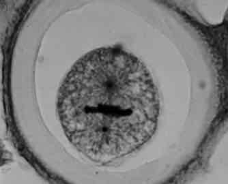
What does the following image depict?
Anaphase 1: Chromosomes begin separating within the ascaris ovary
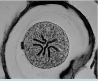
What does the following image depict?
Telophase 1:Chromosomes are completely separated within the ascaris ovary
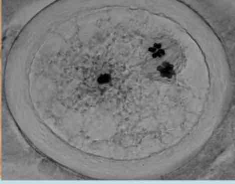
What does the following image depict?
Prophase 2: the extrusion of the first polar body (lower left), the haploid chromosomes will progress through meiosis 2. Following meiosis 2 the 2nd polar body will be extruded. Takes place within the Ascaris ovary.
What does the following image depict?
Pronuclear Fusion: female pronucleus (haploid) migrate to the center of the ovum. These two pronuclei will fuse to a single nucleus (diploid). Taking place within the ascaris ovary
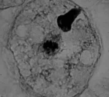
What does the following image depict?
Triploidy: Two sperm simultaneously enter the egg. Almost always lethal in humans. Takes place within the ascaris ovary
What does the following image depict?
Prophase of Mitosis: Chromosomes are within the center of the cell and condense.taking place in ascaris ovary
What does the following image depict?
Metaphase of mitosis: chromosome line up linearly. Takes place within the ascaris ovary
What does the following image depict?
Polar metaphase: top down view of mitosis, ascaris ovary
What does the following image depict?
Anaphase of mitosis in ascaris ovary
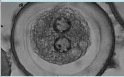
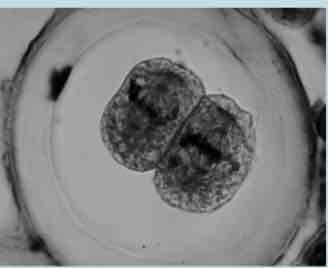
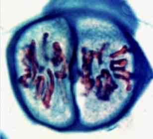
What does the following image depict?
Metaphase of the 2 cell stage in ascaris ovary
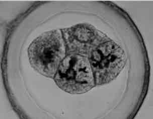
What does the following image depict?
Prophase of the four cell stage, ascaris ovary
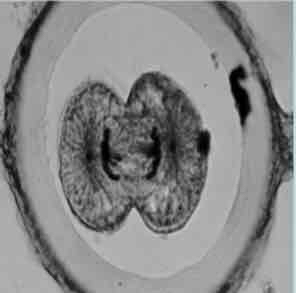
What does the following image depict?
Telophase of mitosis: Cleavage furrow partitions one cell to two, ascaris ovary
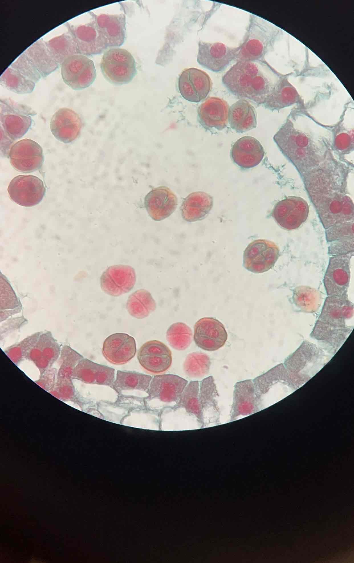
What does the following image depict? Provide name and location
Cross section of Lily anther
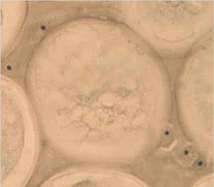
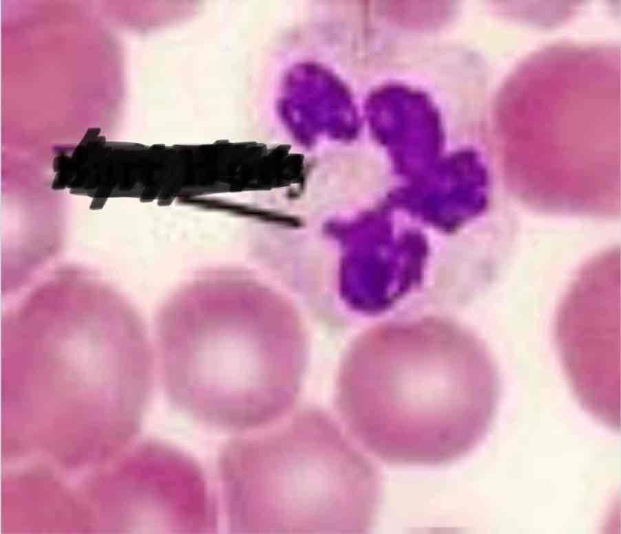
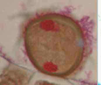
What does the following image depict? Provide name
Neutrophil of female, Barr body
What does the following image depict? Provide name and location
Banded polytene chromosome from salivary chromosomes of Drosophilia, fruit fly
What does the following image depict? Provide name, ploidy #,location
prophase 1 in the anthers of stamens of lily plants, diploid
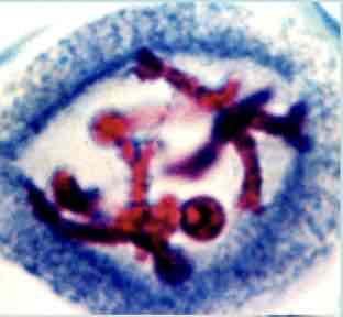
What does the following image depict? Provide name, ploidy #, location
Metaphase 1 in the anthers of stamens of lily plants, diploid
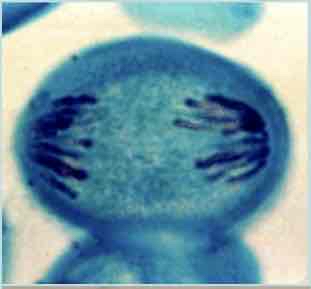
What does the following image depict? Provide name, ploidy # location
Anaphase 1 in the anthers of stamens of lily plants, diploid
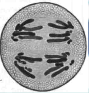
What does the following image depict? Provide name, ploidy #, location
Telophase 1 in the anthers of stamens of lily plants, diploid
What does the following image depict? Provide name, ploidy #, location
Prophase 2 in the anthers of stamens of lily plants, haploid
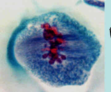
What does the following image depict? Provide name, ploidy #, location
Metaphase 2 in the anthers of stamens of lily plants, haploid
What does the following image depict? Provide name, ploidy #, location
Anaphase 2 in the anthers of stamens of lily plants, haploid
What does the following image depict? Provide name, ploidy #, location
Telophase 2 in the anthers of stamens of lily plants, haploid
What does the following image depict? Provide name and location
Drosophilia salivary chromosome, polytene chromosomes
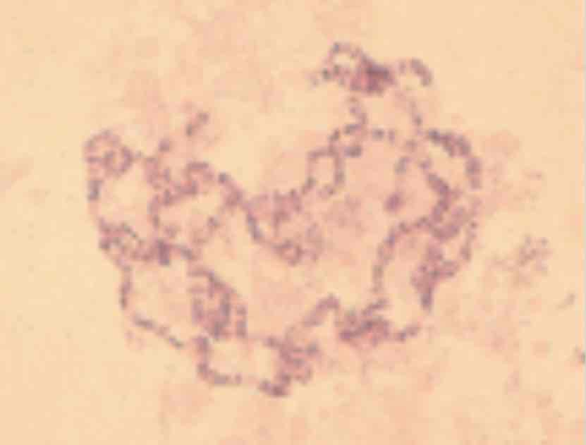
What does the following image depict? Provide name
Barr Bodies
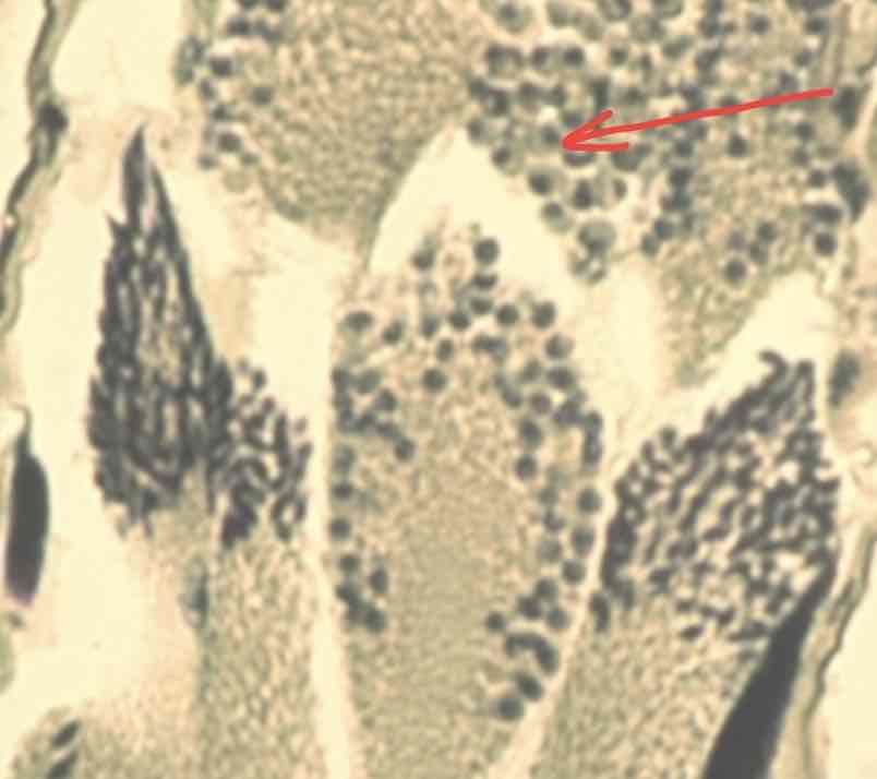
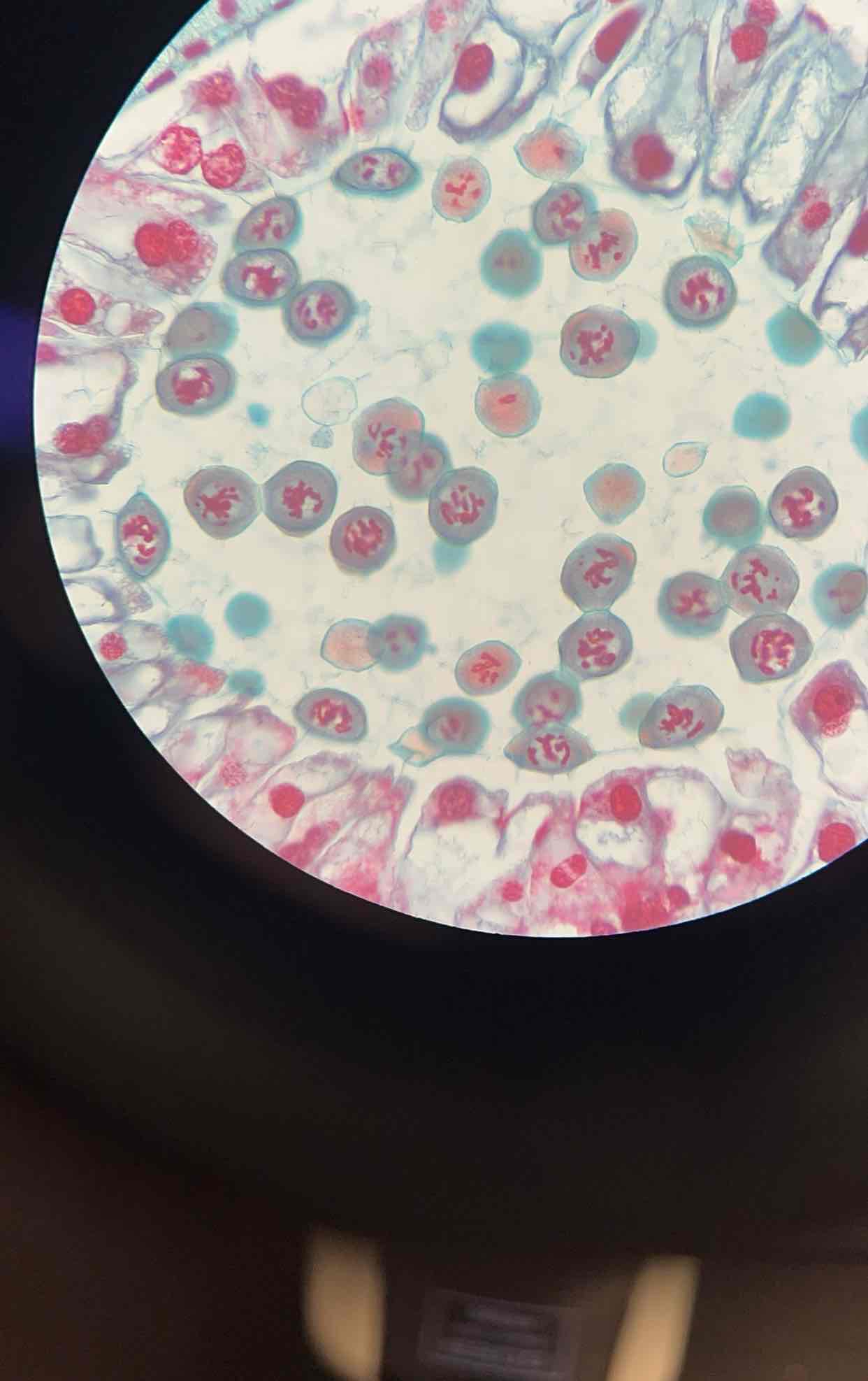
What does the following image depict? Provide name and location
Late prophase 1 and metaphase 1 in the anthers of stamens of lily plants
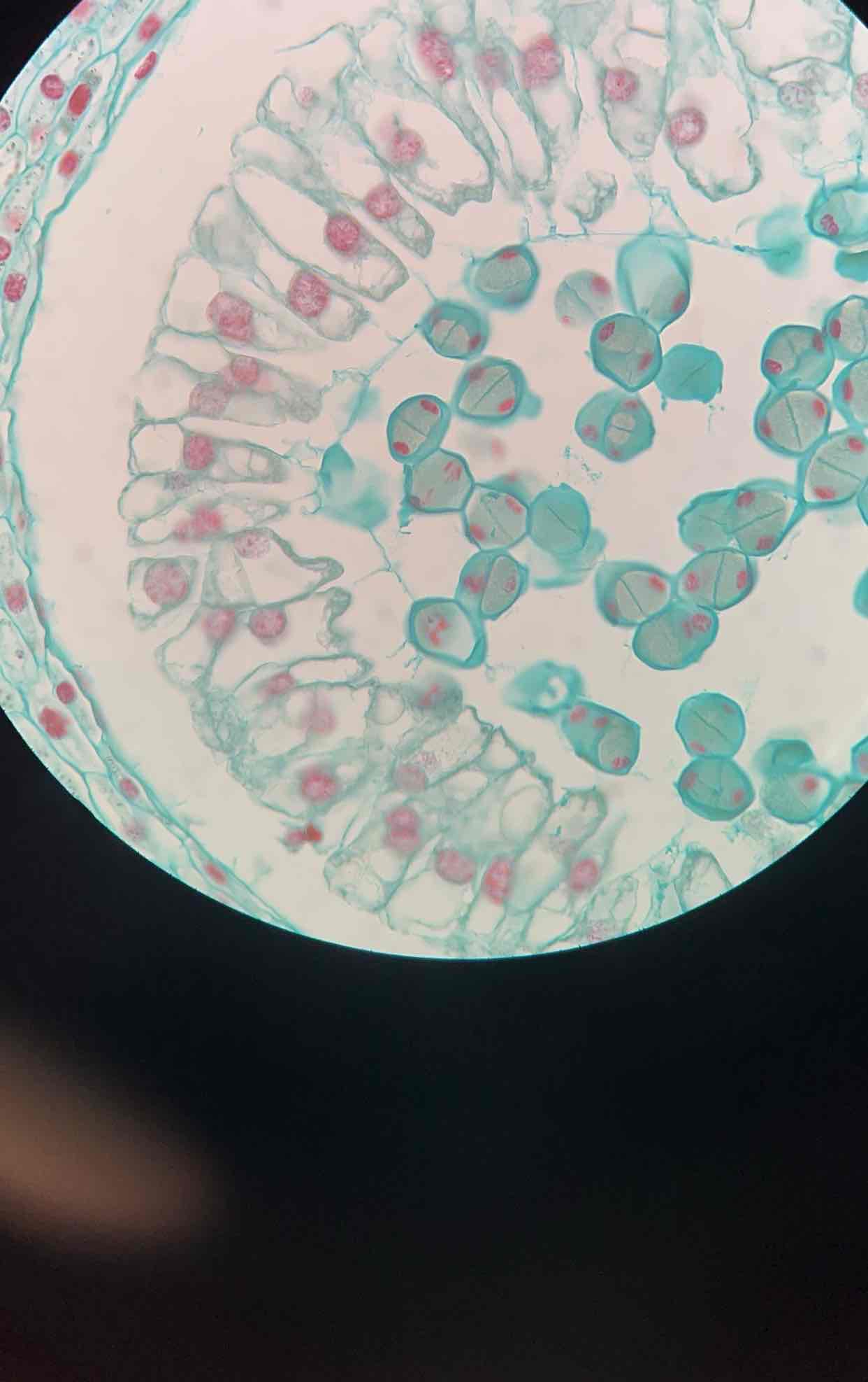
What does the following image depict? Provide name and location
microsporogenesis in the lily anther telophase 1
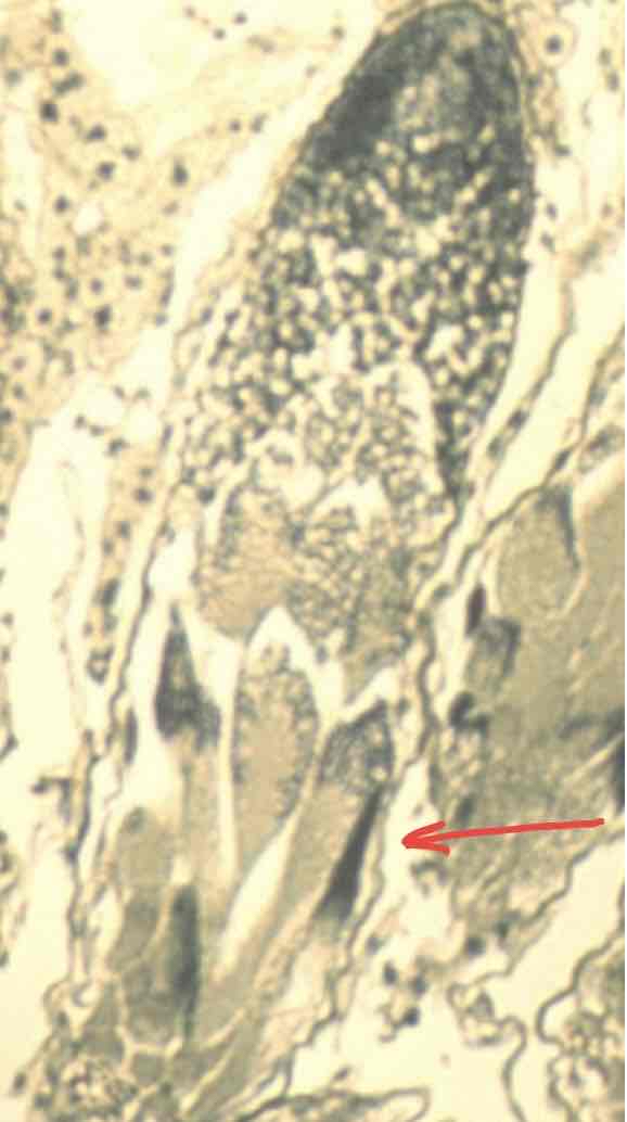
What does the following image depict? Provide name and location
microsporogenesis in the lily anther, early anaphase 1
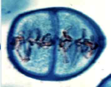
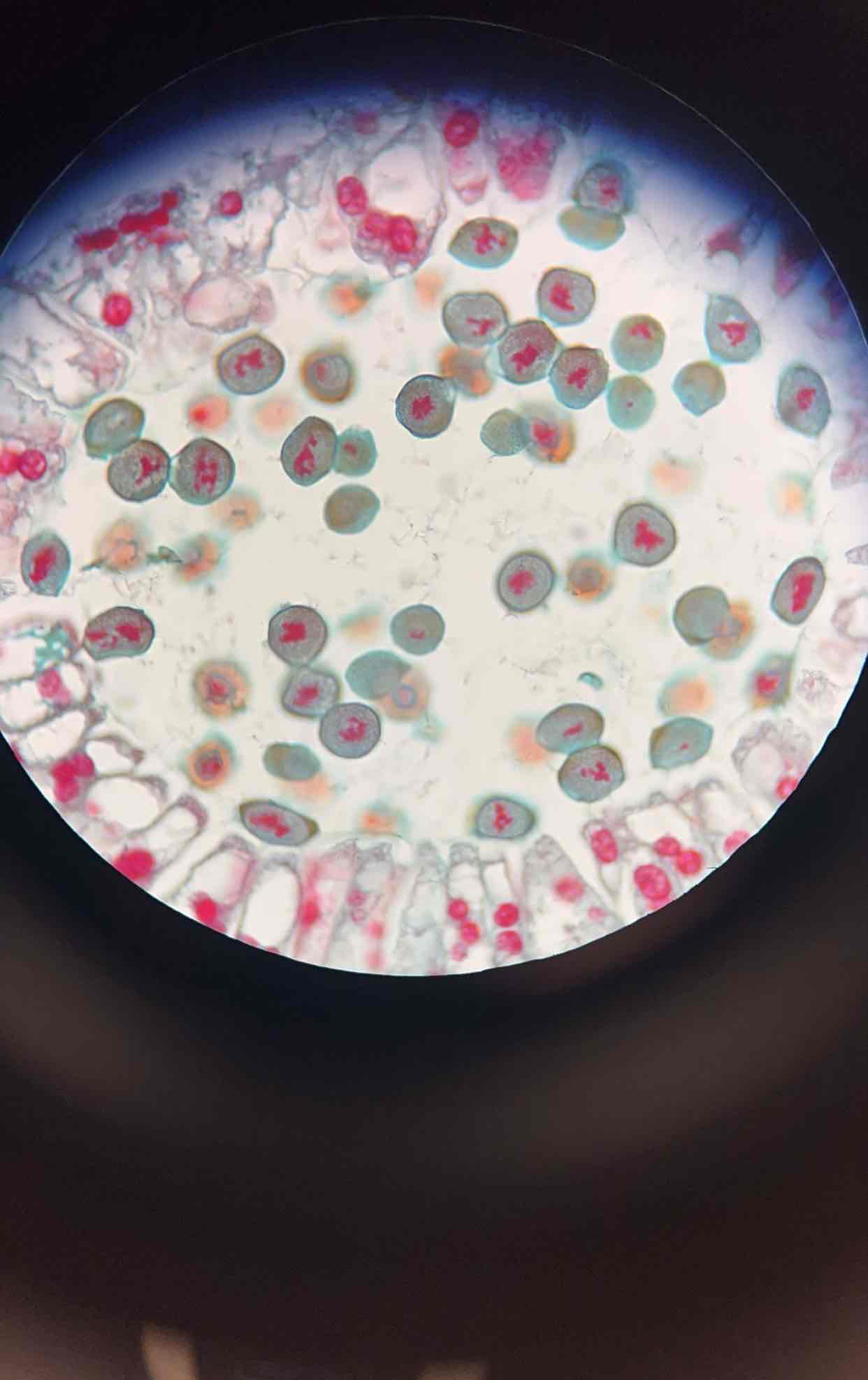
What does the following image depict? Provide name and location
Anaphase 2, metaphase 2
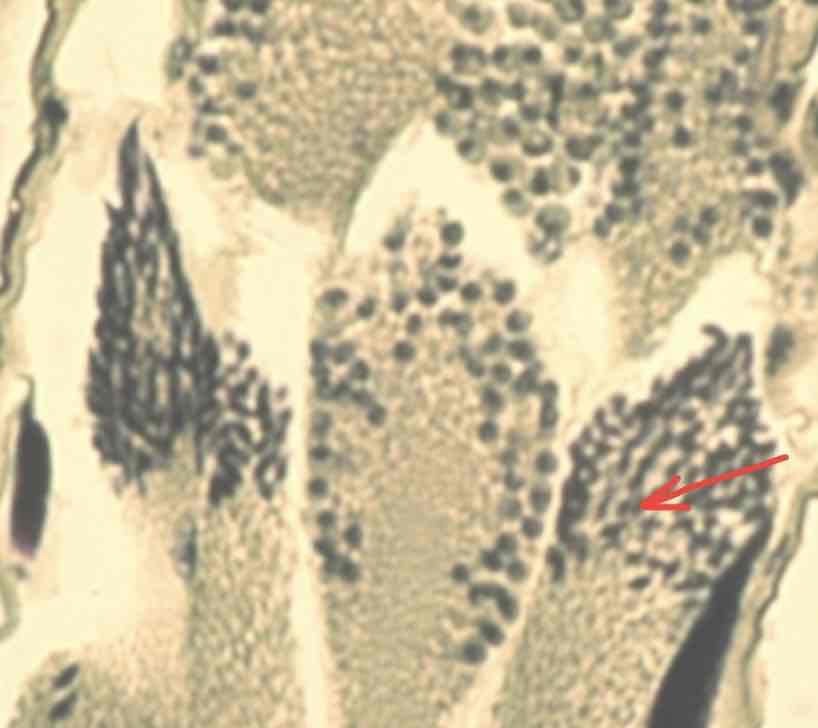
What does the following image depict? Provide name and location
Lily microsporogenesis, anaphase 1 right next to prophase 2
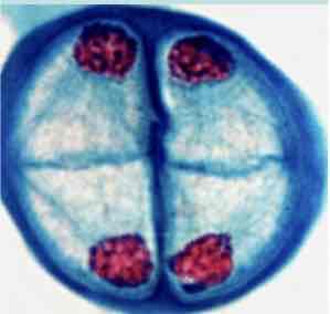
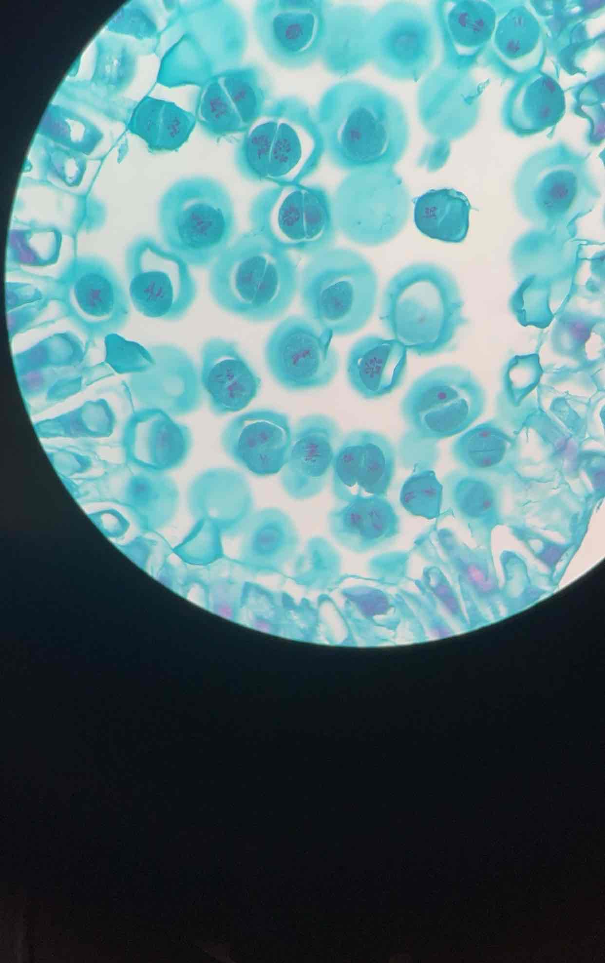
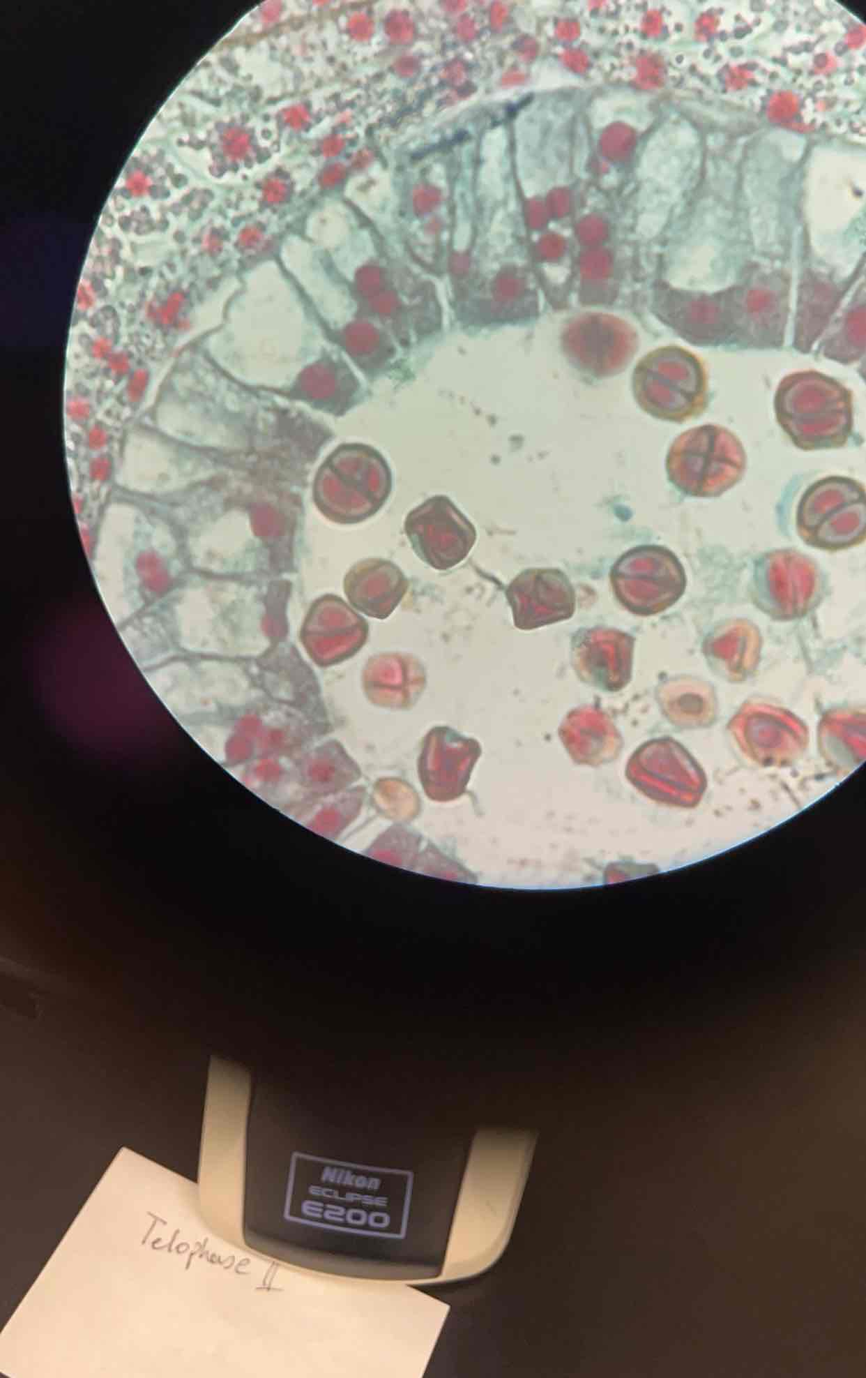
What does the following image depict? Provide name and location
Telophase 2