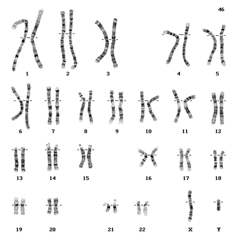
Human chromosome and chromosomal abnormalities
1/38
There's no tags or description
Looks like no tags are added yet.
Name | Mastery | Learn | Test | Matching | Spaced |
|---|
No study sessions yet.
39 Terms
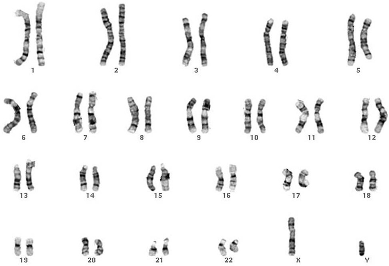
karyotype
the full set of chromosomes in a cell
diploid vs haploid
Somatic cells contain two copies of each chromosome (homologous pairs), except the sex chromosomes (X and Y) in males.
Somatic cells are diploid: chromosome complement = 2n (46 in humans).
Gametes are haploid: chromosome complement = n (23 in humans).
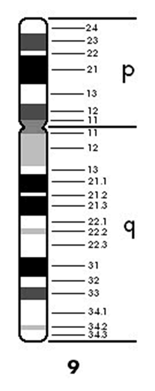
Chromosome “anatomy”: human chromosomes
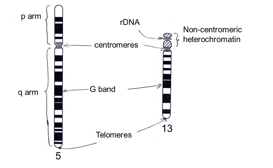
p arm - ‘petite’ smaller arm
q arm ‘queues are long’ - longer arm
G band: region that takes up Giemsa stain, giving characteristic pattern
•Telomeres: specialised repeated DNA sequences, protect ends of chromosomes.
•rDNA: DNA encoding ribosomal RNA (rRNA)
gene map positions
Gene map positions: are given with respect to numbered G bands.
e.g. 9q34.1, the location of a proto-oncogene (ABL1) associated with chronic myeloid leukemia, denotes sub-band 1 of band 4 in region 3 of the long arm of chromosome 9
The number increases as the distance from the centromere increases. For example, 14q21 represents a gene on the long arm of chromosome 14, at position 21
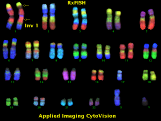
what technique is used to help visualise chromosomes?
Fluorescence in situ hybridisation (FISH) can be used to visualise chromosomes
•Specific cloned DNA sequences tagged with fluorescent chemicals are hybridised (base-paired) to the chromosome DNA sequence.
•Chromosomes viewed under fluorescence microscope: image computer enhanced.
•Chromosome painting (using many probes) can detect small structural abnormalities in the chromosomes.
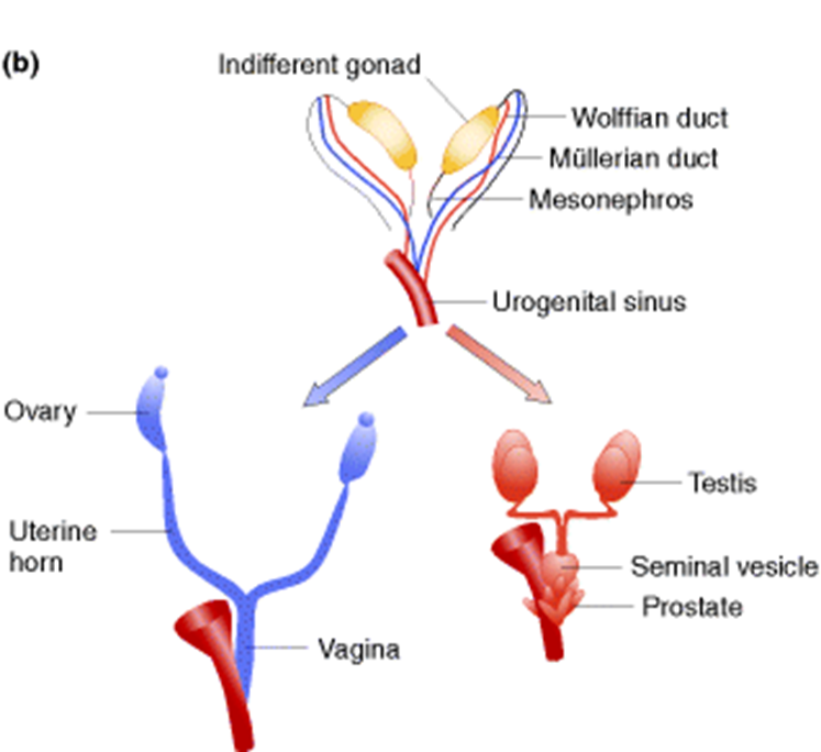
Genetics of mammalian sex determination
Female: XX
Male: XY
Y chromosome carries few genes but presence/absence of Y determines sex.
what gene determines testis determining factor?
SRY gene on Y chromosome encodes testis determining factor.
Leydig cells
Testis determining factor (SRY gene) initiates testis formation in males.
Leydig cells of testis secrete testosterone, an androgenic (male determining) steroid hormone, which activates male-specific gene expression
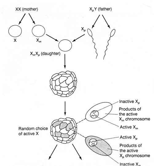
(Lyonisation) and barre bodies
X Chromosome Inactivation (Lyonisation)
Dosage compensation in females
One (randomly chosen) X chromosome is inactivated in each cell the early embryo - further copies of that cell will have their same X chromosome inhibited
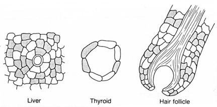
X inactivation persists through cell division, so the adult female is a genetic mosaic.
The inactive X chromosome is condensed (heterochromatin) and visible as a Barr body under the microscope.
Chromosomal abnormalities are a significant cause of genetic disease
Fate of 1 million implanted human zygotes
Chromosomal abnormalities are found in:
0.6% of live births
50% of spontaneous abortions

polyploidy
Polyploidy: multiple sets of chromosomes
In humans.
Triploidy: 3n
15 - 20% spontaneous abortions
1 in 10,000 live births. Die within 1 month
Tetraploidy: 4n
5 % spontaneous abortions
Very rare live births. Short lived.
Aneuploidy:
–the condition of having an abnormal number of chromosomes
•Gene dosage- the number of chromosomes
–important for normal development, so aneuploidy causes abnormalities.
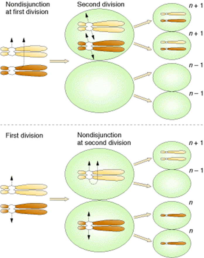
what causes aneuploidy?
Non-disjunction: failure of homologous chromosomes (meiosis 1) or sister chromatids (meiosis 2) to separate at anaphase.
nullisomy
2n-2
Loss of one homologous pair of chromosomes.
No viable human examples
monosomy
Loss of a single chromosome.
One viable human example (Turner syndrome)
2n-1
Trisomy
2n +1
One extra chromosome.
Several viable human syndromes
state two sex chromosome abnormalities
Klinefelter syndrome
turner syndrome
47XXX females and 47XYY males are also found: few if any abnormalities.
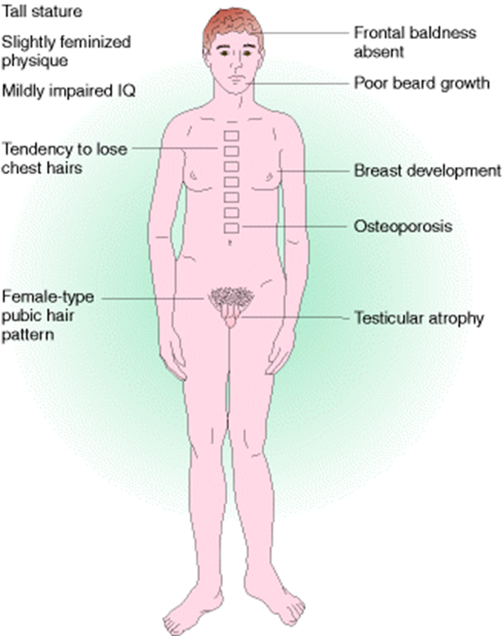
Klinefelter syndrome
47,XXY karyotype
(or more rarely: 48,XXYY; 48,XXXY; 49,XXXXY)
Male
1 in 1,000 male births
Common cause of male infertility
Tall stature, some learning difficulties, underdeveloped testes, 50% some breast development, wide hips .
Testosterone can improve secondary sexual characteristics
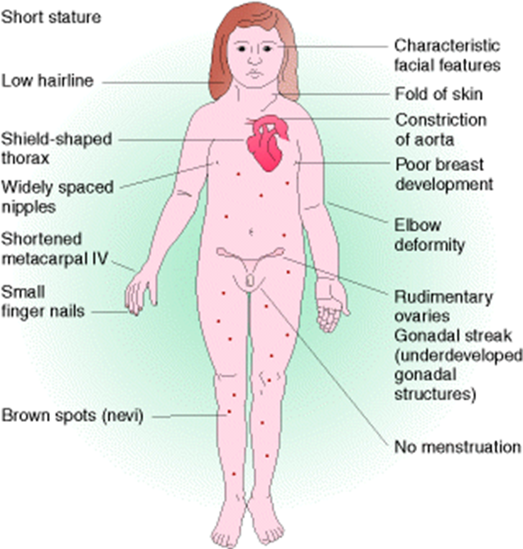
turner syndrome
XO - missing X chromosome (45,X) karyotype
Female
1 in 5,000 female births, but > 90% of cases are spontaneously aborted.
Few noticeable defects until puberty: do not develop secondary sexual characteristics.
Mainly infertile.
list some autosomal abnormalities
(All autosomal monosomies and most autosomal trisomies are lethal before birth.)
Three autosomal trisomies can survive birth
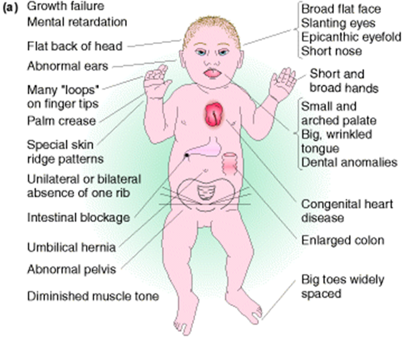
Down syndrome
Edwards syndrome
Patau syndrome
Down syndrome
trisomy chromosome 21
15 per 10,000 births
Many developmental abnormalities, low IQ.
Only autosomal trisomy to survive to adulthood, but life expectancy low
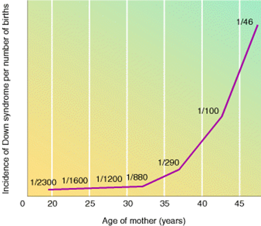
maternal age and prevalence of down syndrome
Patau syndrome
trisomy chromosome 13
1 per 5,000 births
show severe multiple abnormalities: most infants die within a few weeks of birth.
Edwards syndrome:
trisomy chromosome 18
1 per 5,000 births
show severe multiple abnormalities: most infants die within a few weeks of birth.
Why is trisomy 21 the only human autosomal trisomy to survive to adulthood?
Why are numerical/autosomal sex chromosome abnormalities better tolerated than numerical autosomal abnormalities?
Few genes on Y chromosome. All but one X chromosome inactivated
So why do Turner syndrome and Klinefelter syndrome individuals have any abnormalities?
Pseudo-autosomal region on X chromosome is not included in X inactivation: \ gene dosage of genes in this region is abnormal.
structural chromosomal abnormalities can be induced by…5
radiation
viruses
chemicals
transposable elements
errors in crossing over
Classification of structural chromosomal abnormalities
Arise from breaks in chromosomes
Gene dosage important, so most large deletions cause disease in heterozygote and are lethal in homozygote.
deletion, inversion, translocation, duplication
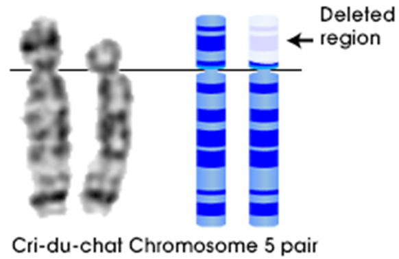
Cri-du-chat syndrome
Part of short arm of one copy of chromosome 5 deleted (5p-).
1 in 20,000 - 1 in 50,000 live births
Mental retardation and physical abnormalities.
Cat-like cry.
inversion mutations- Pericentric
includes centromere
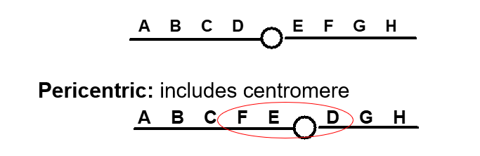
inversion mutations - Paracentric:
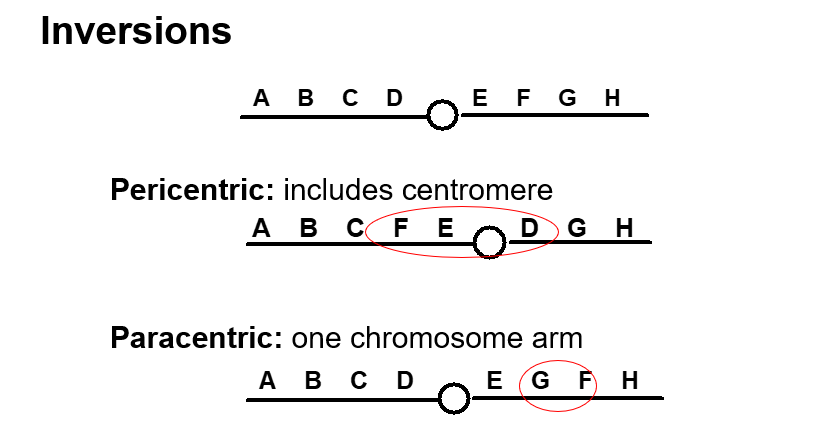
‘Balanced’ - in relation to inversion mutations
no loss of material, but breaks may occur within genes or regulatory regions.
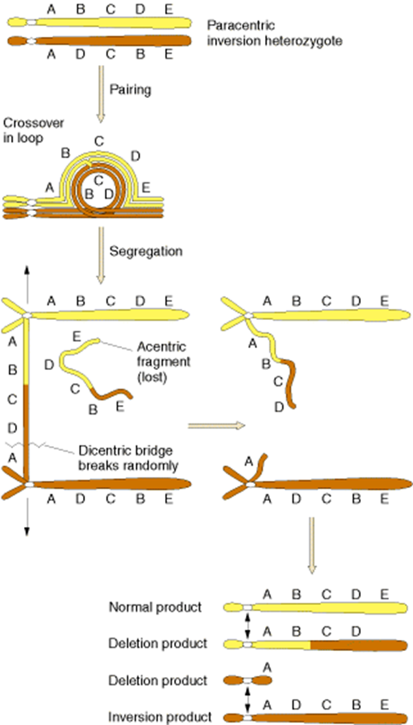
Consequences of inversion
In heterozygotes duplications and deletions can arise when recombination takes place within an inversion during meiosis.
So an individual with a large inversion may be normal, but may have children with severe chromosomal abnormalities
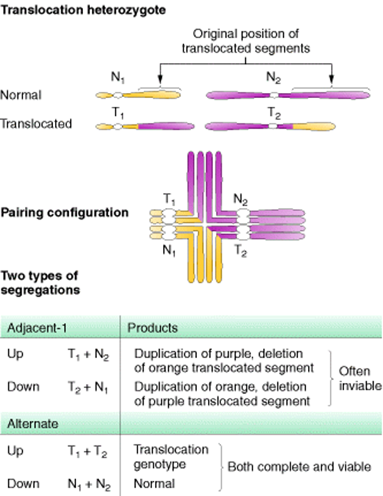
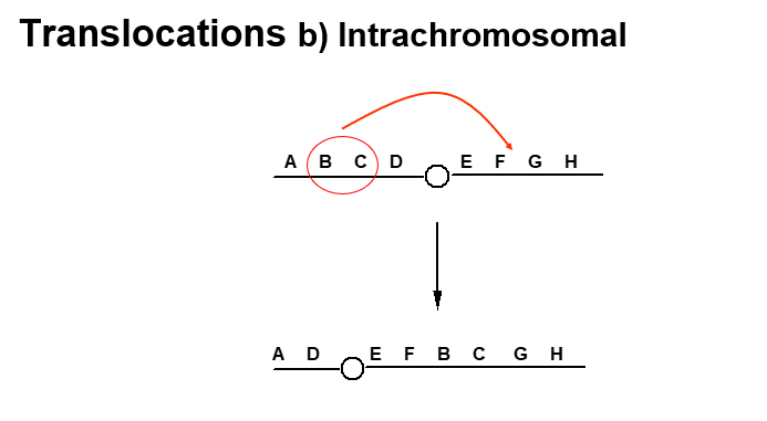
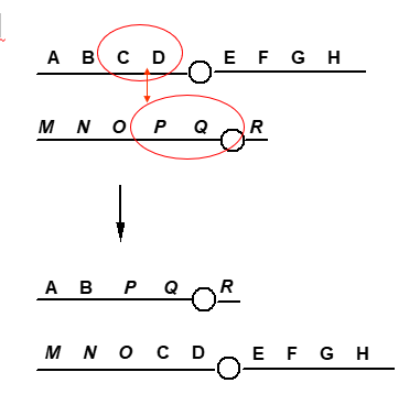
Translocations
Change in position of chromosome segment
can be either interchromosomal or intrachromosomal
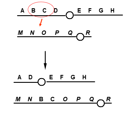
interchromosomal translocations
reciprocal (this slide), non reciprocal (first slide)
intrachromosomal translocation
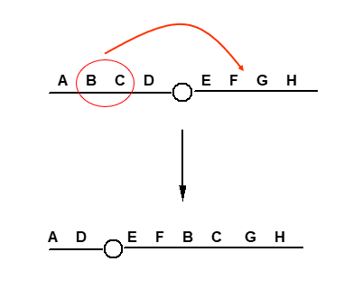
translocation vs inversion
The main difference between a translocation and an inversion is that a translocation moves a piece of DNA from one chromosome to another - or with intrachromosomal translocation its a chop and change , while an inversion reverses a segment of DNA within a chromosome
consequences of translocation
In translocation, heterozygotes segregation at meiosis can lead to genetically unbalanced gametes.
So as with inversions, an individual with a translocation may be normal, but may have children with genetic abnormalities.
Translocations are balanced, but genes at break point may be affected.