Special Sense - Vision
1/19
There's no tags or description
Looks like no tags are added yet.
Name | Mastery | Learn | Test | Matching | Spaced | Call with Kai |
|---|
No analytics yet
Send a link to your students to track their progress
20 Terms
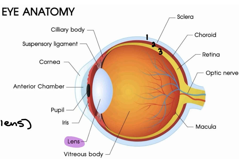
Structure of the eyeball
3 layers/tunics (superficial → deep)
1. Fibrous tunic
2. Vascular tunic (Uvea)
3. Sensory tunic (Retina)
Lens
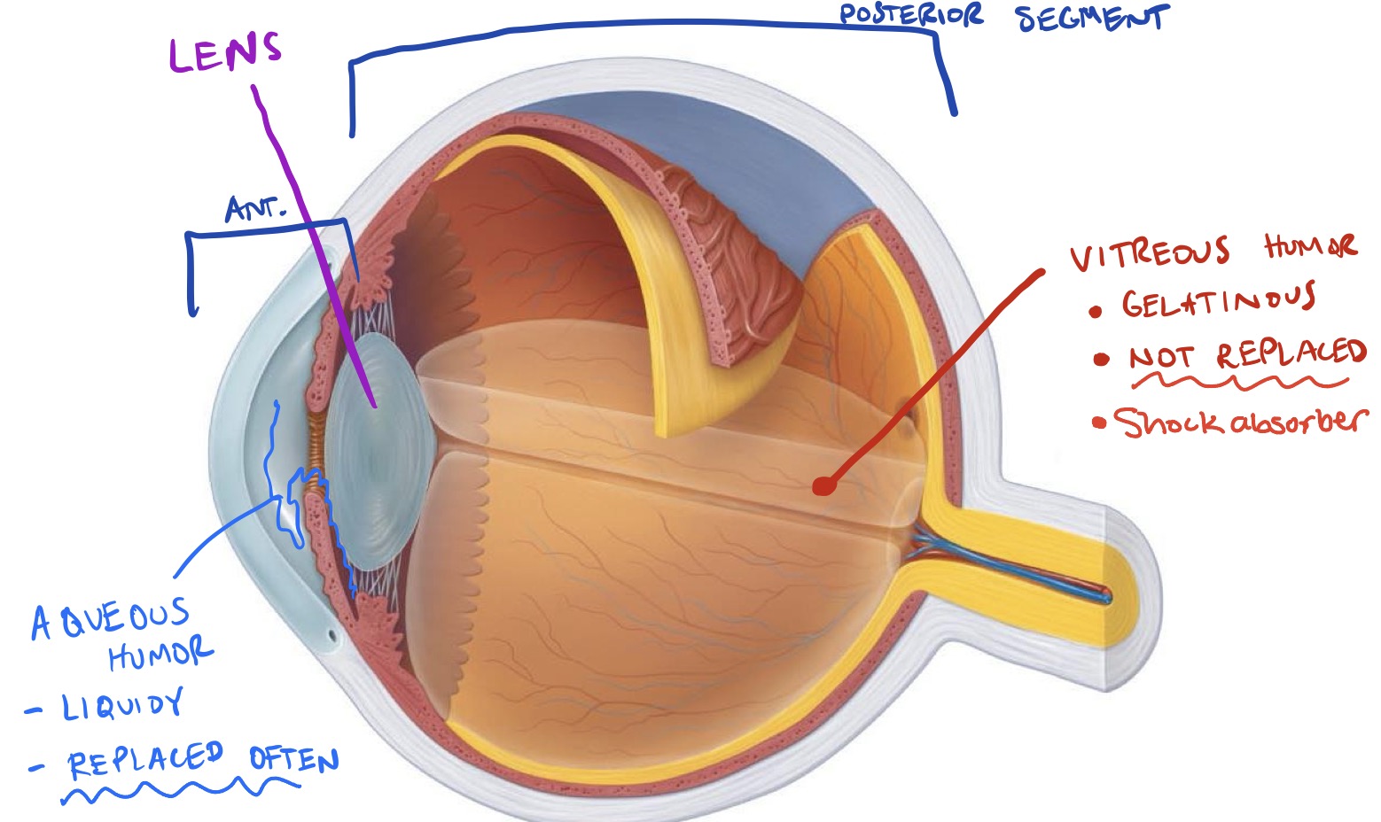
Internal cavity (ant. & pos. to lens)
Anterior segment → aqueous humor
Posterior segment → vitreous humor
Fibrous tunic
Composed of:
Sclera
Cornea
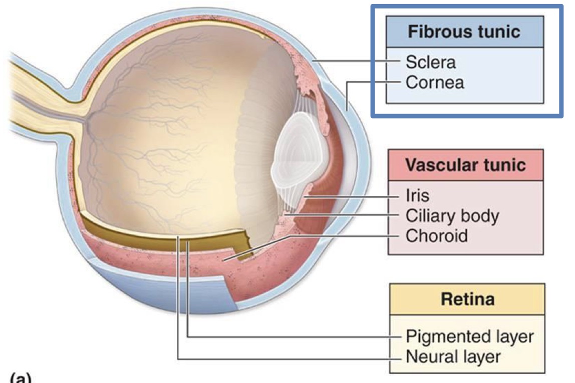
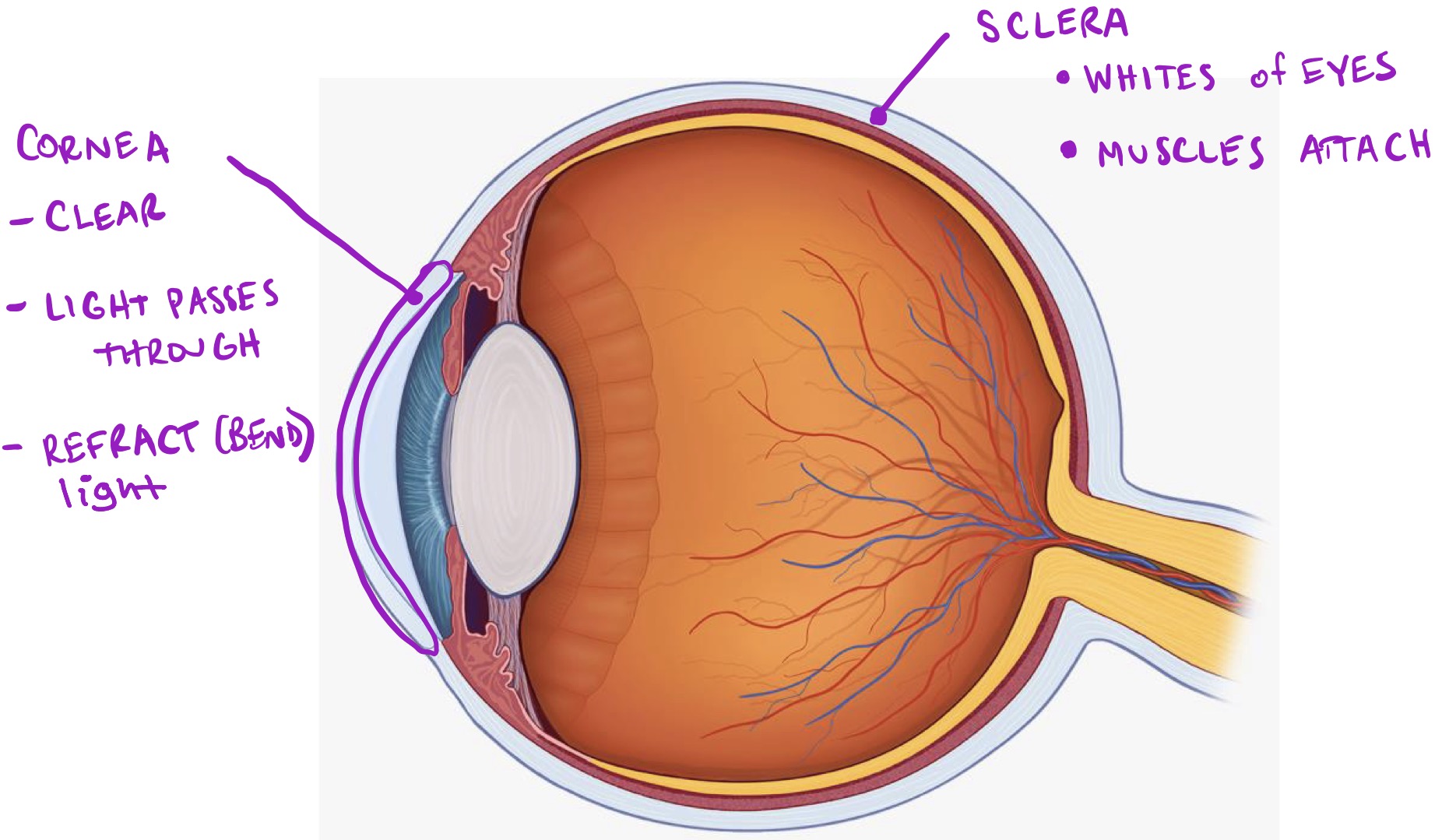
Fibrous tunic - labeled
Protective; superficial
Sclera:
Whites of eyes
Muscles attach
Cornea:
Clear
Light passed through
Refract (bend) light
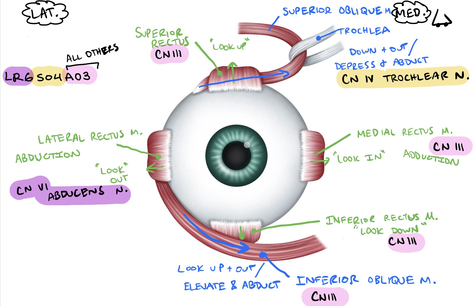
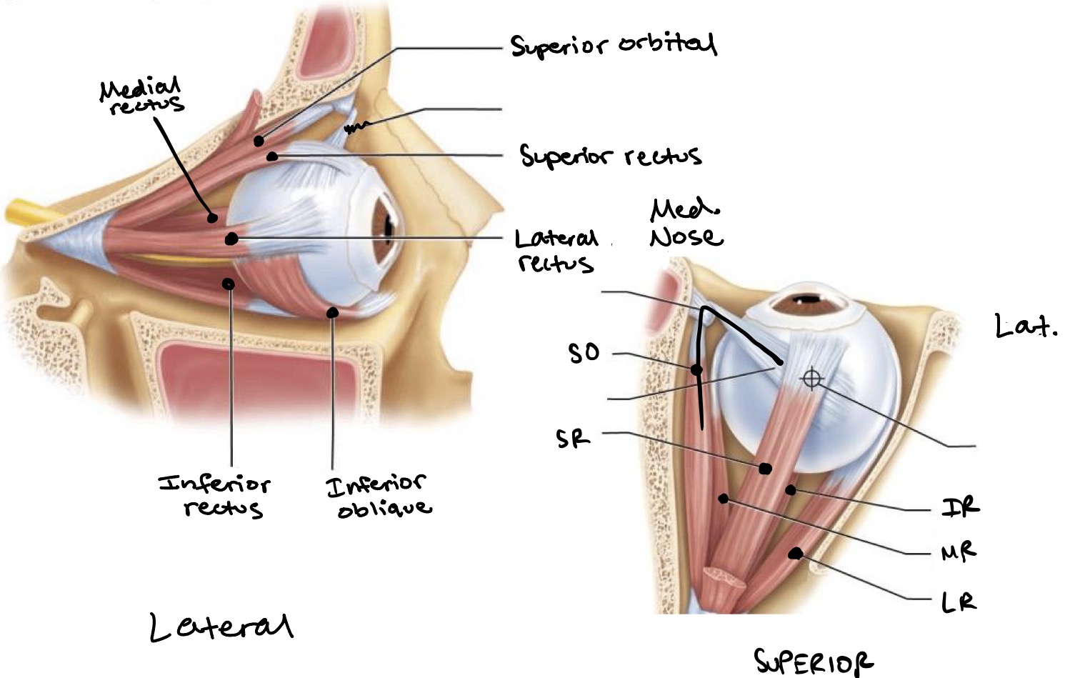
Extrinsic muscles of the eye
Attach to the fibrous tunic
Originate on surface of orbit (bone) & insert on sclera
6 muscles:
Superior rectus muscle
A: Elevation of eyeball
I: Oculomotor nerve (CN lll)
Inferior rectus muscle
A: Depression of eyeball
I: Oculomotor nerve (CN lll)
Medial rectus muscle
A: Adduction of eyeball
I: Oculomotor nerve (CN lll)
Lateral rectus muscle
A: Abduction of eyeball
I: Abducens nerve (CN Vl)
Superior oblique muscle
A: Depression & abduction of eyeball
I: Trochlear nerve (CN lV)
Inferior oblique muscle
A: Elevation & abduction of eyeball
I: Oculomotor nerve (CN lll)
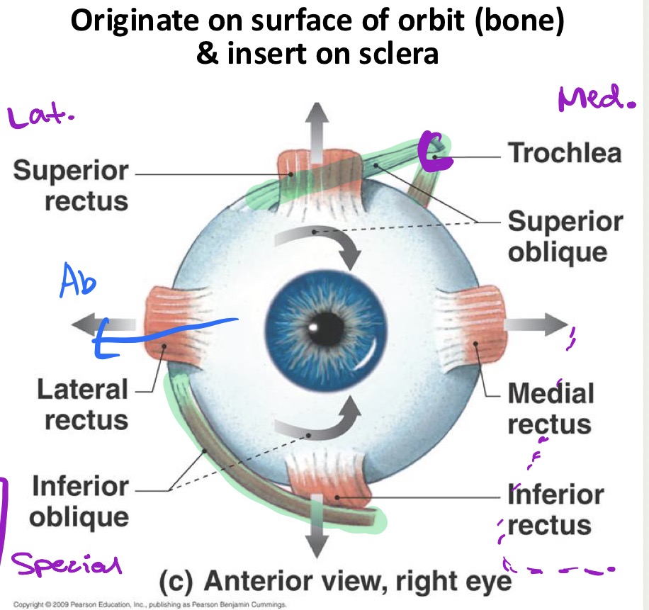
Extrinsic muscles of the eye - labeled
To help remember:
LR6SO4AO3
Lateral Rectus → CN Vl (6) / Abducens N.
Superior Oblique → CN lV (4) / Trochlear N.
All Others → CN lll (3) / Oculomotor N.
Extrinsic muscles of the eye - lateral & superior views
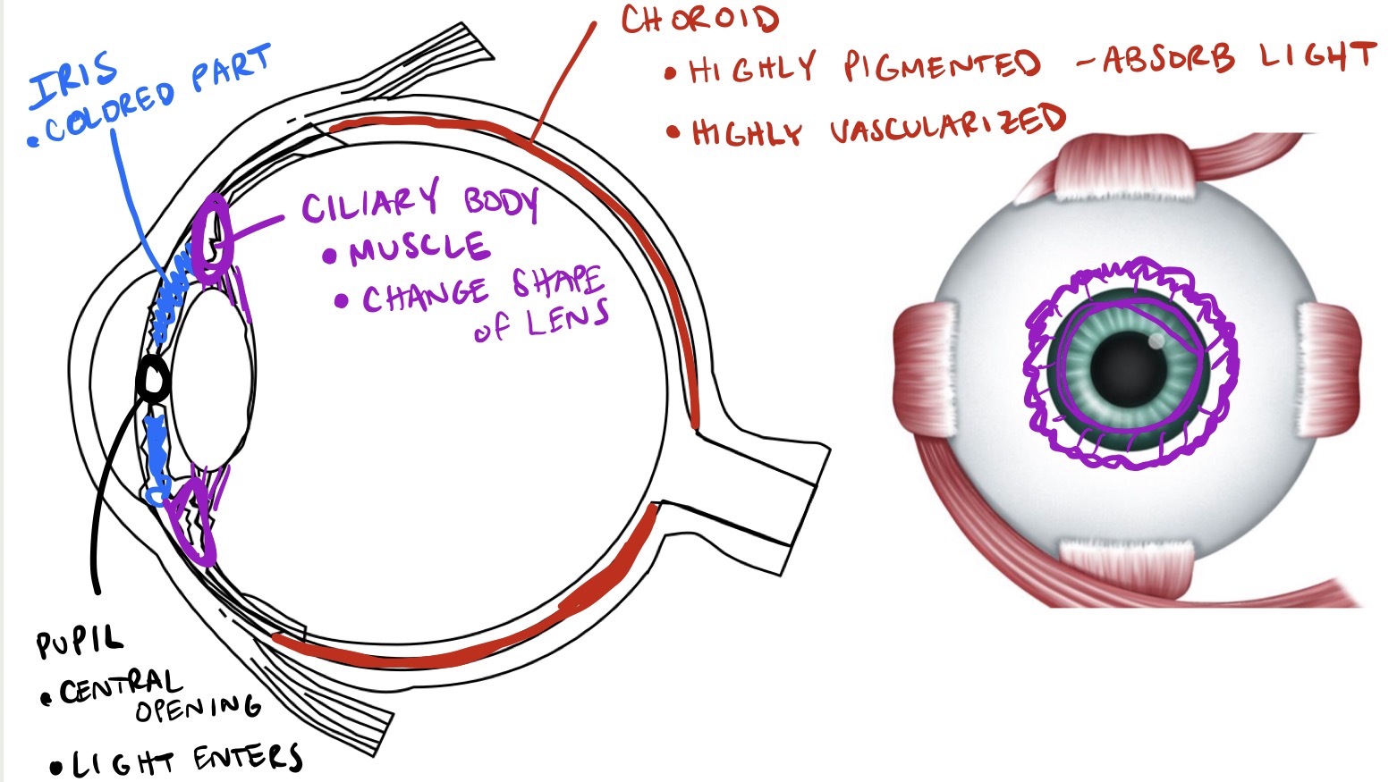
Vascular Tunic (Uvea)
Composed of:
Choroid
Ciliary body
Iris → central opening = pupil
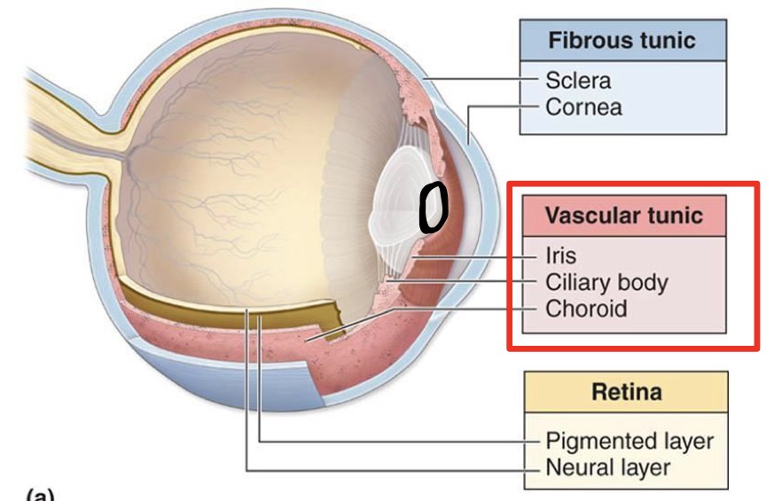
Vascular Tunic - labeled
Choroid:
Highly pigmented - absorbs light
Highly vascularized
Ciliary body:
Muscle
Change shape of lens
Iris:
Colored part
Pupil:
Central opening
Light enters
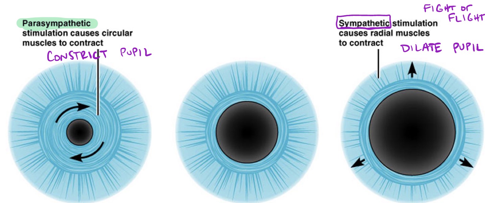
Oculomotor nerve (CN lll) - parasympathetic
Iris = change pupil diameter
Parasympathetic:
Stimulation causes circular muscles to contract, which constricts the pupil
Sympathetic:
Stimulation causes radial muscles to contract, which dilates the pupil
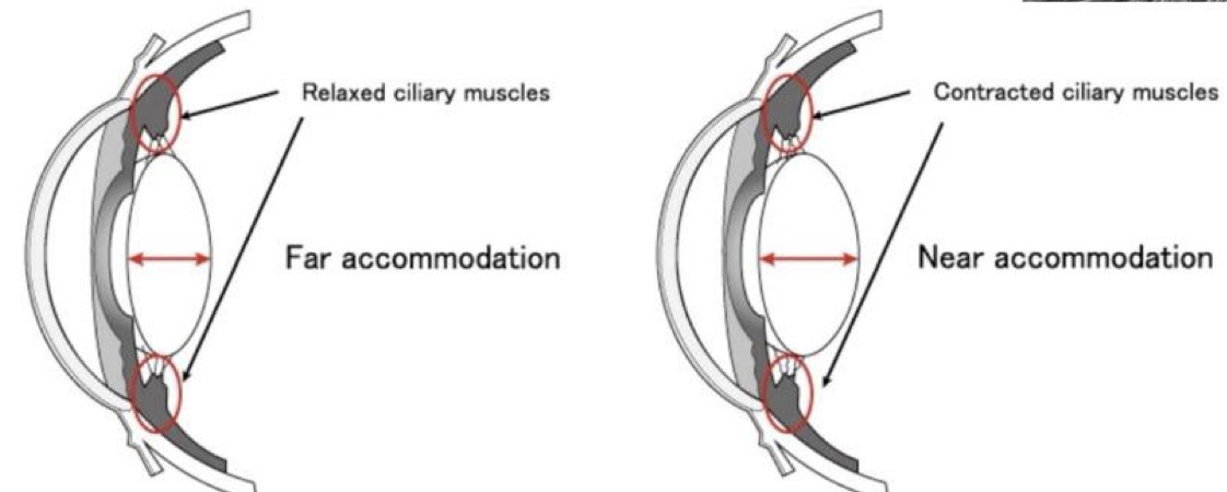
Lens
Used to focus light on the retina by changing shape
Relaxed ciliary muscles = far accommodation = narrower lens
Contracted ciliary muscle = near accommodation = wider lens
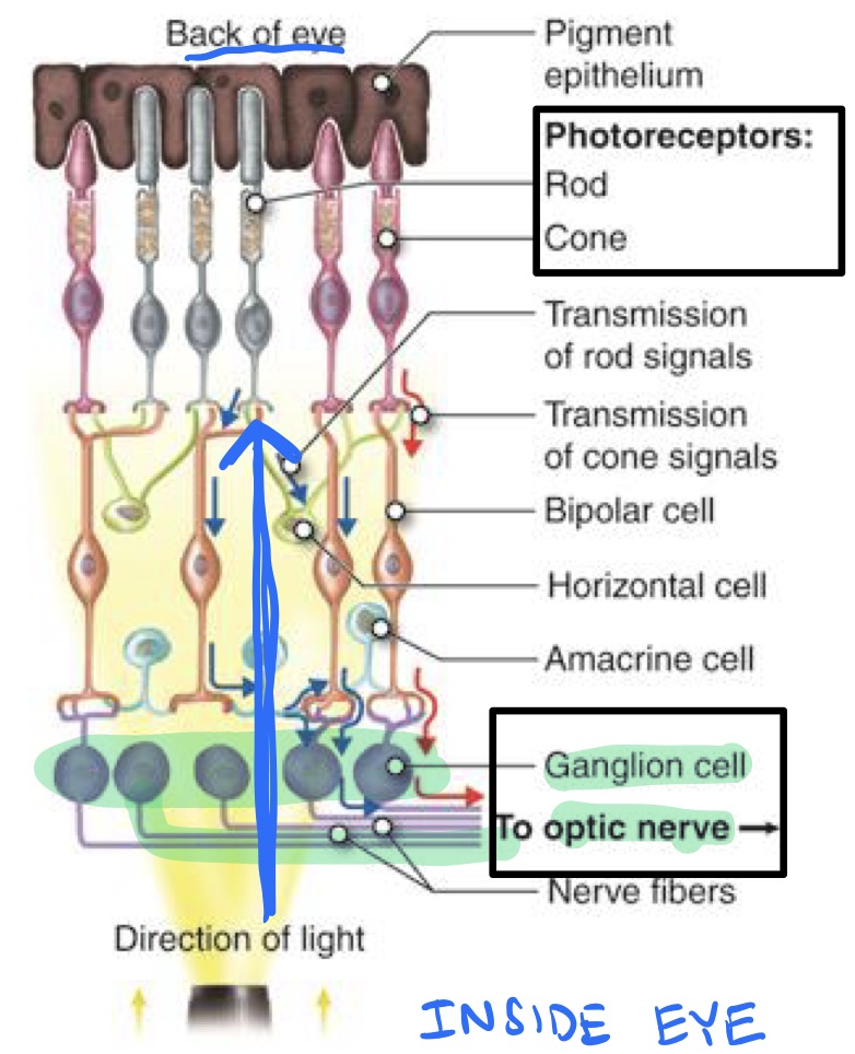
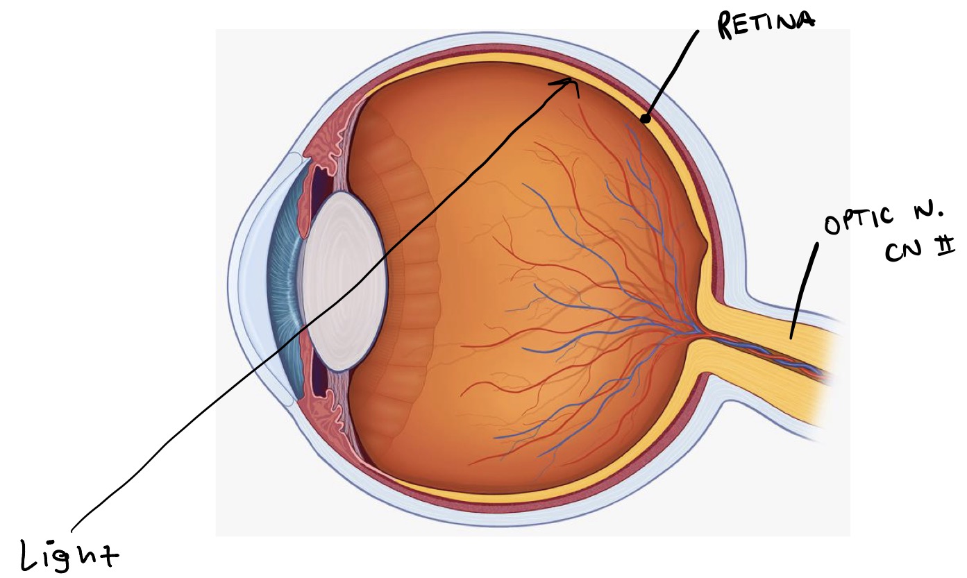
Retina (sensory tunic)
Composed of photoreceptors
Optic nerve (CN ll) is made up of the axons of the retinal ganglion cells
Sensory Tunic (Retina) - labeled
Interior cavity
Anterior segment:
Anterior to lens
Filled w/ aqueous humor
Liquidy; replaced often
Posterior segment:
Posterior to lens
Filled with vitreous humor
Gelatinous
Not replaced
Shock absorber
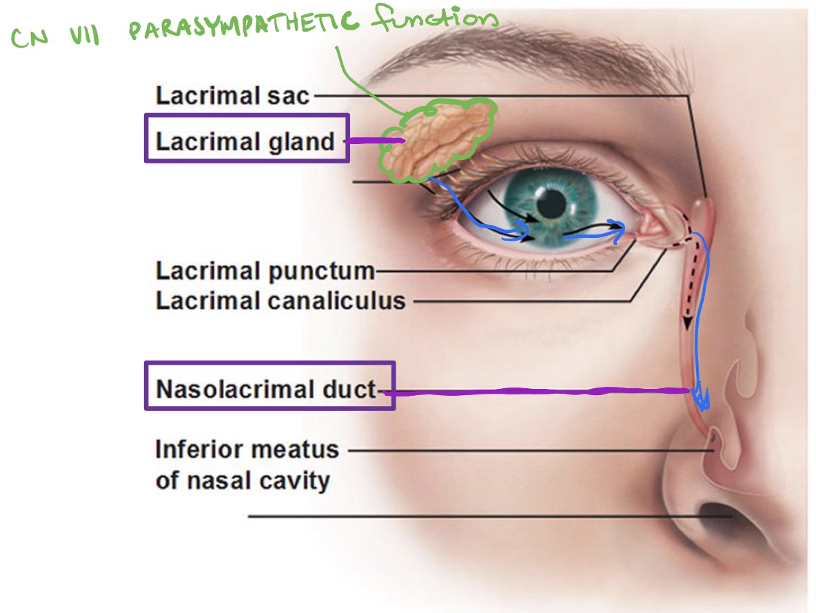
Lacrimal Apparatus
Lacrimal gland
CN Vll - Parasympathetic function
Nasolacrimal duct
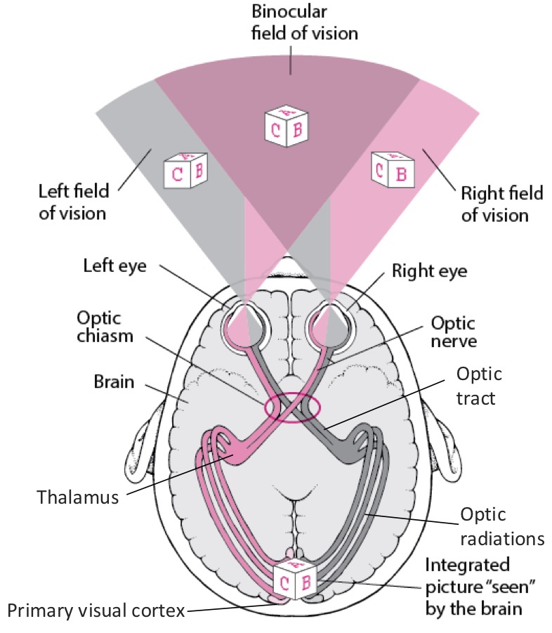
Vision
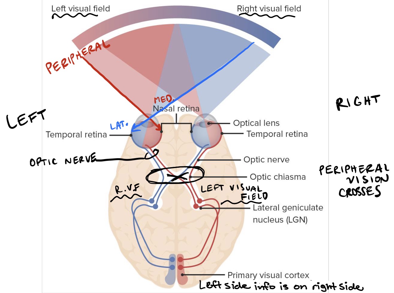
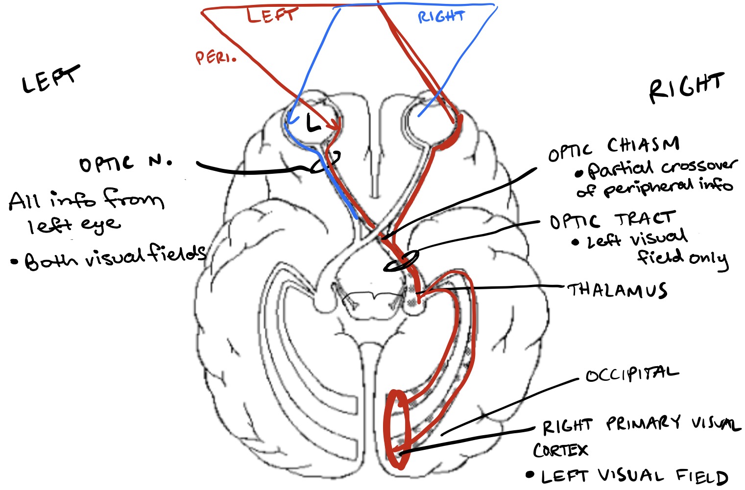
Visual pathway (right side example)
Optic nerve coming from left eye: all info from left eye (both visual fields)
Optic chiasm: partial crossover of peripheral info
Optic tract on right side: left visual field only
Right primary visual field: left visual field
Visual pathway - order summary
Photoreceptors
Ganglion cells (axons for the optic nerve / CN ll)
Optic chiasm (partial crossover of peripheral info)
Optic tract
Thalamus
Primary visual cortex
The visual info from the left eye travels in which structure?
Left optic nerve
The visual info from the left visual field travels in which structure?
Right optic tract
Peripheral vision crosses over in which structure?
Optic chiasm