apicomplexa
1/103
There's no tags or description
Looks like no tags are added yet.
Name | Mastery | Learn | Test | Matching | Spaced |
|---|
No study sessions yet.
104 Terms
Sarcocystis hominis
Sarcocysts are microscopic in muscles of cattle
Sarcocystis suihominis
Sarcocysts are macroscopic in muscles of swine
Major species affecting humans
Sarcocystis hominis & Sarcocystis suihominis
Pigs and cattle
Inffective host
Sarcocyst
IS CONTAIN SPOROZOITES
Tissue biopsy samples
Diagnostic Stages:
Bradyzoites (within sarcocyst)
Stool sample
Diagnostic Stages:
Oocysts & Sporocysts
Raw meat consumption
Infective Stages:
Bradyzoites (within sarcocyst)
Fecal contamination of food and water
Oocysts & Sporocysts
Vasculitis and myositis
Invasive form – rare in • Sarcosporidiosis or Sarcocystosis
Nausea, abdominal pain and diarrhea
48 hrs
Intestinal form of Sarcosporidiosis or Sarcocystosis
Mild 48hrs
Intestinal form disease hrs
Intestinal Coccidians
• Apicomplexans that affect animals as well as humans
• Both sexual and asexual reproduction in one host
Sporulated / mature oocyst
Infective stage Intestinal Coccidians
Ingestion
MOT Intestinal Coccidians
Gastroentritis
Disease manifestation Intestinal Coccidians
Human and animals
Cryptosporidium parvum (C. parvum)
Oocysts (Thick-walled)
Infective & Diagnostic Stage:Cryptosporidium hominis (C. parvum)
C. hominis
formerly known as C. parvum genotype I
C. parvum
formerly known as C. parvum genotype II
human
C. hominis
Infective Stage: Mature (sporulated) oocysts
Diagnostic Stage: unsporulated oocysts
Cyclospora cayetanensis
Infective Stage: __________
Diagnostic Stage: __________
Infective Stage: Mature (sporulated) oocysts
Diagnostic Stage: Unsporulated oocysts
Cystoisospora (Isospora) belli
Infective Stage: _________
Diagnostic Stage: __________
Cryptosporidium > Cyclospora > Cystoisospora
Arrange into its sizeCystoisospora cryptosporidium Cyclospora
Gastroenteritis/Diarrhea usually self-limiting
Severe diarrhea among immunocompromised
(especially C. hominis)
• Autoinfection can occur in C. hominis
AUTOFLUORESCENCE
important criterion for identifying C. cayetanensis oocysts
Bluish green circles under UV light
C. cayetanensis oocysts
Bluish green circles under UV light
Trimethoprim-Sulfamethoxazole
Drug use for c. Cayetanesis
Nitazoxanide
TMP-SMX/Trimethoprim-Sulfamethoxazole
C. belli
Toxoplasma gondii
Worldwide distribution affecting a wide variety of mammals including humans
Cosmopolitan distribution
Members of Family Felidae
Cats
What is the definitive host of toxoplasma gondii
Cats
Both definitive and intermediate host in toxoplasma gondii
rodents, birds, pigs, man
Intermediate host in toxoplasma gondii?
Toxoplasma gondii
Originally described from North African Rodents (Gundi)
Tachyzoites, Bradyzoites and Oocyst
Infective stage in toxoplasma gondii?
• Ingestion of infected undercooked meat= bradyzoites
• Consumption of food or water contaminated with cat feces=oocytes
• Blood transfusion or organ Transplantation=Tachyzoites, Bradyzoites
• Vertical transmission=Tachyzoites
MOT of toxoplasma Gondii
Tachyzoites= Blood & Body fluids
Bradyzoites= Tissue Consumption\
Oocysts= Fecal Contamination of Food & Water
Infective Stages:
Tachyzoites= __________
Bradyzoites= __________
Oocysts= __________
Diagnostic Stages:
Tachyzoites= Blood & body fluids samples
Bradyzoites= Tissue biopsy samples
Oocysts= Stool sample
Diagnostic Stages:
Tachyzoites(masmibilis)= ___________
Bradyzoites= ___________
Oocysts= ___________
oocyt
can be infective/ diagnostic in humans in toxoplasma gondii
Tachyzoites toxoplasma gondii
- quickly multiplying forms
- responsible for initial spread and tissue destruction
- Blood & body fluids
Bradyzoites Toxoplasma Gondii
- slower developing form
- characteristic of older infection
- Tissue
Periodic Acid Schiff
What stain used for tissue BradyzoitesPOSITIVE
Tachyzoites toxoplasma gondii
actively multiplying
Bradyzoites Toxoplasma Gondii
slow growing morphologic appearance: similar to tachyzoites
28-152 um in length, 22-123 um wide
Shape of Bradyzoites Toxoplasma Gondii
crescent shape, often more rounded on one end
Shape Tachyzoites toxoplasma gondii
3-7 × 2-4 um
Size Tachyzoites toxoplasma gondii
One
Number of Nuclei of Tachyzoites toxoplasma gondii
contains a variety of organelles that are not readily visible
Other features of Tachyzoites toxoplasma gondii
Smaller than tachzyzoites
Size of Bradyzoites toxoplasma gondii
One to two contrctile vacoules
cytoplasm may contain food vacoules and bacteria
Small cytotome present
Layer of cilia around organism
Number of nuclei of Bradyzoites toxoplasma gondii
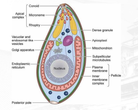
identify parts
• May exhibit flu like symptoms
• Encephalitis is usually manifested
• Other manifestations include retinochoroiditis,lymphadenopathy, splenomegaly
• Congenital Defects
DISEASE AND PATHOGENESIS of TOXOPLASMA GONDII
immunocompromised PERSONS
• Clinical Manifestations appear among______ in toxoplasma gondii
TRUE
TRUE OR FALSE: Infections are usually asymptomatic
Giemsa Stained Tissue imprints
Examination of Toxoplasma Gondii
Resistant to staining with Methylene Blue
adding live tachyzoites and complement to patient serum
if patient has antibodies, they will bind to the tachyzoites
binding will activate complement which will induce lysis of parasite
Sabin Feldman Test
- Unstained –______
- Stained – ______
Diagnosis of toxoplasm gondii
- Unstained – POSITIVE
- Stained – NEGATIVE
Sabin Feldman Test
IgM antibody detection
TORCH testing
• Molecular Techniques
Diagnosis in toxoplasma gondii
TO – Toxoplasma
R – Rubella
C – CMV
H – Herpes Simplex Virus 1&2
TORCH TESTING MEANING?
• Pyrimethamine and Sulfadiazine
Treatment of Toxoplasma gondii which is combination for 1 month
cook meat(150 or 66c); proper hygiene; disinfect and clean daily cat litter pans; pregnant women should avoid contact with cats
Prevention of toxoplasma gondii
Two; 10-18 µm long (each)
Sarcocystis spp. Mature Oocyst Number & Size of each sporocyst
Transparent
Appearance of Sarcocystis spp. Mature Oocyst:
Oval
Shape of Sarcocystis spp. Mature Oocyst:
Four sausage-shaped sporozoites
Contents of each sporocyst of Sarcocystis spp. Mature Oocyst:
Unicellular with granular cytoplasm
Oocyst cell wall appearance Sarcocystis spp. Mature Oocyst:
Clear, colorless, double layered
Young oocyst of Sarcocystis spp.
Two sporocysts, each containing four sausage-shaped sporozoites
Mature oocyst of Sarcocystis spp.
History of Recent ingestion of raw or undercooked meat
Muscle Biopsy – definitive diagnosis
• Stool Exam – detection of sporocyst
• Flotation Techniques
• PCR
Diagnosis and Treatment of Sarcocystis
Rarely Required (asymptomatic)
albendazole, metronidazole; co-trimoxazole
Treatment of Sarcocystis
Thorough Cooking of Meat; Freezing of meat
Prevention of Sarcocystis
Muscle Biopsy
definitive diagnosis for Sarcocystosis
Stool Exam
detection of sporocyst Sarcocystis
4-6um
Size of Cryptosporidium parvum oocyt
Roundish
Shape of Cryptosporidium parvum oocyt
None
Number of Sporocyt Cryptosporidium parvum oocyt
Four(small)
Number of Sporozoites
thick cell wall
one six dark granules may be visible
Cryptosporidium parvum
Infective Stage: Mature (sporulated) oocysts
Infective Stage of Cyclospora cayetanensis: _____
common if rasberry and basil leaves
Unsporulated oocysts
Diagnostic Stage: _______
affected humans
7-10 µm in diameter
Size of Cyclospora cayetenensis Mature Oocyst
Two
Number of in sporocysts Cyclospora cayetenensis Mature Oocyst
Each sporocyst contains two sporozoites
Contents of sporocysts in Cyclospora cayetenensis Mature Oocyst
25-35 um long, 10-15 um wide
Cystoisospora (Isospora) belli size range?
transparent
Appearance of Cystoisospora (Isospora) belli mature oocyt
Oval
Shape of Cystoisospora (Isospora) belli mature oocyt
Two layered,colorless and smooth
Cell wall of Cystoisospora (Isospora) belli mature oocyt
Unicellular with granular layer
Developing sporoblast of Cystoisospora (Isospora) belli mature oocyt
Two sporoblast
young of Cystoisospora (Isospora) belli mature oocyt
Two sporocytes, each containing four sausage shaped sporozoites
Mature oocyst of Appearance of Cystoisospora (Isospora) belli mature oocyt
• S/S: Gastroenteritis/Diarrhea usually self-limiting
Severe diarrhea among immunocompromised (especially C. hominis
Pathogenesis of cyrtispora belli
• Autoinfection can occur in C. hominis
C.hominis and C. belli
Modified kinyoun Positive
C. cayetanesis
Modified kinyoun variable
C. cayetanensis
AuramineRhodamine: Bright Yellow Green
C. hominis
AuramineRhodamine:Yellow Green
C. belli
AuramineRhodamine:Bright Yellow Green