(1) fundamental principles of imaging modalities
1/41
There's no tags or description
Looks like no tags are added yet.
Name | Mastery | Learn | Test | Matching | Spaced | Call with Kai |
|---|
No analytics yet
Send a link to your students to track their progress
42 Terms
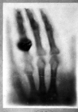
who is wilhelm conrad roentgen?
discovered the first x-ray image in 1896
what was the first x-ray picture of?
his wife anna bertha’s hand
how does radiography work?
x-ray source on one side of patient & x-ray detector on the other side
short duration (<1.2 second) pulse of x-rays is emitted by the tube
most rays interact with the patient
some pass throught the patient and reach the detector
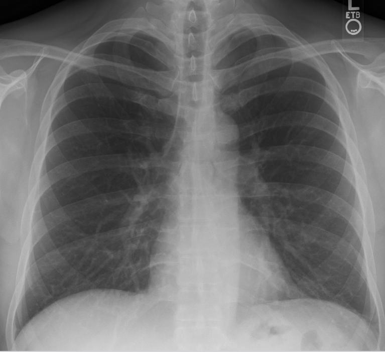
what causes a heterogeneous pattern of x-rays reaching detector?
different attenuation patterns of tissue
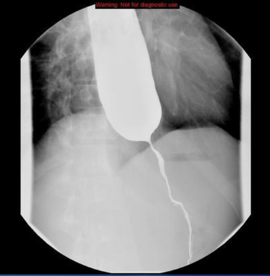
how does fluoroscopy work?
continuous/pulsed x-ray beam passed through body to create real-time, moving images on a monitor
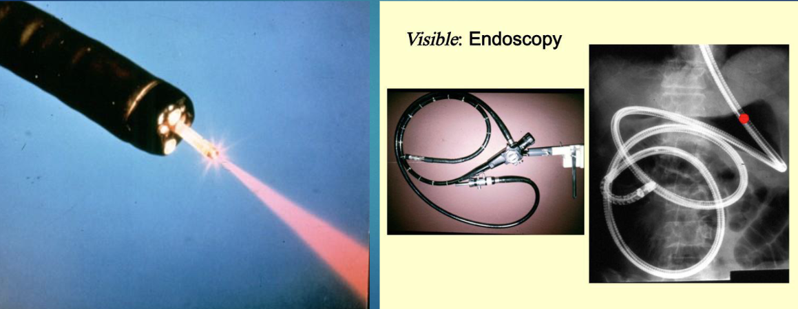
what is endoscopy?
non surgical procedure where doctor uses a thin flexible tube equipped with light and camera (endoscope) to examine the inside of the body
digestive tract usually
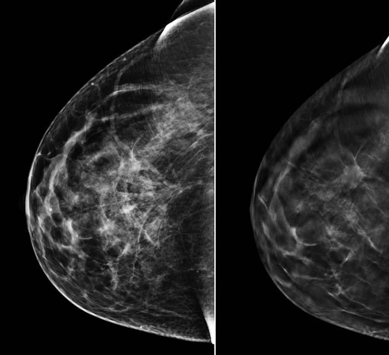
what is mammography?
specialized low dose x-ray image of breast
special detector used to accentuate contrast in breast
what do you need to see in breast mammography?
glandular and fatty tissues
skin line of breast
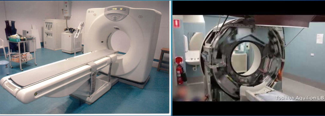
when did computed tomography become clinically available?
1970s
what is computed tomography?
passes x-rays through body at large number of angles
done by rotating x-ray tube around patient
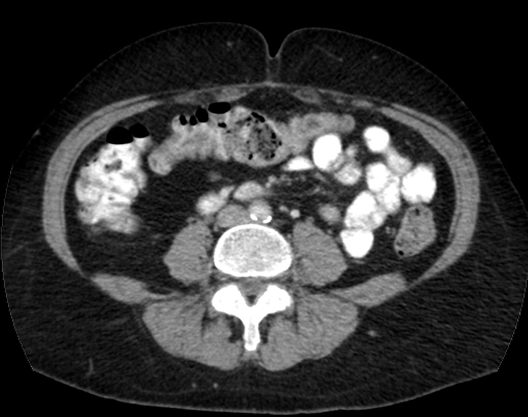
how are these images produced in CT?
computer synthesizes the data into tomographic images
tomographic images eliminate superposition of anatomical structures
you get unobstructed view of the anatomy
the radiation burden in the US has increased due to
CT
½ of all medical radiation is due to CT
what is an angiogram?
type of vascular interventional radiography
dye that is visible by an x-ray machine is injected into blood vessels of the heart
x-ray rapidly takes a series of images (angiogram)
how do stents appear on xray?
bright, metallic, tubular/cylindrical mesh structures
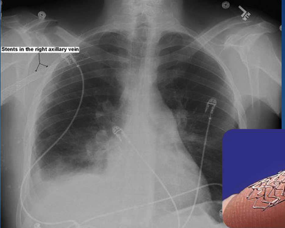
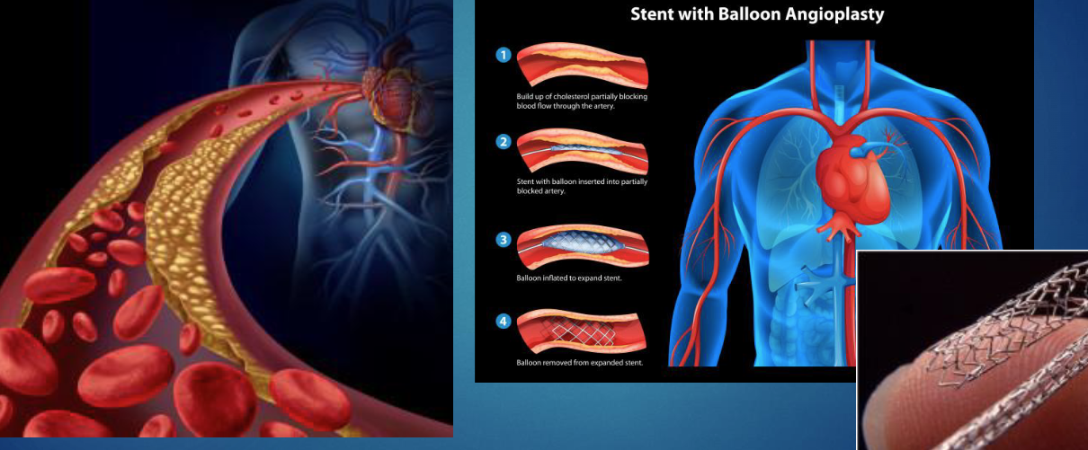
what is cardiac balloon angioplasty and stenting?
when a balloon tipped catherter is inserted in artery
balloon is inflated to flatten plaque & expand artery
mesh tube/stent is placed to keep artery open
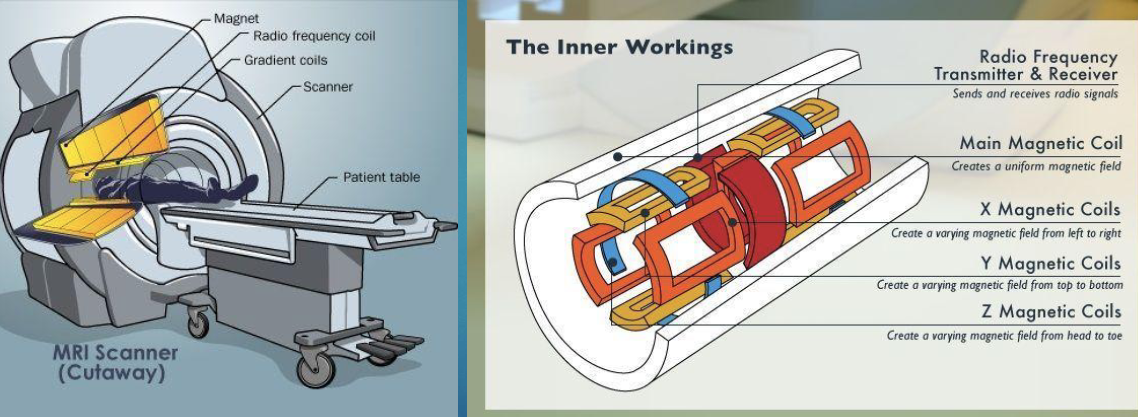
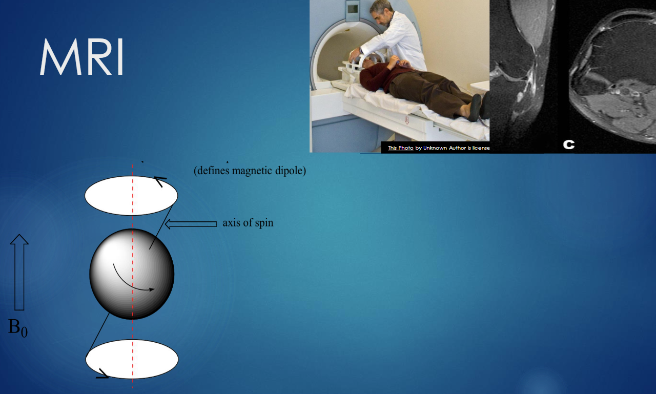
what is MRI?
radiation free medical imaging technique that uses powerful magenetic fields (10,000-60,000) times stronger than earth’s magnetic field
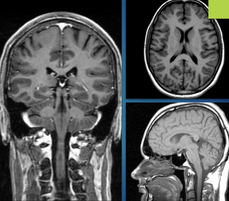
how does MRI work?
it uses the magnetic resonance properties of protons
nucleus of hydrogen atom (which are abundant in biological tissues)
produces 3D detailed images
what is nuclear medicine?
FUNCTIONAL imaging
shows how organs and tissues work (physiology)
not for structural images
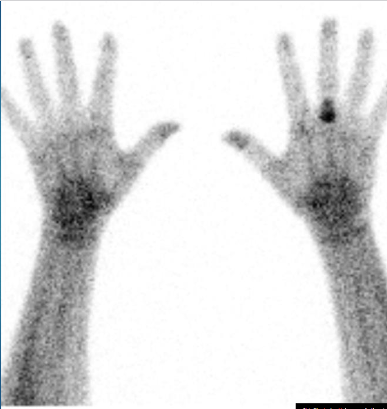
how does nuclear medicine work?
uses tiny amounts of radioactive materials that are injected, inhaled or swallowed → therefore the patient is the source of radiation
produces EMISSION images as opposed to transmission images
this is because the radioisotopes emit their energy from inside the patient
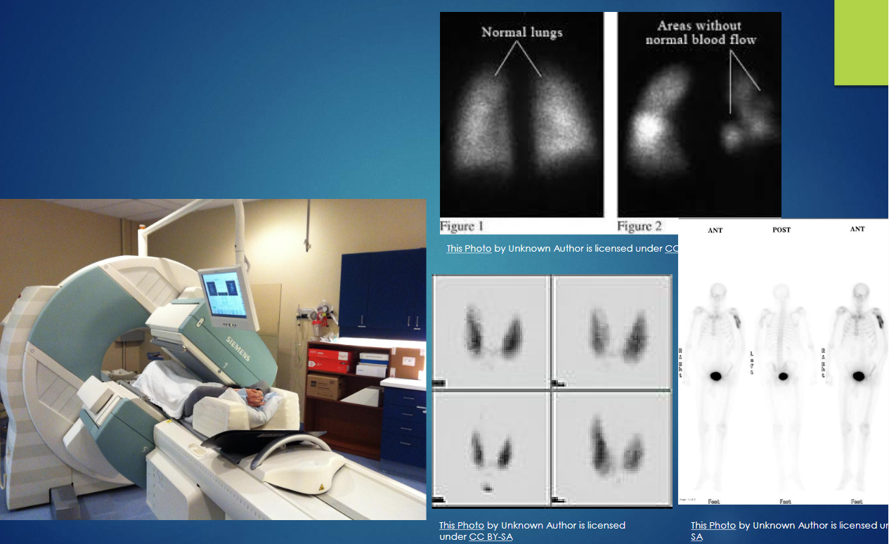
what are the types of nuclear medicine imaging?
PET, SPECT, PET/CT
what is SPECT?
single photon emission computed tomography
3D nuclear imaging test
computer is used to create cross sectional pictures of parts of the body
good for stress test imaging
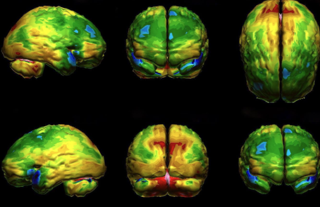
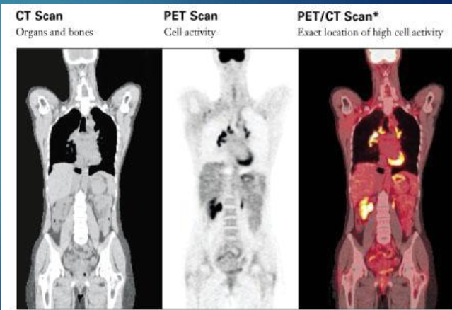
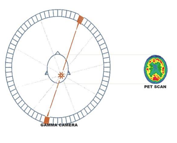
what is PET?
positron emission tomography
nuclear imaging that allows visualization of physiologic proceses in body
imaging, characterization, quantification of bio processes, stages → all happen at cellular and subcellular levels of living subjects
what is the difference between SPECT and PET scans?
PET
higher resolution
better sensitivity
faster scan times
SPECT
more widely available
less expensive
uses tracers with longer half lives
what is the speed of sound in tissue?
1540 cm/s
what is radiation? types?
energy that travels thorugh space or matter
electromagnetic (EM)
particulate
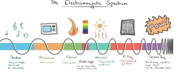
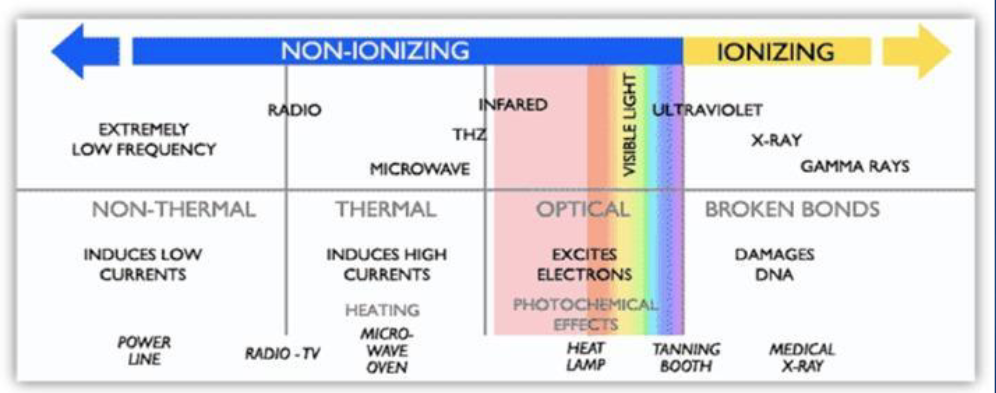
what is ionization vs non ionization?
non ionizing
any type of EM radiation that does not carry enough energy to change atoms/molecules
ionizing
high energy EM waves
removes electrons from atoms → can break chemical bonds in molecules/ change basic make up of atoms in cells
what is a sound wave?
MECHANICAL wave of pressure
*requires a medium to propagate
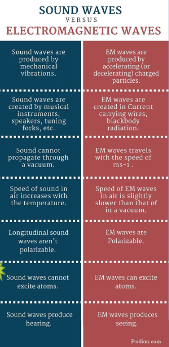
what are EM waves?
form of energy emitted and absorbed by charged particles transmitted as oscillating electric and magnetic fields
can travel through vaccum

which cells are more sensitive to radiation effects?
actively dividing cells
bone marrow
lymph glands
gonads
who is the most susceptible to radiation effects?
children and developing fetuses
what is the ALARA principle?
as low as reasonably achievable
minimize time of exposure
double distance between body and radiation source (reduced radiation exposure by factor of 4)
shielding : use absorber materials such as plexiglas for beta particles and lead for x-rays/gamma rays
what is the best distance from radiation source?
6ft
when is organogenesis?
betwen 8-15 weeks of pregnancy
this is when fetus is more sensitive to radiation induced effects
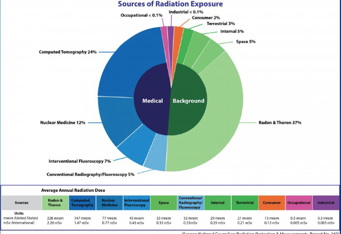
most medical sources of radiation exposure come from
computed tomography (24%)

most background radiation exposure comes from
radon and thoron (37%)
what are mild anaphylactoid reactions to contrast media?
mild urticaria (hives)
mild pruritis (itchy skin)
what are intermediate anaphylactoid reactions to contrast media?
more severe urticaria (hives)
hypotension
mild bronchospasm
what are severe anaphylactoid reactions to contrast media?
severe bronchospasm
alryngeal edema
unconsciousness
convulsions
pulm collapse
cardiac arrest
what increases risk of anaphylactoid reactions to contrast media?
history of asthma (10x)
history of atopy (10x)
genetic tendency to develop allergic diseases
previous anaphylactoid reaction to iodinated contrast media
what is contrast induced nephropathy (CIN)
refers to greater than 25% reduction of renal function occuring within 3 days of contrast medium injection
most cases resolve in 1-2 weeks
what are risk factors for CIN?
pre-existing impaired renal function
dehydration
sepsis
age >60 years
what are safety issues for MRI?
metallic items
medical equipment
implanted items in patients
nephrogenic systemic sclerosis