Anatomy and Physiology neurons
1/42
There's no tags or description
Looks like no tags are added yet.
Name | Mastery | Learn | Test | Matching | Spaced | Call with Kai |
|---|
No analytics yet
Send a link to your students to track their progress
43 Terms
Neuron
transmit information (at synapse, using neurotransmitters)
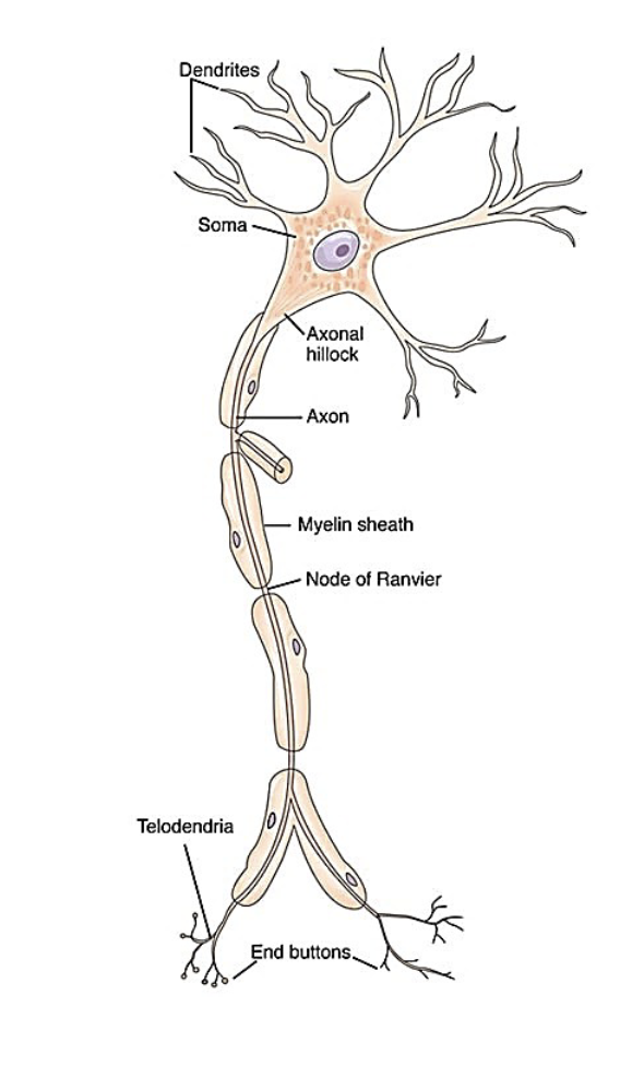
neuron anatomy
1. dendrite - receives information
via neurotransmitters (motor or
sensory signals) from synapse of
previous neuron
2. soma - the cell body - groups of
these form gray matter (also can
receive signals from synapses)
3. axon hillock - junction of the
axon and soma
4. axon - transmits information via
action potentials
5.myelin sheath - fatty wrapping that
insulates the axon; speeds up neural
conduction - white matter
6. nodes of Ranvier - area between
myelin segments that allows increased
velocity of conduction
7. telodendria - ends of the axon
8. terminal boutons or end buttons -
contain synaptic vesicles which
release neurotransmitters for
information transfer to the next neuron
Glial Cells
support neurons, provide support & nutrients to
neurons
- facilitate long-term memory
1.Astrocytes – form blood brain
barrier - adhere to blood capillaries
to transports nutrients; prevent
toxins passing from the
cerebrovascular (blood) system to
neurons
2.Schwann cells - myelin cells of the
PNS
3.Oligodendrocytes - myelin cells of
CNS
4.Microglia - clean up abnormal
areas of tissue (lesions) in the brain
(phagocytosis
Nervous System
Central Nervous
System (CNS):
1. Brain - cerebral
cortex, cerebellum,
brain stem,
subcortical structures
(hypothalamus,
thalamus,
basal ganglia)
2. Spinal Cord
Peripheral
Nervous System
(PNS):
1. Spinal nerves -
31 pairs
2. Cranial nerves -
12 pairs part of the
“system” (2 of them
actually originate
from cerebrum, but
are considered part
of PNS)
3. Sensory
receptors
Nervous system functions
Conducts activities within cerebral cortex of the brain
• Cognitive functions - e.g., thought, emotion, memory, language
2. Communicates with rest of the body
• Efferent – motor pathways: command signals are carried away from the brain via nerves or tracts to be executed by the body
• Afferent – sensory or somatic pathways: sensory signals
from the internal and external environment are received and
carried towards the brain for evaluation
Sensory (Afferent) Information - Levels & Types
Levels of sensory information:
1. Superficial senses - in skin & mucous membranes -
temperature, pain, touch
2. Deep senses - in muscles, tendons, ligaments & joints -
tension, length, pain, pressure, vibration, and joint position
• Types of sensory information:
1. Somatic - pain, temperature, and mechanical stimulation
(pressure, stretch, and vibration)
2. Kin(a)esthetic - sense of the body in motion
3. Special senses - vision, hearing, taste, smell, and touch
Periferal Nervous system
somatic (voluntary skeletal muscles)
autonomic (smooth muscle+glands automatic)
Parasympathetic - restores the body to a state of calm when it is stressed
Sympathetic - “fight or flight” response - getting the body ready for potential physical or mental activity that occur during times of stress or danger
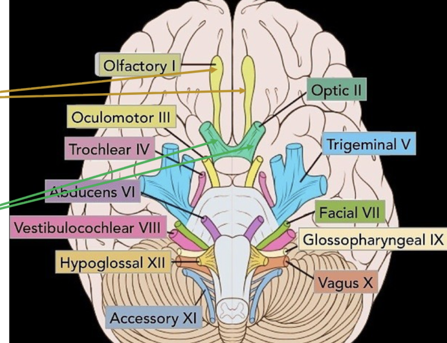
Cranial Nerves
from brainstem, the origination or starting point of each nerve is called its nucleus (same innervate multiple) Midbrain: III Oculomotor and
IV Trochlear nerve nuclei
Pons: V Trigeminal, VI Abducens,
VII Facial, and VIII Vestibulocochlear
nerve nuclei
Medulla: IX Glossopharyngeal,
X Vagus, XI Accessory,
and XII Hypoglossal nerve nuclei
Cranial nerve I - Olfactory
nerve nuclei are located in
the olfactory tract bulbs
• Cranial nerve II - Optic
nerve nuclei are located in
the optic nerve
8 cervical nerves ≠ 7 cervical vertebrae
Cranial Nerve Classifications
• By type:
I. Efferent - convey motor input from the brain to muscles and glands
II. Afferent - bring sensory input to the brainstem
I. Somatic - transmit information from skin, muscles, tendons, joints to
the CNS
II. Visceral - transmit information from organs (part of your body that
performs a specific function) to the CNS
III.Mixed or both efferent & afferent - some cranial nerves have both functions
• By function:
1. “Special” - specific to cranial nerves that 1) perform senses (vision, hearing,
smell, taste); or 2) muscles that originated from the branchial arches of an
embryo including the pharynx, larynx, soft palate, face and mandible
2. “General” - specific to 1) functions that can occur throughout the body; or
2) combine the functions of both cranial and spinal nerves
Cranial Nerve Path Mnemonic
Some Say Marry Money But My Brother Says Big Brains Matter More
sensory sensory motor motor both motor both sensory both both motor motor
On on on they traveled and found voldemort guarding very ancient horcuxes
olfactory optic oculomotor trochlear trigeminal abducens facial vestibulocochlear glossopharyngeal vagus accessory hypoglossal
Cranial nerves
Information from all other cranial nerves is processed in the thalamus first.
I. Olfactory (not true cranial nerve due to olfactory bulb processing) - sensory (sensors in epithelium of nasal cavity send the signal to the nuclei in the olfactory bulbs)
II. Optic- sensory (visual info from retina, medial retina crosses information to
the opposite hemisphere at the optic chiasm, while lateral part remains on the side its on)
III. Oculomotor- motor (for pupil restriction and upward movement)
IV. Trochlear- motor (rotates eye down)
V. Trigeminal - both (sensory feelings in Ophthalmic branch, Maxillary branch, Mandibular branch. motor mandibular branch for chewing muscles)
VI. Abducens- motor (moving eye back and forth
VII. Facial- both (sensory feeling in 2/3 front of tongue, motor upper face movement from both hemispheres lower face from opposite, middle ear reflex, lacrimal tears and salivary glands)
VIII. Vestibulocochlear- sensory hearing and balance
IX. Glossopharyngeal- both (sensory taste of back 1/3 of tongue and touch/pain/temperature sensation from posterior 1/3 of tongue, fauces, pharynx
and eustachian tube. motor saliva production laryngeal and pharyngeal elevation)
X. Vagus both (sensory pain, touch, temperature, taste sensation from behind ear to the pharyngeal-laryngeal area to the cardiac, trachea, lung and digestive area. motor movement of velum, larynx &
pharynx (part of pharyngeal
plexus), parasympathetic regulation of heart rate breathing speech+swallowing, GI tract peristalsis)
XI. Accessory motor (neck+shoulder muscles, cranial and spinal, pharyngeal
plexus with vagus velum pharynx tongue)
XII. Glossopharyngeal motor (extrinsic intrinsic parts of tongue)
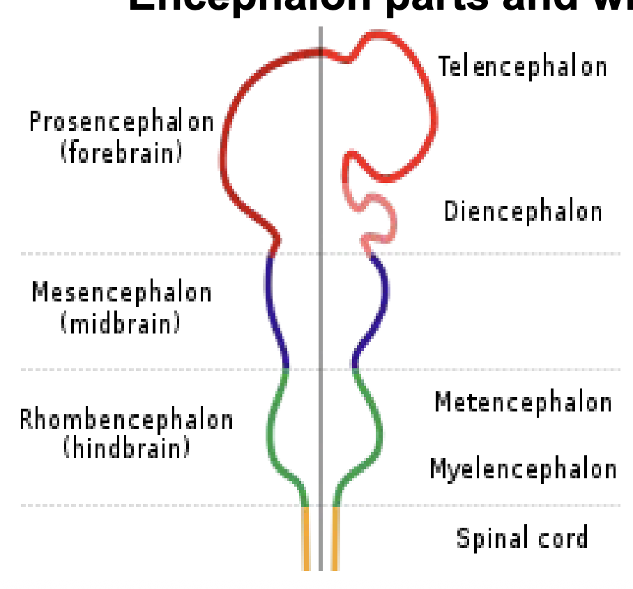
Nervous system development-
encephalon develops from the neural tube of an embryo at 4 weeks of development, brain and spinal cord
FOREBRAIN - Prosencephalon
• Telencephalon - lobes, white
matter, basal ganglia, olfactory
tract
• Diencephalon - thalamus,
hypothalamus, pituitary gland and
optic tract
MIDBRAIN - Mesencephalon
HINDBRAIN - Rhombencephalon
• Metencephalon - pons and
cerebellum
• Myelencephalon - medulla
oblongata
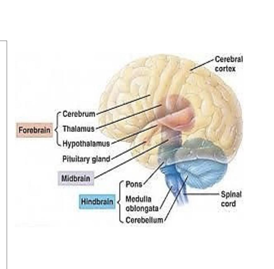
Forebrain, Midbrain, Hindbrain
1. Cerebrum - thought, learning, speech,
reading, writing, emotions, muscle
functions
2. Thalamus - relay station for all sensory
information except smell; sleep,
wakefulness, consciousness, learning &
memory
3. Hypothalamus - coordinating station
for homeostasis; releases chemical
messages to the pituitary, regulates
emotional response, body temperature,
heart rate, appetite, thirst, sexual
behavior
4. Pituitary gland - master control gland
that secretes hormones for growth,
metabolism, and reproduction
Midbrain- auditory and visual processing
(part of the brainstem)
Hindbrain
1. pons - message station
between cerebrum, cerebellum
and spinal cord; eye and body
movement, sleep and arousal
(part of the brain stem)
2. medulla oblongata - control
center for heart and lungs;
unconscious acts-breathing,
swallowing, circulation, (part of
brain stem) note spinal cord
enters skull at the medulla
3. cerebellum - coordinates and
regulates muscular activity
The Brain Stem
Functions:
1. major body functions (e.g.,
breathing, consciousness,
heart rate, sleep)
2. connects the cerebrum to the
spinal cord
• Contains:
1. nuclei of most of the cranial
nerves - not all
2. sensory and motor nerve
pathways (see next slides)
Cerebrum
80% of brain uppermost, 2 hemispheres 4 lobes
1) meninges (brain fluid suspension position, prevent brain movement trauma)
)dura mater - two tough outer
layers with space in between,
adheres to bone
• the foldings of this layer
separate the cerebral right
and left hemispheres and
the cerebellum
2)arachnoid mater/layer -
spider-like covering where the
blood vessels pass
3)pia mater - thin inner
covering where all the major
arteries and veins serve the
surface of the brain with blood
flow
2) cerebrospinal fluid (nutrient delivery and waste removal, cerebrum to the cerebellum+spinal cord)
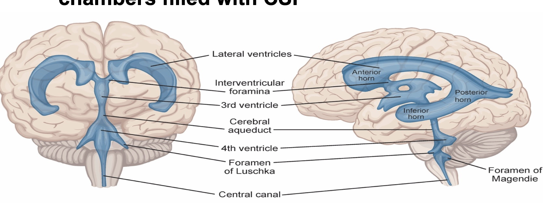
3) ventricles (open spaces to cushion and float the brain, 2 lateral, a third, and a fourth interconnected chambers with cerebrospinal fluid produced by choroid plexus)
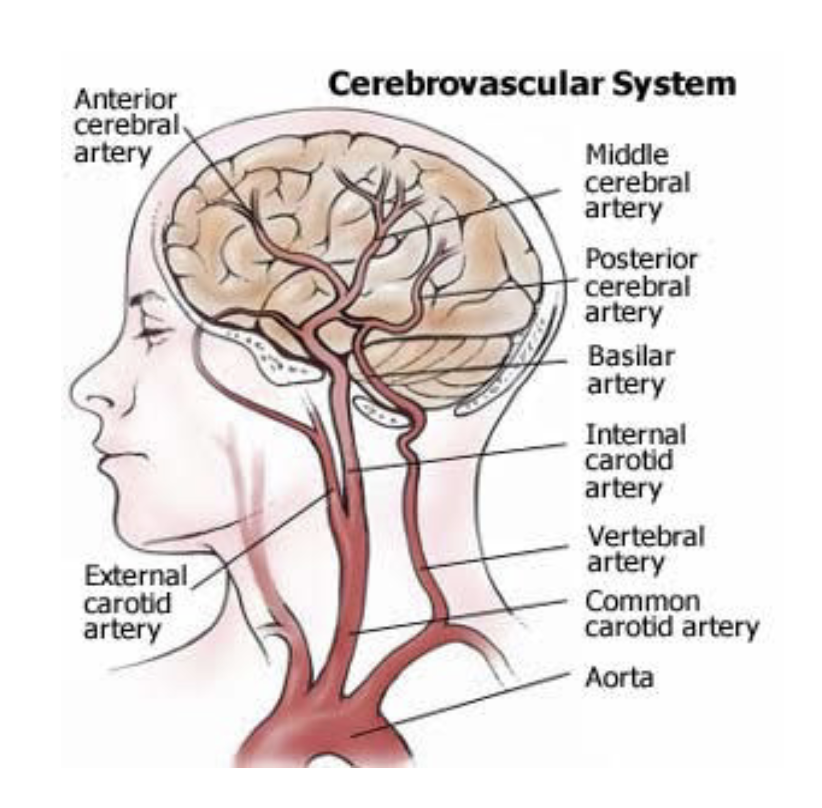
Cerebrovascular System
blood from to heart to brain,
Arteries- transport oxygen and nutrient rich blood to brain
Veins- bring deoxygenated blood back to heart to be reoxygenated
1) Cerebral arteries - provide blood
flow in the anterior, middle and
posterior cerebrum
2) Carotid arteries - circulate blood
from heart
1) Internal carotids (2) - to brain
2) External carotids (2) - to face
and neck
3) Vertebral arteries (2) - circulate
blood from heart to the back of the
neck - merge to form the basilar
artery at level of pons
4) Basilar artery – forms where the two
vertebral arteries join at the base of
the skull.
• carries oxygenated blood to the
cerebellum, brainstem, and occipital
lobes
Circle of Willis
Joining of the two internal carotid and basilar artery (supplied by the two vertebral arteries) form a circle, circulating constantly in case of cerebrovascular accident and equalizes pressures
Spinal Cord function & makeup
pairs of spinal nerves that provide nerves that serve the limbs and trunk of the body, has
gray matter - cell bodies - bundles
of nerve fiber tracts that
communicate information to and
from the brain
• white matter - fatty wrapping -
myelination to protect the axon
pathways
Afferent and efferent nerve
impulses, blood and cerebrospinal
fluid run through the spinal cord via
multiple tracks 4
Spinal Cord Anatomy
-continuous with the caudal end of
medulla oblongata at the Foramen
Magnum
-spans from the cervical area to the
lumbar vertebrae at the Conus
Medullaris
-there are individual spinal nerves
below that point called the Cauda
Equina
pinal cord is covered dura mater adheres to the vertebrae, cerebrospinal fluid flows in the
subarachnoid space in between the arachnoid space and the pia mater, pia mater follows the inner surface of the
cord
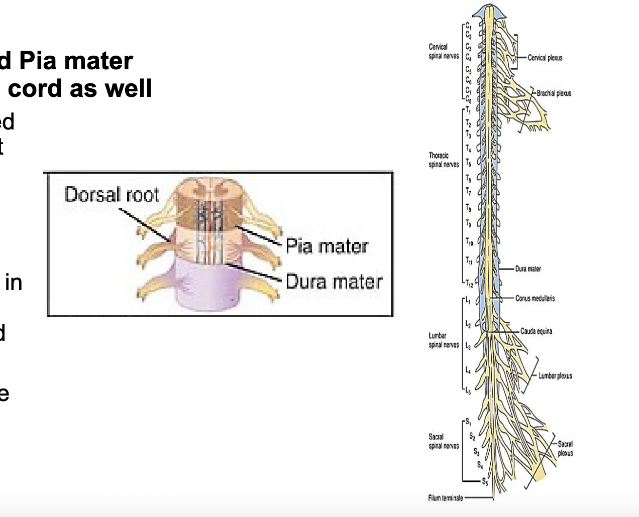
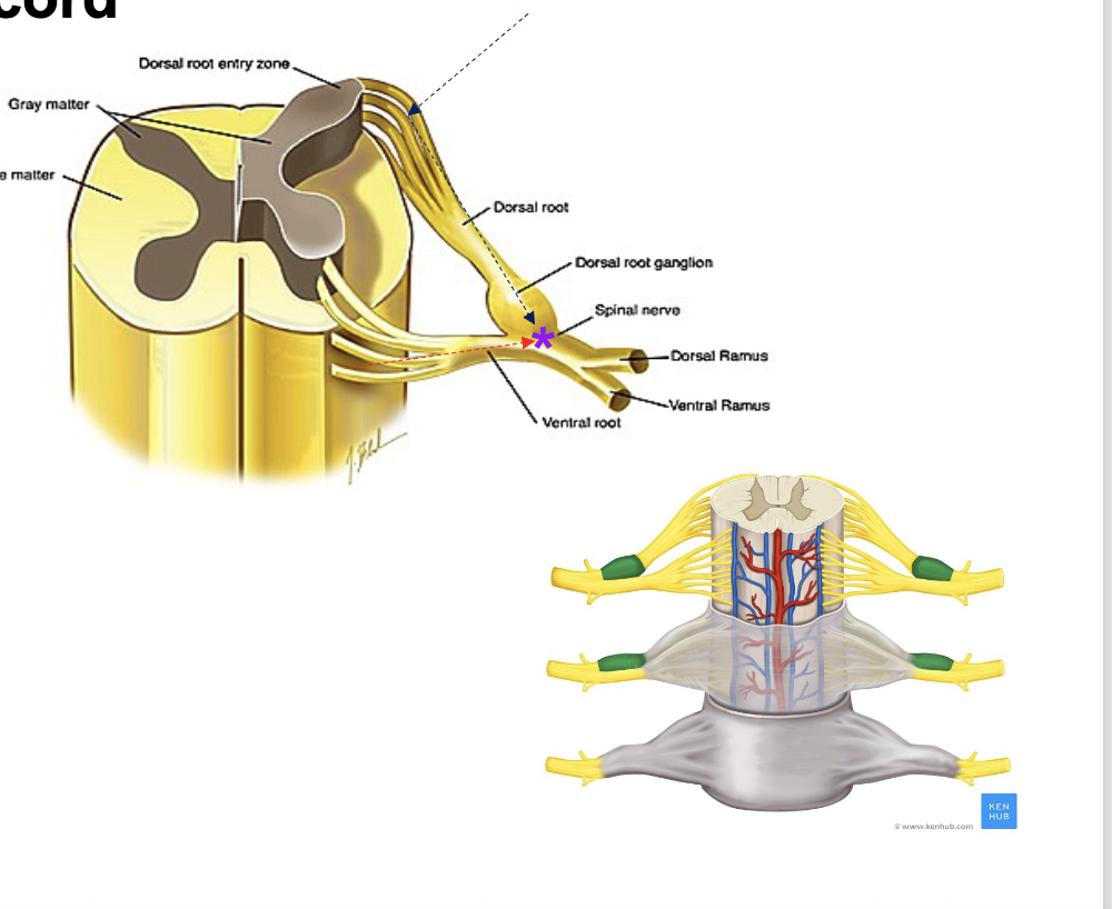
Roots
sensory information enters
from outside the spinal cord at
the dorsal roots (afferent
fibers) and combine into the
dorsal root ganglia
motor information leaves the
spinal cord the ventral roots
(efferent fibers)
both combine to be spinal nerves then divide to serve afferent or efferent information
to the anterior and posterior
portions of the body
Motor Activation path
1. upper motor neurons (UMN) - efferent fibers that come from upper brain levels and bring commands to the rest of the body by synapsing with
• 2. lower motor neurons (LMN) at the spinal cord to activate or inhibit muscle
-motor activation nerve locationoriginate within the spinal cord from the ventral root fibers
All Sensory Responses - nerve location
cell bodies (clusters of neurons) are in the dorsal root ganglia which are clusters of neurons that originate just outside the spinal cord
Sensory response
1.Sensory neurons transmit information from sensory receptors to the central nervous system (CNS)
2. Relay neurons (interneurons) transmit information within the CNS as part of the decision- making process
3. Motor neurons then transmit information from the CNS to effectors (muscles or glands), in order to initiate a muscle or glandular response
• This is for deep kinesthetic senses and special senses
Spinal reflex sensory response/dorsal root response
• involuntary response, other type of sensory response
• lower motor neuron response only - quick sensory to motor
response, stays within the spinal cord
• no upper motor neurons response, does not process in the cerebrum
• reflex arc = the neural pathway that controls the action reflex
• Example of reflexive responses:
• Withdrawal reflex - to pain such as touching something hot
• Patellar or the knee jerk reflex - leg kicks forward when hit in a
certain area (think doctor’s exam when tapping your knee
Spinal Reflex Arc is a neural
pathway that controls a
reflex.
• Sensory neurons do not pass
directly into the brain but
synapse in the spinal cord
• This is for superficial somatic
responses (temperature, pain,
and touch)
Spinal cord
nerve pathways at the levels of the 1. brain stem, 2. cerebrum, & 3. spinal cord
midbrain- cerebral peduncles - link the brain stem to the thalamus and cerebrum, for passage of motor pathways
pons- cerebellar peduncles – link the cerebellum to the midbrain and the medulla for passage of motor pathways
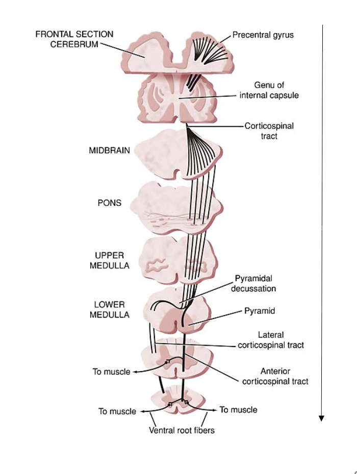
medulla- pyramidal decussation - point where motor commands that originated in one hemisphere of the cerebrum, cross over in the anterior of the medulla to serve the other side of the body via the spinal cord (e.g., left hemisphere to right side of body)
corticobulbar tract
Pathway that carries motor commands (upper motor neurons) from the pre-central gyrus (motor strip) of the cerebrum to the cranial nerves in the brainstem [“bulb”]
![<p><span><span>Pathway that carries motor commands (upper motor neurons) from the pre-central gyrus (motor strip) of the cerebrum to the cranial nerves in the brainstem [“bulb”]</span></span></p>](https://knowt-user-attachments.s3.amazonaws.com/d40c6c3f-ccdc-4b8c-850e-9b9375177a77.png)
corticospinal tract
Pathway that carries motor commands
(upper motor neurons) from the pre-
central gyrus (motor strip) of the right or
left side (hemisphere) of cerebrum to
the spinal nerves (lower motor
neurons) to provide nerve innervation
to skeletal muscles
• fiber bundles decussate (cross over)
through the pyramids of the medulla
before coursing down through the
spinal cord to the skeletal muscles of
the opposite side of the body
20
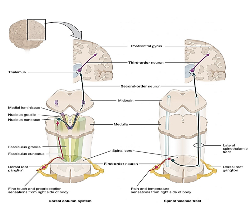
dorsal columnar tract and Spinothalamic Tract
dorsal columnar tract
Pathway that carries information from the spinal cord to the thalamus to be processed
• Fine touch and proprioception (awareness where body parts are without looking at them, and where they are relative to each other)
Spinothalamic Tract
Pathway that carries information from the spinal cord to the thalamus to be processed
• Pain and temperature sensations
Gray matter (cell bodies) and white matter (myelin) differences between brain and spinal cord
Brain
• Gray - outer layer - cerebral cortex (sensory & motor processing, as well is higher brain functions)
• White - deep inner layer (brain tracts between hemispheres or to the spinal cord)
• Spinal cord
• White - outer layer (sensory and motor tracts)
• Gray - deep inner layer (reflex processing)
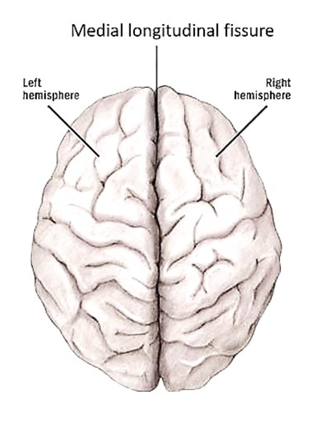
Cerebral Cortex and hemispheres
Sheet of neural tissue that is outermost layer of the cerebrum
• Brain functions that are carried out in the cortex are referred to as “cortical”
• It has two hemispheres - left and right, which are separated by the central or medial longitudinal fissure
Hemispheres
Left - dominant for language and speech (Broca’s and Wernicke’s areas are there) - controls right side of the body
• Right - artistic and creative aspects - controls left side of the body
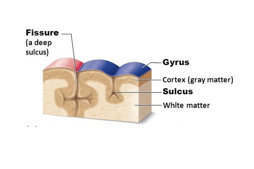
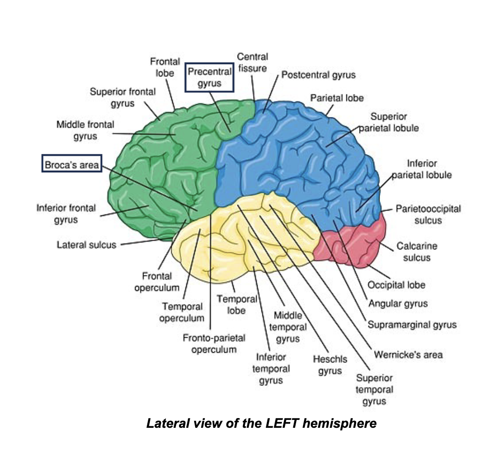
Terms for the landmarks of the Cortex
gyrus (gyri) - bumps
• sulcus (sulci) - grooves or folds
• fissure = deep groove
• these landmarks form the borders of the lobes and other regions of the cerebrum
Landmarks of the Cortex
Central or Medial
Longitudinal Fissure
• Central Sulcus (Fissure of Rolando - Rolandic Fissure)
• Precentral Gyrus (Motor Strip)
• Post Central Gyrus (Primary Somatosensory
Cortex, Sensory Strip)
• Lateral Sulcus (Fissure of Sylvius - Sylvian Fissure)
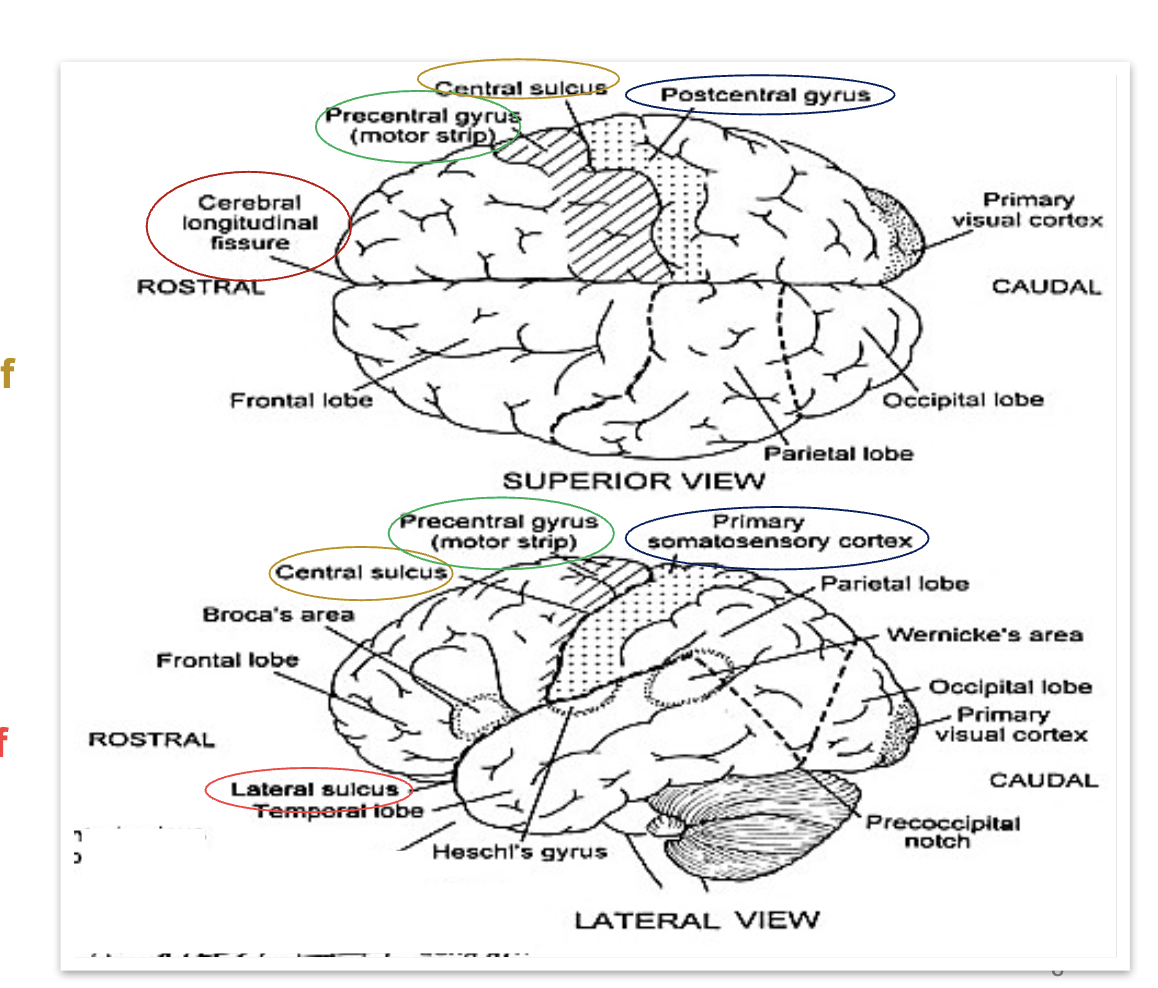
Lobes of the Cortex
Frontal Lobe
• Functions: memory; emotion; intellect; and motor functions: planning, inhibition, and issuance of motor commands
• Important regions within the lobe:
• Precentral Gyrus (Motor Strip) - initiation of voluntary movement
• Broca’s Area - speech production
Parietal Lobe
• Functions: somatic sense
• Important regions within the lobe:
• Postcentral gyrus (Sensory strip) - primary site of sensory input
• Inferior parietal lobule - integrates vision, hearing, and somatic sense
• Supramarginal gyrus - phonological processing
• Angular gyrus - receives input from visual, auditory, and somatic centers
Occipital Lobe
• Functions: visual stimulation and processing
Temporal Lobe
• Functions: auditory reception, auditory
processing
• Important regions within the lobe:
• Heschl’s Gyrus – primary auditory cortex (processing of all incoming sound, including elementary processing of speech sounds)
• Wernicke’s Area -language processing
Subcortex
• parts of the brain and nuclei that are
inferior or lie below the cerebral cortex or
cerebrum of the brain
• Brain functions that occur in the subcortex
are referred to as “subcortical”
• Contains:
1. Deeper sections of the forebrain:
1) Basal ganglia - control voluntary
motor movement, muscle tone, as
well as executive functions and
emotions
2) Diencephalon: thalamus,
hypothalamus, pituitary gland and
optic tract)
2. Limbic structures (see later slide for
ones not also part of the forebrain)
3. Brain stem - midbrain, pons, medulla
oblongata
4. Cerebellum
Limbic Lobe
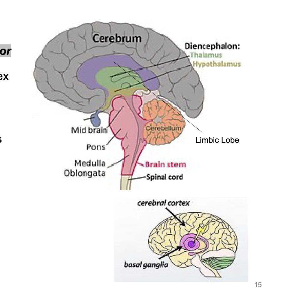
Limbic lobe and limbic system
Limbic Lobe
• Function: communicates with the limbic system = group of interconnected structures that facilitate memory retrieval and storage, establish emotional states, and link conscious intellectual functions of the cortex with unconscious autonomic functions of the brain stem
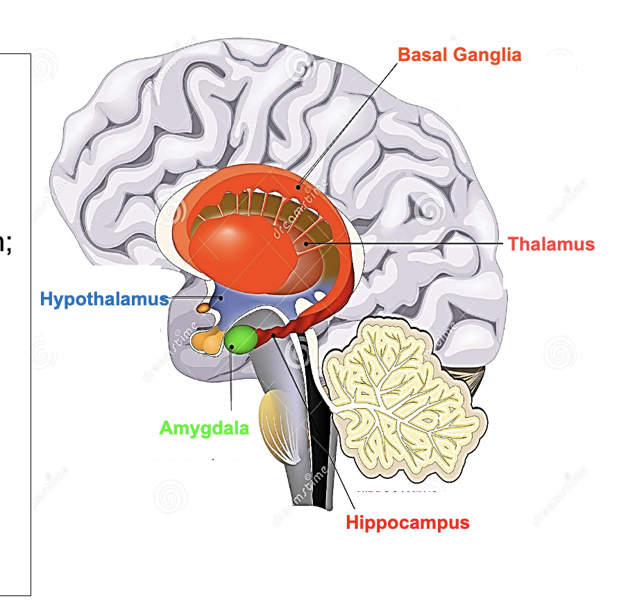
Limbic System parts
i.Hypothalamus - center of limbic system
that links endocrine (managing hormones)
& peripheral nervous system (somatic &
autonomic systems) for homeostasis
ii. Amygdala - detects and regulates
emotions such fear, threat, and aggression;
also tied to memory and decision-making
iii. Hippocampus - memory and spatial
navigation
iv. Thalamus - relays sensory information
to process emotion, memory, sexual
arousal and learning
v. Basal Ganglia - procedural learning,
habit and conditional learning, and
emotions
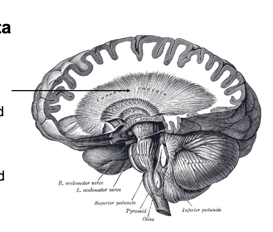
Corona Radiata
Made of: projection fibers - myelinated nerve fibers or tracts allowing communication to and from the cortex with other areas of the CNS
• Contains afferent and efferent pathways
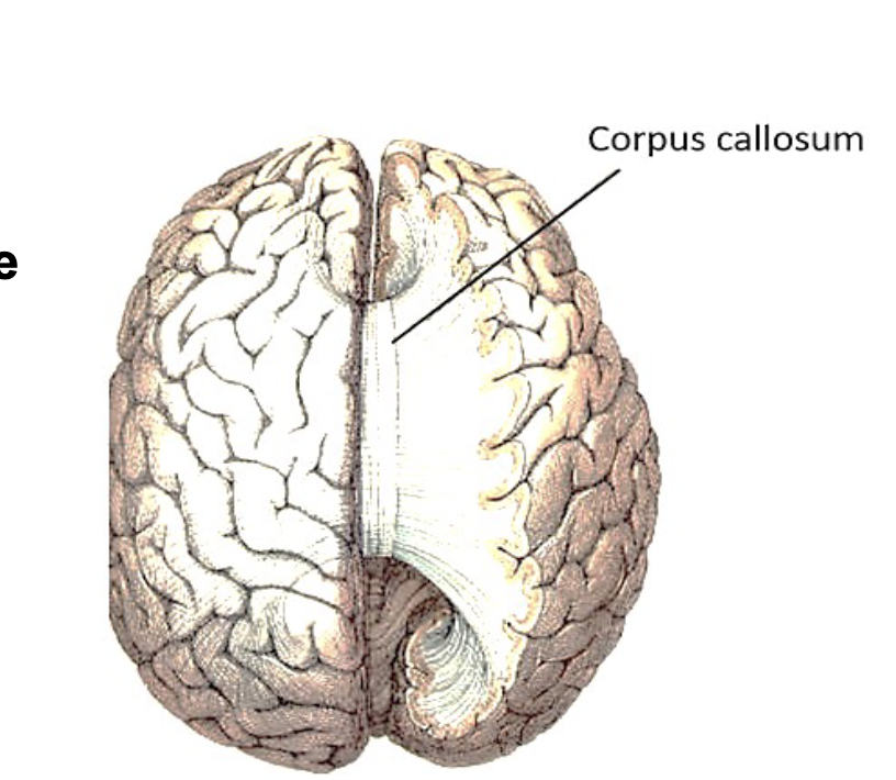
Corpus Callosum
• Made of: commissural fibers - myelinated nerve fibers or tracts allowing communication from the one hemisphere to the other hemisphere
• Even though control of muscles and glands is mostly contralateral (= from opposite of brain), many brain functions require combination of processes from both the left and right hemispheres of the brain
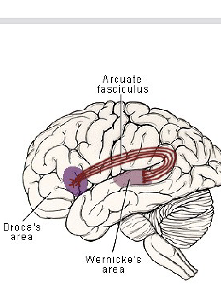
Arcuate Fasciculus
• Made of: association fibers - myelinated nerve fibers or tracts allowing communication between regions in the same hemisphere
• Important neural pathway in the left hemisphere connecting the expressive speech (Broca's) and receptive language (Wernicke’s) areas
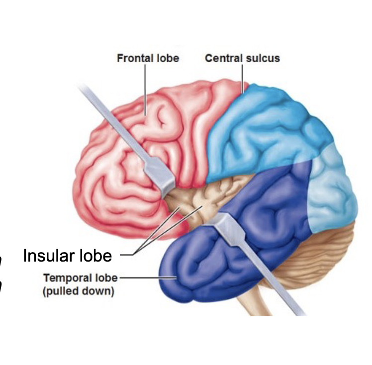
Insular Lobe/Insula, Insular cortex/Island of Reil
Deep within the lateral sulcus of the
brain in a part of the brain called the Operculum which overlaps with temporal, parietal and frontal lobes
• It is an integration hub that connects cortical and subcortical brain regions
• Functions: perception, motor speech planning, taste perception, perception of self, processing of emotion, development of compassion or empathy
Brain lesions
Lesion = a region in an organ or tissue that has suffered damage through injury or disease
1) hemorrhage- rupturing of blood
vessel or a “bleed”
2) thrombosis- blockage due to foreign object such as a local blood clot
3) embolism- blood clot that came from elsewhere in the circulatory system
4) aneurysm- ballooning of blood vessel walls which can sometimes be repaired. If an aneurysm breaks, the result is a hemorrhage.
Organic Etiology
-aging, genetics, smoking, diet, tumor, autoimmune disease, toxin, radiation/chemical exposure, vascular conditions, plaque
causes & signs of a brain lesion
Cerebrovascular accident (CVA) or Stroke
• Lesions: block (thrombosis or embolism) or brain bleed (hemorrhage)
interrupts blood flow to neural tissue leading to ischemia (lack of blood supply
= cell death)
Transient Ischemic Attack (TIA) or Mini-Stroke
• blockage is
temporary
• blood flow returns
on its own
• person still needs
to be checked for
any residual brain
damage
Traumatic Brain Injury (TBI)
2 TBI types and traumas to the brain
open head injury skull penetration
closed head injury no skull penetration
Blunt force trauma- open penetration - fractures skull
• Blunt force trauma - Coup injury - frontal lobe
• Contrecoup injury - caused by the rebound after initial impact - occipital lobe
• Rotational movement of the brain
Results of brain trauma
Immediate:
1. contusion - bruising of the brain due to bleeding & swelling
• Bleeding - hematomas can occur superficial or deep to the duramater
2. Tearing of axons of the Corona Radiata
3. Brain stem Impact = Loss of consciousness or coma
• Lasting:
1. Death
2. Permanent brain tissue death/damage
3. Loss of function
Loss of Function 1: Left Hemisphere Lesions
I. Aphasia = loss of ability to understand and/or to express speech
i. Fluent Aphasia - produces fluent speech, but does not make sense
• Wernicke’s Aphasia
• Conduction Aphasia - also trouble with word finding and repetition
ii. Non-fluent Aphasia - nonfluent speech
• Broca’s Aphasia
• Global Aphasia - damage in both Wernicke’s and Broca’s Area
II. Dysarthria = paralysis, muscular weakness, and discoordination of the speech musculature resulting in poor respiration, phonation, resonation, and articulation
III. Apraxia = deficit in motor planning existing without muscular weakness or paralysis that can affect oral movement and speech-can also have apraxia in body
Loss of Function Right Hemisphere & Other Lesions
Right Hemisphere Damage
• Poor attention, memory, organization, problem solving, reasoning, regulation
• Poor pragmatics
• Anosognosia - reduced awareness of deficits
• Visual neglect - aspects of visual stimulus are ignored (typically on the left)
II. Frontal Lobe lesion
• Impaired judgment or planning/goal setting
• Struggle with social interaction
• Loss of intellect
• Difficulty controlling emotions
• Motor functions
III. Hippocampal lesion
• Impaired short and long term memory