BIO163: Chapter 17-18 (Runyan)
1/141
There's no tags or description
Looks like no tags are added yet.
Name | Mastery | Learn | Test | Matching | Spaced | Call with Kai |
|---|
No analytics yet
Send a link to your students to track their progress
142 Terms
Blood
Life-sustaining transport vehicle of the cardiovascular system
Functions of Blood
1. Transportation
–Delivering O2 and nutrients to body cells
–Transporting metabolic wastes to lungs and kidneys for elimination
–Transporting hormones from endocrineorgans to target organs
2. Regulation
–Maintaining body temperature by absorbing and distributing heat
–Maintaining normal pH using buffers; alkaline reserve of bicarbonate ions
–Maintaining adequate fluid volume in circulatory system
3. Protection
–Preventing blood loss
•Plasma proteins and platelets in blood initiate clot formation
–Preventing infection
•Agents of immunity are carried in blood
–Antibodies
–Complement proteins
–White blood cells
Formed elements of blood
Erythrocytes (RBC)
Leukocytes (WBC)
Platelets
Erythrocytes
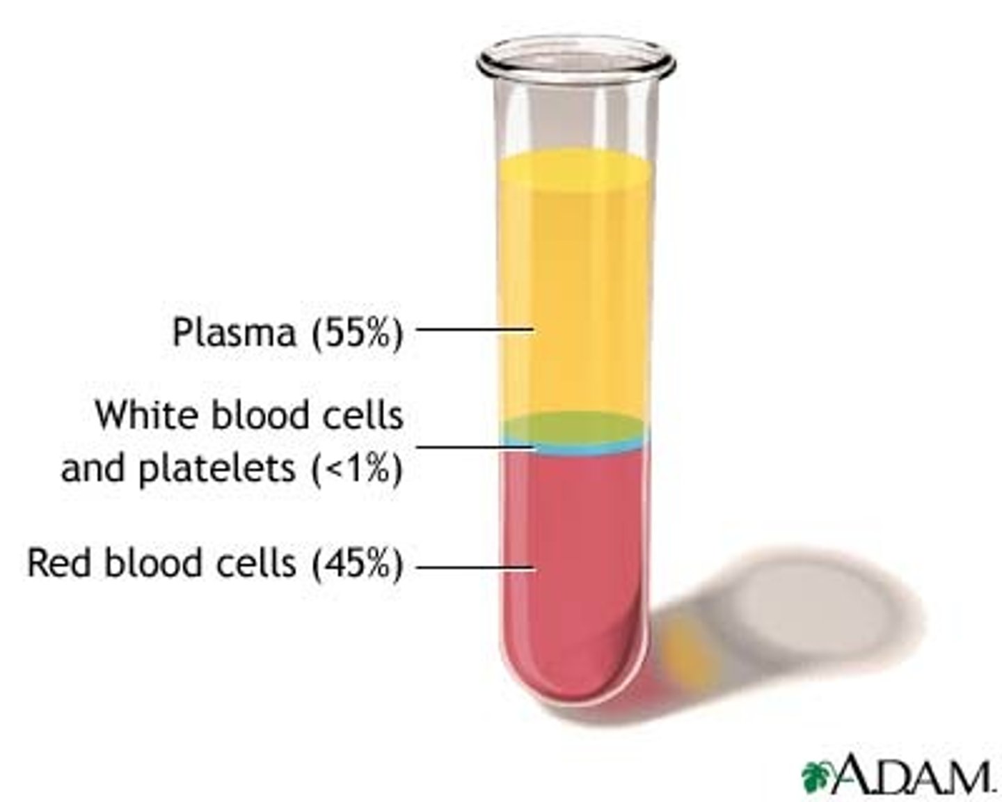
On bottom (~45% of whole blood) of tube

Hematocrit
Percent of blood volume that is RBCs
Normal values of hematocrit
Males: 47% ± 5%
Females: 42% ± 5%
Buffy coat (< 1%)
Thin, whitish layer between RBCs and plasma layers
---> WBCs and platelets
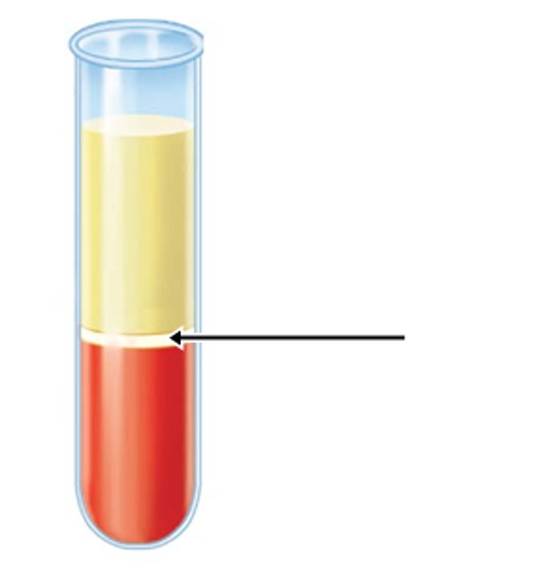
Plasma
On top (~55%) of tube (yellow)
Physical Characteristics and Volume of blood
Blood is a sticky, opaque fluid with metallic taste
- Color varies with O2 content
---> High O2 levels show a scarlet red
---> Low O2 levels show a dark red
- pH 7.35-7.45
- Makes up ~8% of body weight
- Average volume:
Males: 5-6 L
Females: 4-5 L
Blood plasma
Straw-colored sticky fluid
---> About 90% water
Plasma proteins
Most abundant solutes
• Remain in blood; not taken up by cells
• Proteins produced mostly by liver
• Albumin: makes up 60% of plasma proteins
– Functions as carrier of other molecules, as blood buffer, and contributes to plasma osmotic pressure
Formed Elements
- Only WBCs are complete cells
- RBCs have no nuclei or other organelles
- Platelets are cell fragments
- Most blood cells originate in bone marrow and do not divide
Erythrocytes (Red Blood Cells)
- Small-diameter (7.5 μm) cells that contribute to gas transport
---> Cell has biconcave disc shape, is anucleate, and essentially has no organelles
- Filled with hemoglobin (Hb) for gas transport
- Contain plasma membrane protein spectrin (provides flexibility to change shape) and other proteins
- Hemoglobin binds reversibly with O2 and CO2
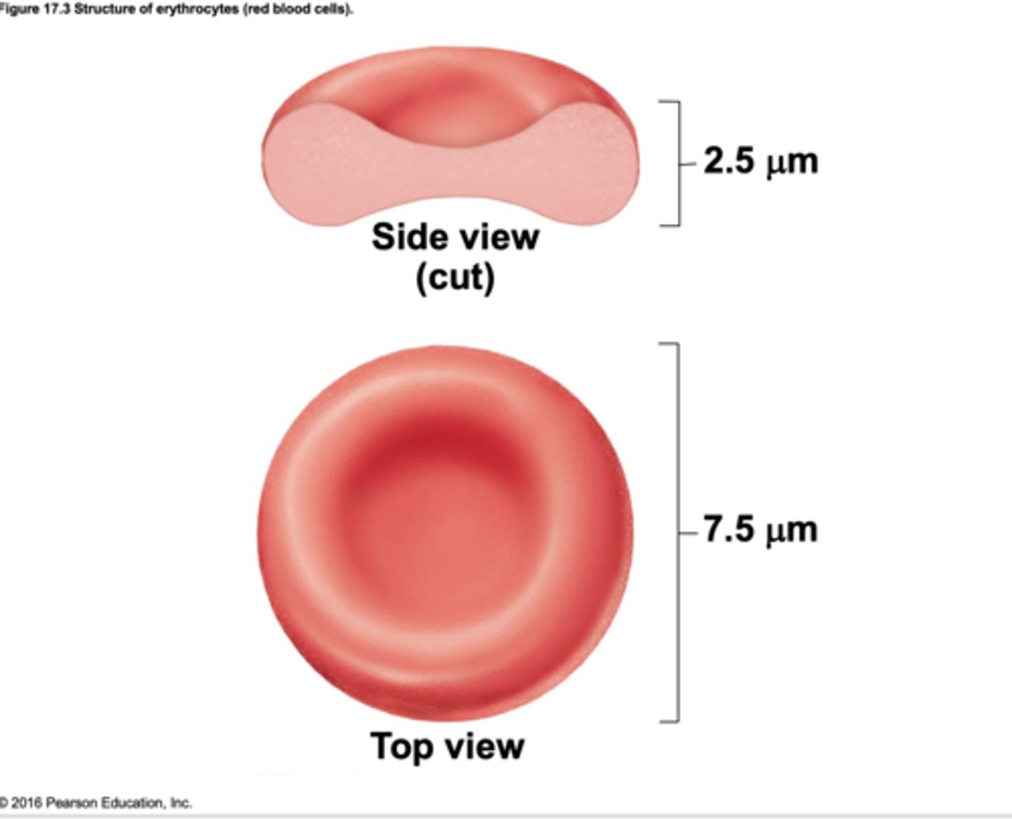
Three features make for efficient gas transport
1- Biconcave shape offers huge surface area relative to volume for gas exchange
2- Hemoglobin makes up 97% of cell volume (not counting water)
3- RBCs have no mitochondria
---> ATP production is anaerobic, so they do not consume O2 they transport
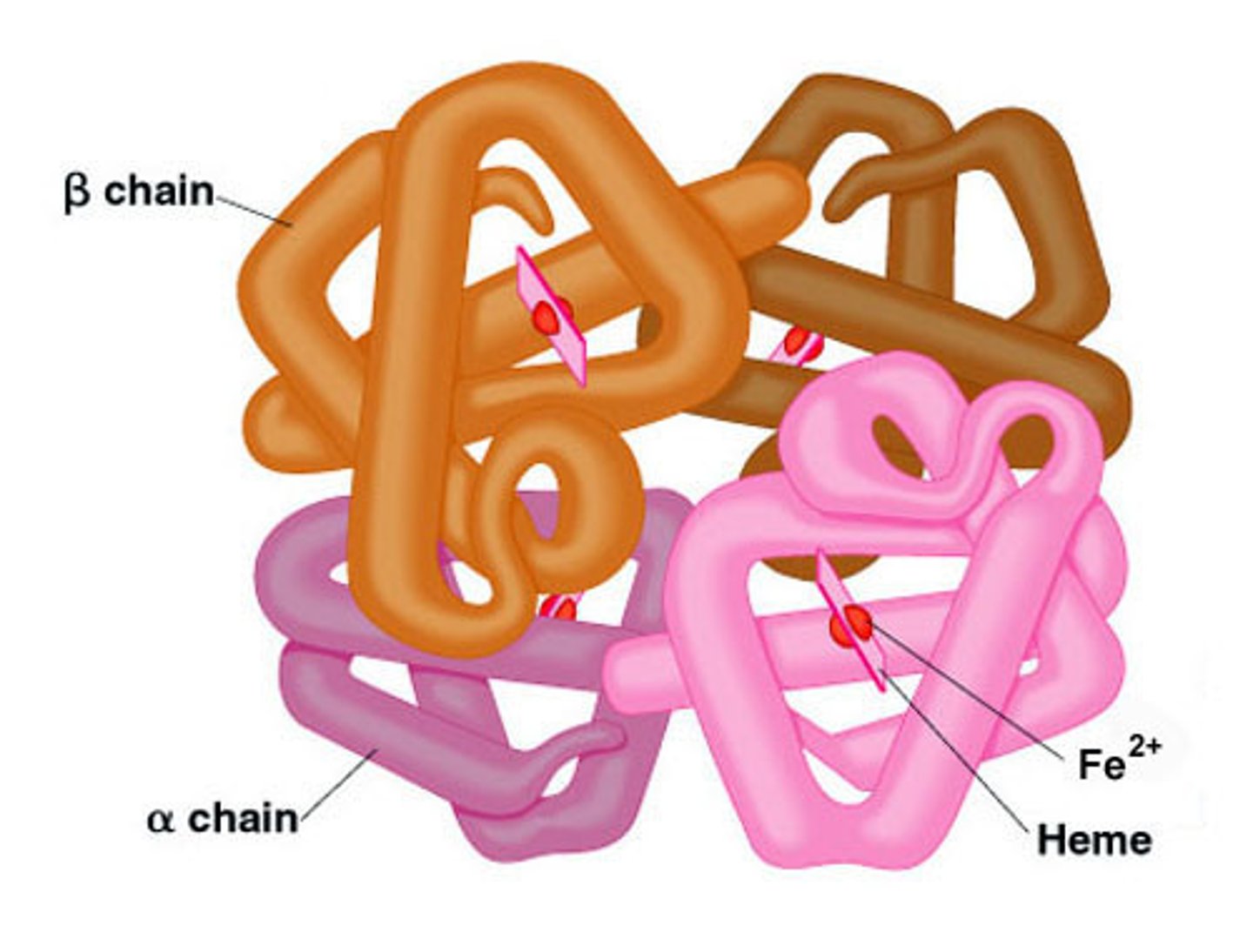
Hemoglobin
Consists of red heme pigment bound to the protein globin
- Each Hb molecule can transport four O2
- Each RBC contains 250 million Hb molecules
Globin
Composed of four polypeptide chains
---> Two alpha and two beta chains
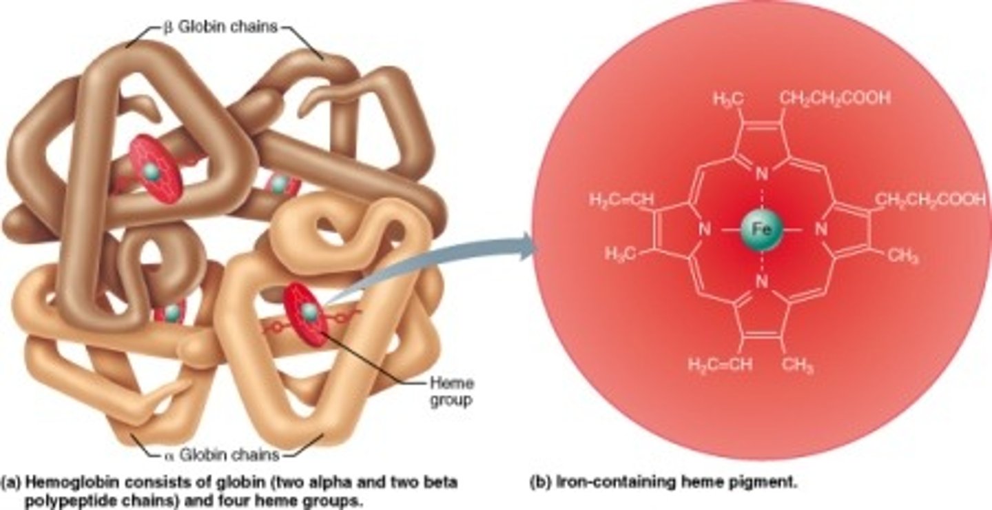
Heme pigment
Bonded to each globin chain
---> Gives blood red color
---> Each heme’s central iron atom binds one O2
Hematopoiesis
- Formation of all blood cells
- Occurs in red bone marrow; composed of reticular connective tissue and blood sinusoids
---> In adult, found in axial skeleton, girdles, and proximal epiphyses of humerus and femur
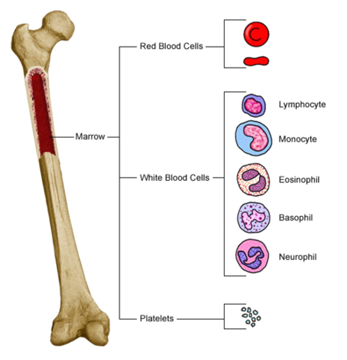
Hematopoietic stem cells (hemocytoblasts)
- Stem cell that gives rise to all formed elements
- Hormones and growth factors push cell toward specific pathway of blood cell development
- Committed cells cannot change
- New blood cells enter blood sinusoids
Stages of Erythropoiesis
1. Erythropoiesis: process of formation of RBCs that takes about 15 days
2. Reticulocyte count indicates rate of RBC formation
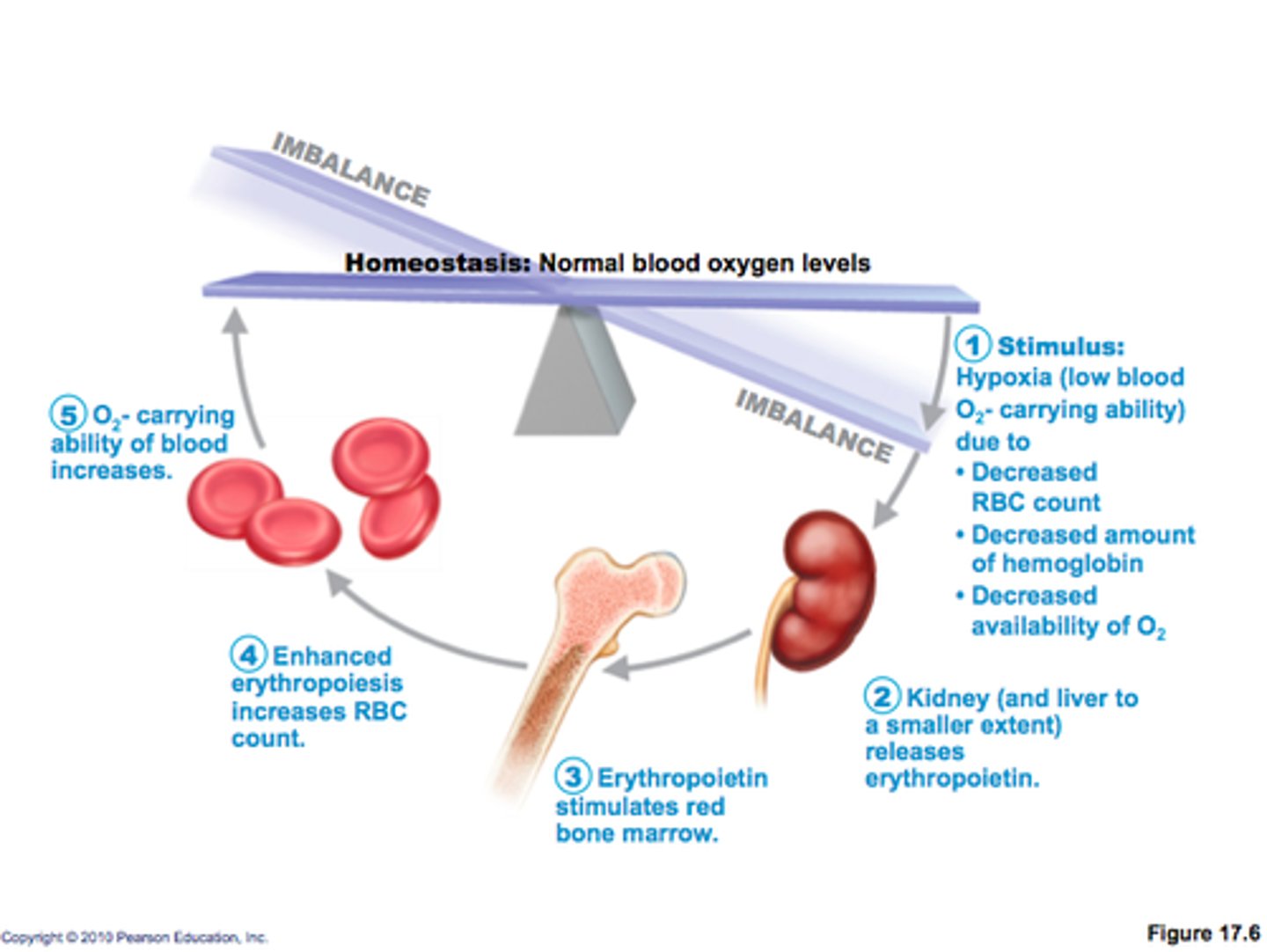
Tissue Hypoxia
Too few RBCs
Increase blood viscosity
Too many RBCs
Hormonal controls & Dietary requirements
Balance between RBC production and destruction depends on
Hormonal Control of Erythropoiesis
Erythropoietin (EPO): hormone that stimulates formation of RBCs
- Always small amount of EPO in blood to maintain basal rate
- Released by kidneys (some from liver) in response to hypoxia
Anemia
- Blood has abnormally low O2-carrying capacity that is too low to support normal metabolism
- Symptoms: fatigue, pallor, dyspnea, and chills
- Three groups based on cause
•Blood loss
•Not enough RBCs produced
•Too many RBCs being destroyed
Hemorrhagic anemias result from blood loss.
- Rapid blood loss (example: severe wound)
- Treated by blood replacement
Chronic hemorrhagic anemia
- Slight but persistent blood loss
---> Example: hemorrhoids, bleeding ulcer
- Primary problem must be treated to stop blood loss
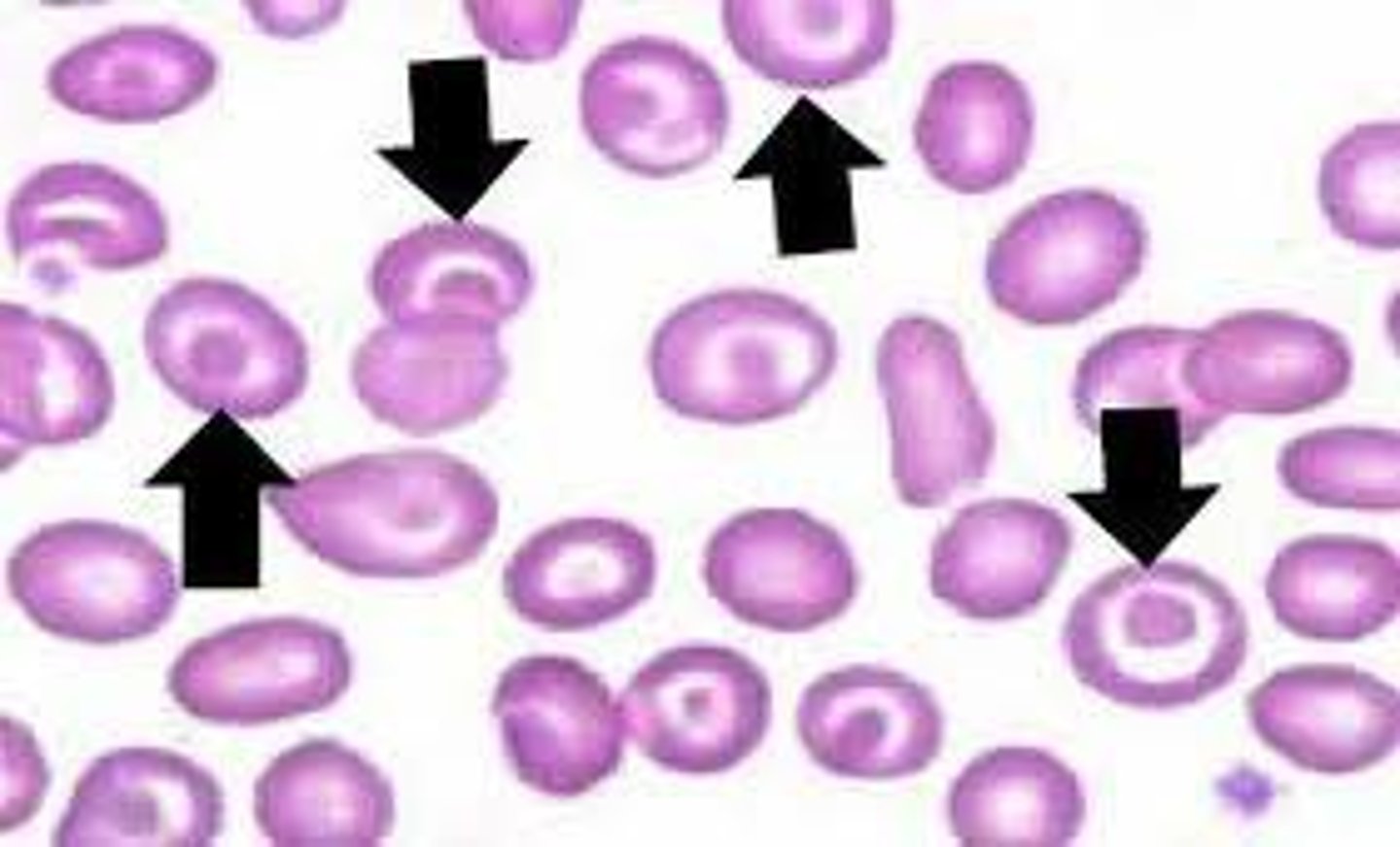
Iron-deficiency anemia
- Caused by hemorrhagic anemia, low iron intake or impaired absorption
- RBCs produced are called microcytes
---> Small, pale in color
---> Cannot synthesize hemoglobin because there is a lack of iron
- Treatment: iron supplements
Pernicious anemia
- Autoimmune disease that destroys stomach mucosa that produces intrinsic factor
---> Intrinsic factor needed to absorb B12
- B12 is needed to help RBCs divide
- Treatment: B12 injections or nasal gel
- Can also be caused by low dietary intake of B12
---> Can be a problem for vegetarians
Renal anemia
- Caused by lack of EPO
- Often accompanies renal disease
---> Kidneys cannot produce enough EPO
Treatment: synthetic EPO
Aplastic anemia
- Destruction or inhibition of red bone marrow by drugs, chemicals, radiation, or viruses
- All formed element cell lines are affected
---> Results in anemia as well as clotting and immunity defects
- Treatment: short-term with transfusions, long-term with transplanted stem cells
Hemolytic anemias
- Premature lysis of RBCs
- Can be caused by:
---> Incompatible transfusions or infections
---> Hemoglobin abnormalities:
»Thalassemias
»Sickle-cell anemia
Thalassemias anemia
- Typically found in people of Mediterranean ancestry
- One globin chain is absent or faulty
- RBCs are thin, delicate, and deficient in hemoglobin
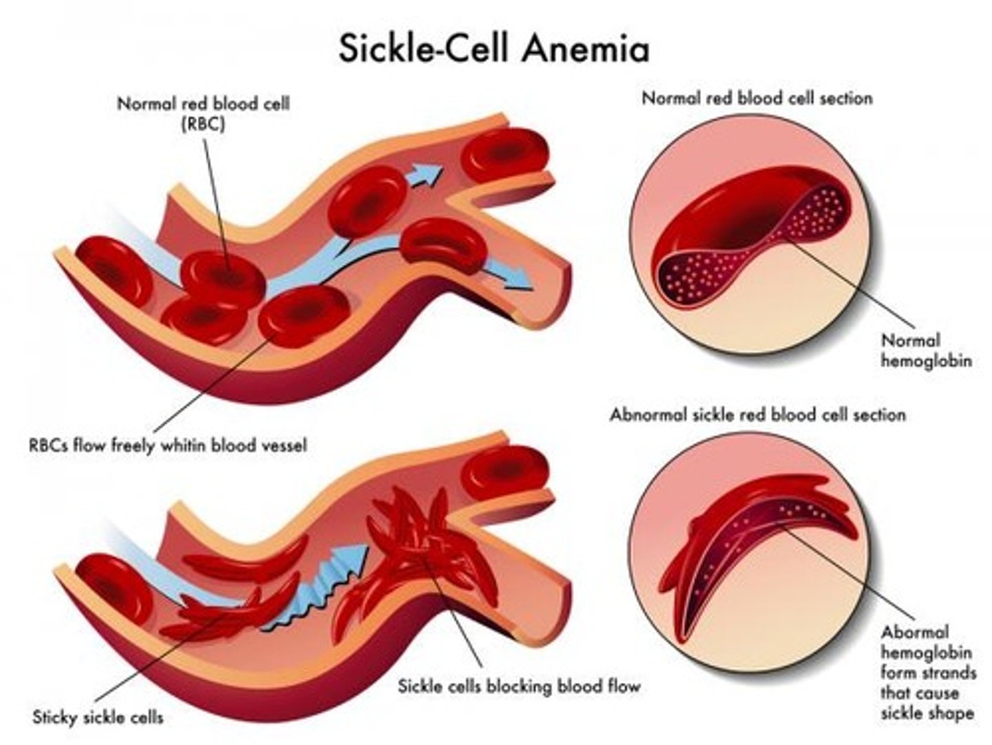
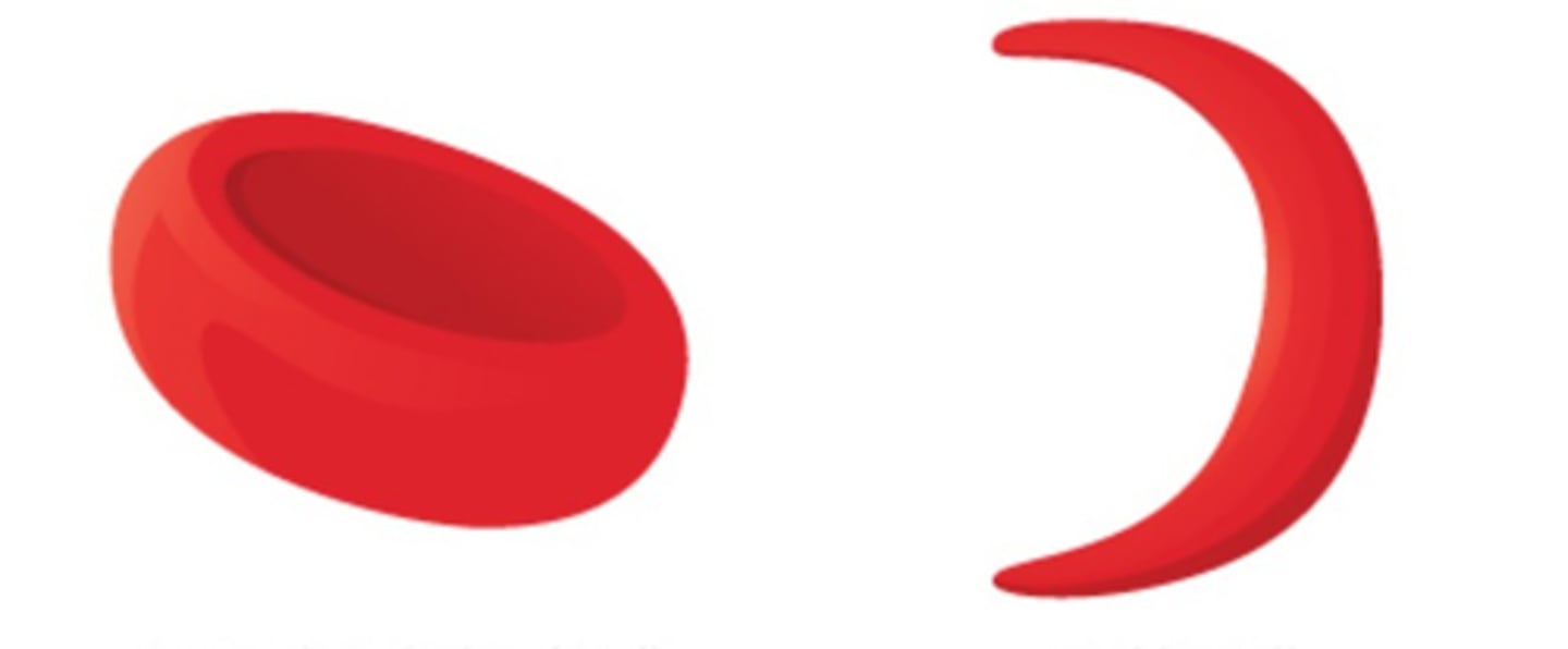
Sickle-cell anemia
- Hemoglobin S: mutated hemoglobin
---> Only 1 amino acid is wrong in a globin beta chain
- RBCs become crescent shaped when O2 levels are low
---> Example: during exercise
- Misshaped RBCs rupture easily and block small vessels
---> Results in poor O2 delivery and pain
Polycythemia
Abnormal excess of RBCs; increases blood viscosity, causing sluggish blood flow
Polycythemia vera
Bone marrow cancer leading to excess RBCs
Secondary polycythemia
Caused by low O2 levels (example: high altitude) or increased EPO production
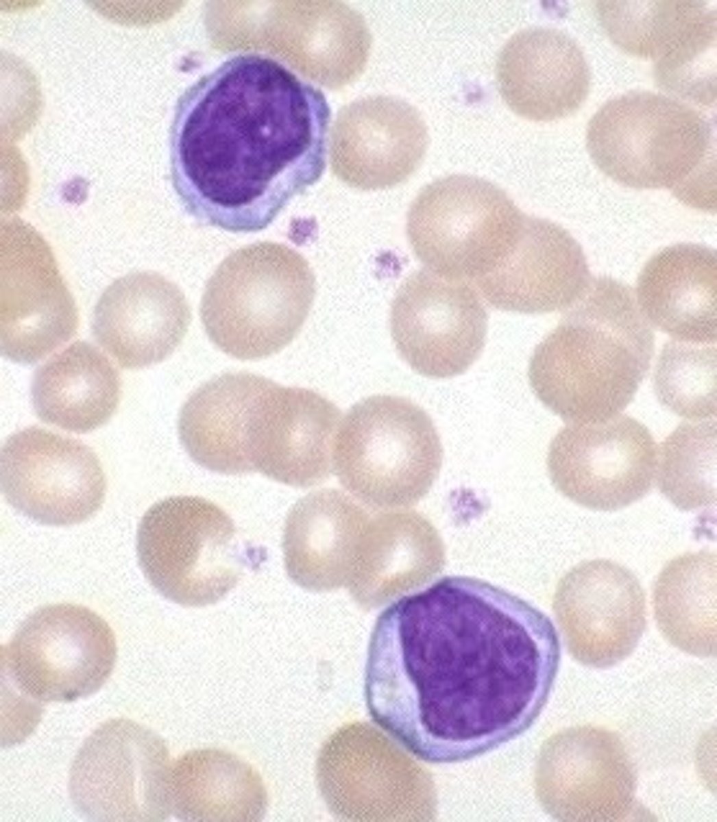
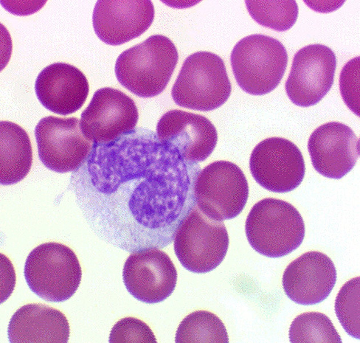
Leukocytes (White Blood Cells)
- Only formed element that is a complete cell with nuclei and organelles
- Make up <1% of total blood volume
---> 4800 to 10,800 WBCs per microliter blood
- Function in defense against disease
---> Can leave capillaries via diapedesis
---> Move through tissue spaces by amoeboid motion and positive chemotaxis
Leukocytosis
WBC count over 11,000 per microliter
---> Increase is a normal response to infection
Leukocytes grouped into two major categories:
1. Granulocytes: contain visible cytoplasmic granules
2. Agranulocytes: do not contain visible cytoplasmic granules
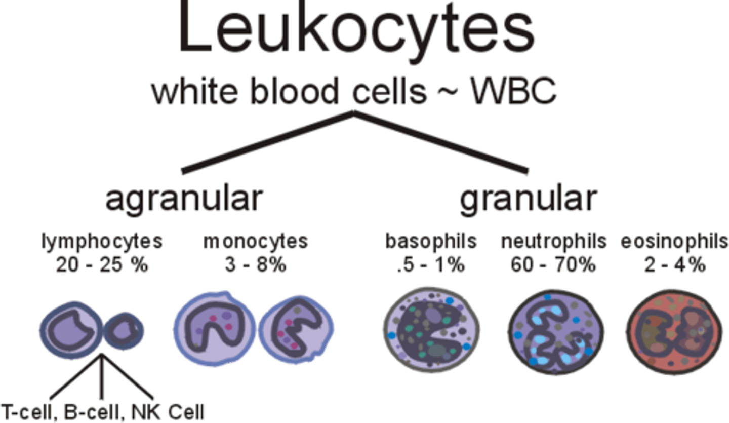
Granulocytes
- Neutrophils, eosinophils, basophils
- Larger and shorter-lived than RBCs
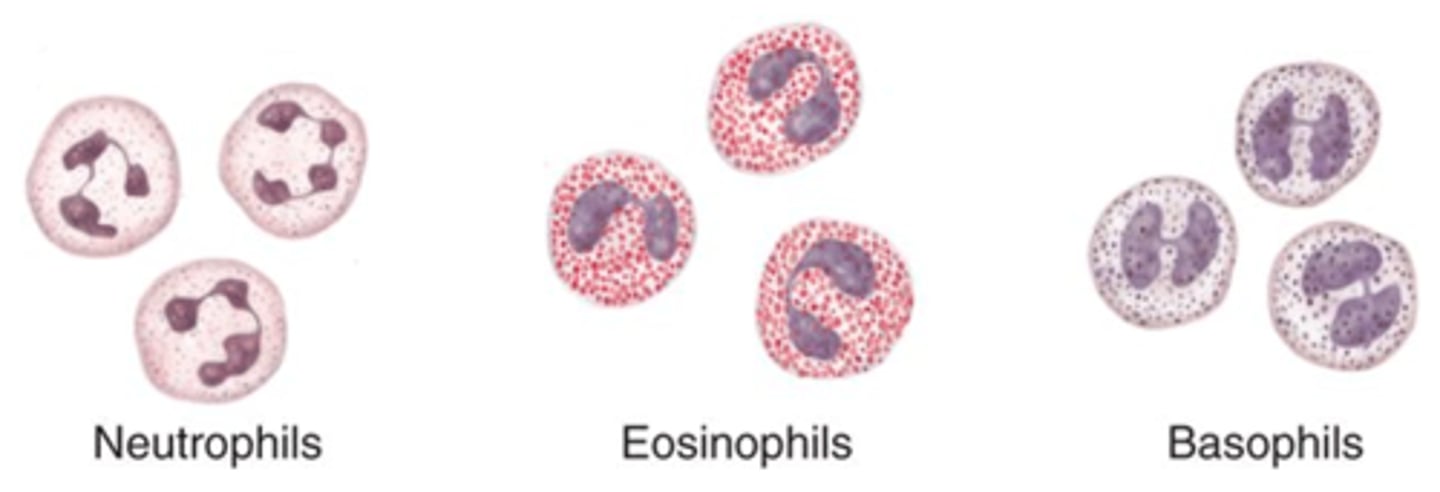
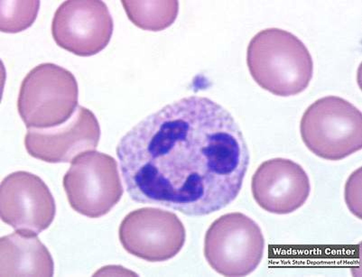
Neutrophils (granulocyte)
Most numerous WBCs
- Very phagocytic - “bacteria slayers”
Eosinophils (granulocyte)
- Red-staining granules contain digestive enzymes
---> Release enzymes to digest parasitic worms
- Play role in allergies and asthma, as well as immune response modulators
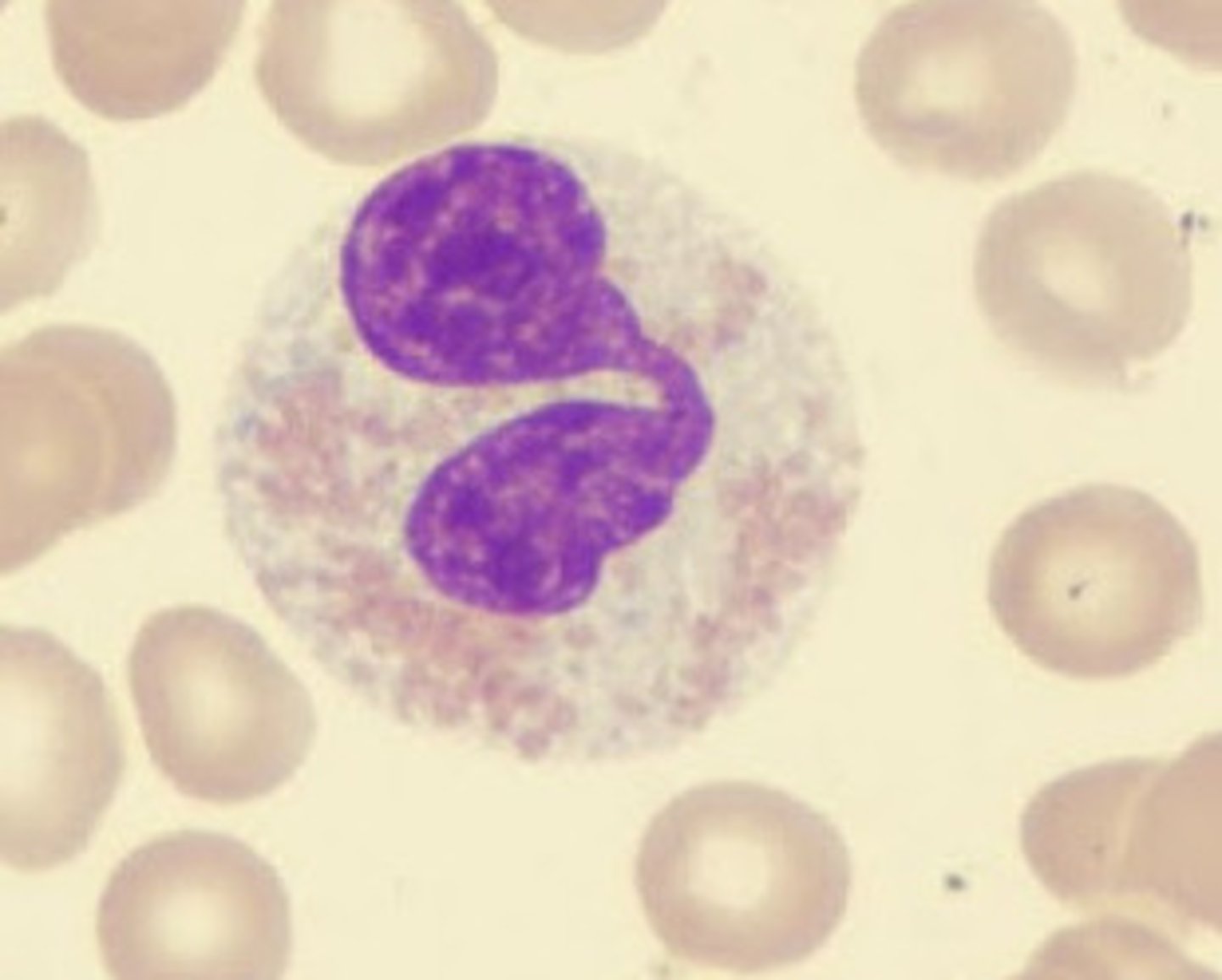
Basophils (granulocyte)
- Rarest WBCs
- Large, purplish black (basophilic) granules contain histamine
---> Histamine: inflammatory chemical that acts as vasodilator and attracts WBCs to inflamed sites
Agranulocytes
Lymphocytes and Monocytes
- Both have spherical or kidney-shaped nuclei
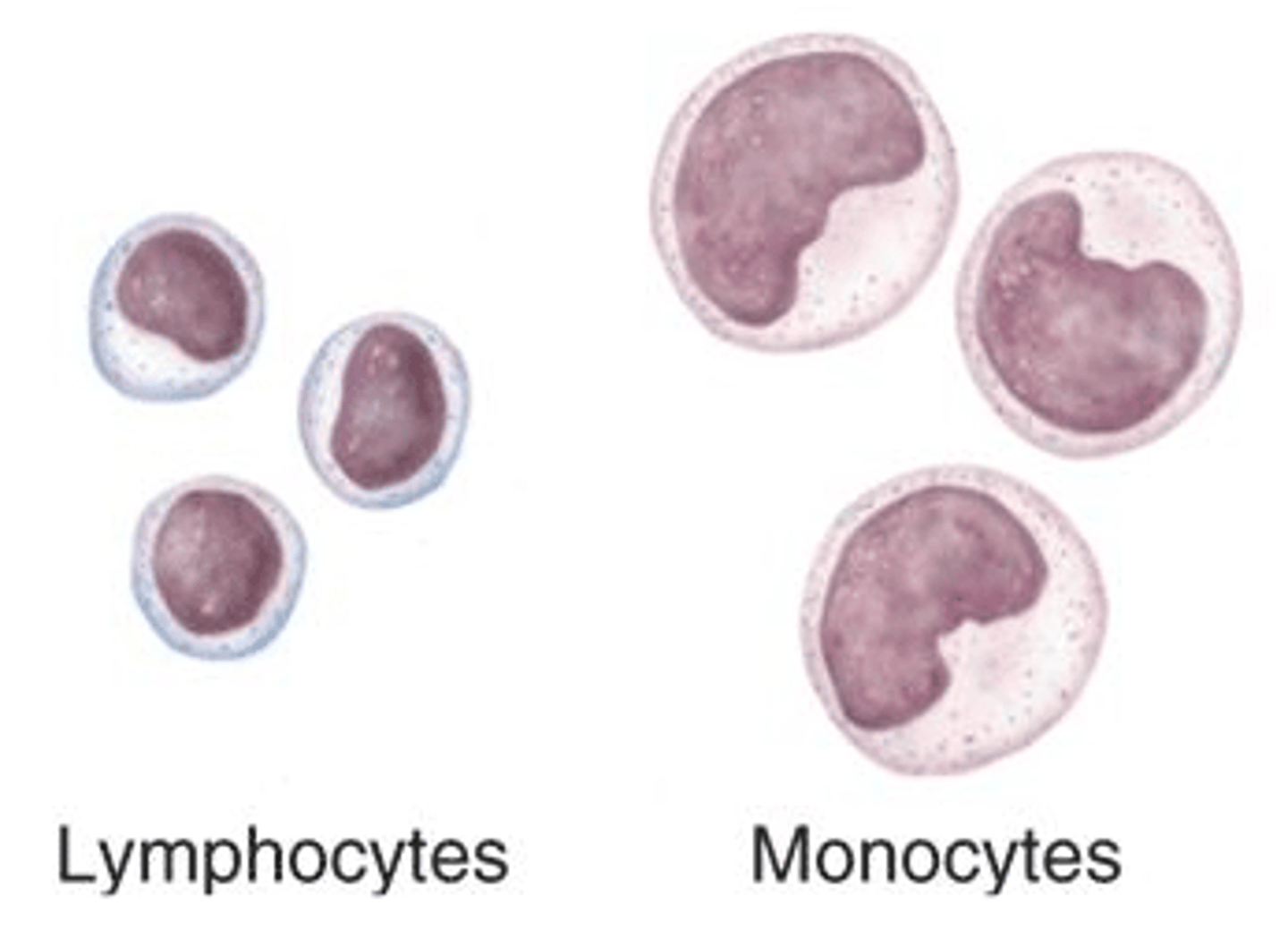
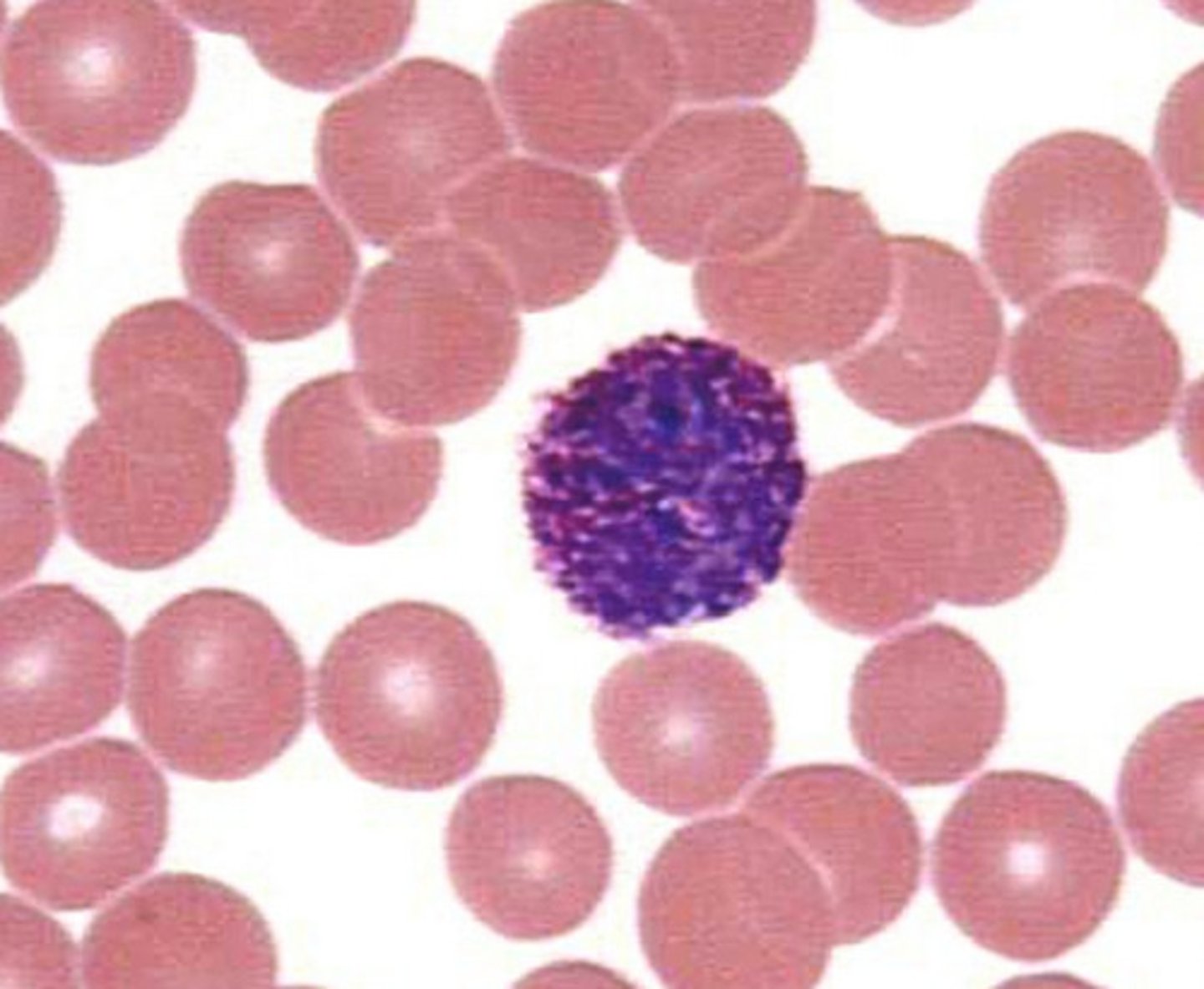
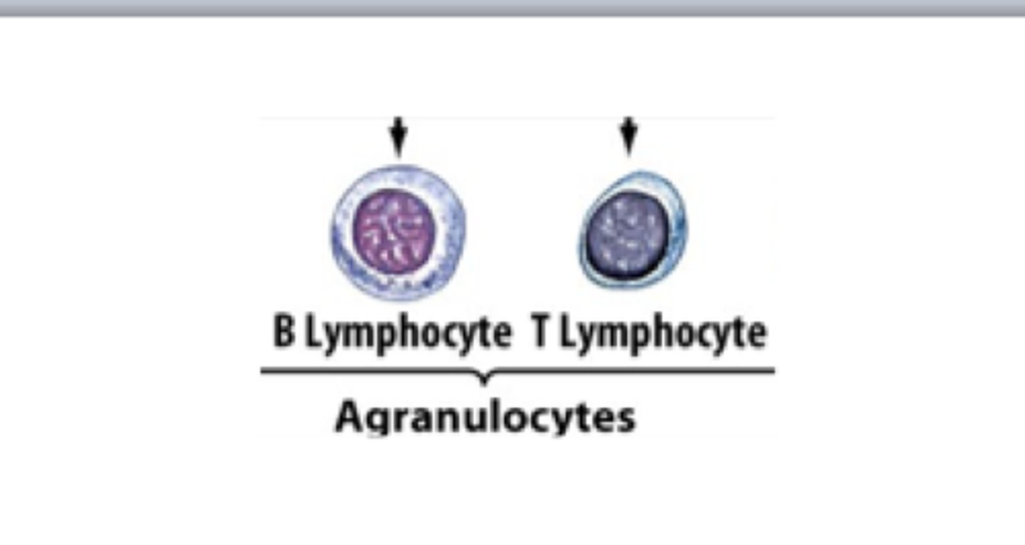
Lymphocytes (Agranulocytes)
- Second most numerous WBC
- Found in lymphoid tissue (example: lymph nodes, spleen), but a few circulate in blood
- Crucial to immunity
Two types of lymphocytes
1. T lymphocytes (T cells) act against virus-infected cells and tumor cells
2. B lymphocytes (B cells) give rise to plasma cells, which produce antibodies
Monocytes
- Largest of all leukocytes
- Leave circulation, enter tissues, and differentiate into macrophages
- Activate lymphocytes to mount an immune response
Leukopoiesis
Production of WBCs
- Stimulated by two types of chemical messengers from red bone marrow and mature WBCs
1. Interleukins are numbered (e.g., IL-3, IL-5)
2. Colony-stimulating factors (CSFs)
- All leukocytes originate from hemocytoblast stem cells that branches into two pathways:
---> Lymphoid stem cells produces lymphocytes
---> Myeloid stem cells produce all other elements
Leukemias
- Cancerous condition involving overproduction of abnormal WBCs
- Named according to abnormal WBC clone involved
---> Myeloid leukemia involves myeloblast descendants
---> Lymphocytic leukemia involves lymphocytes
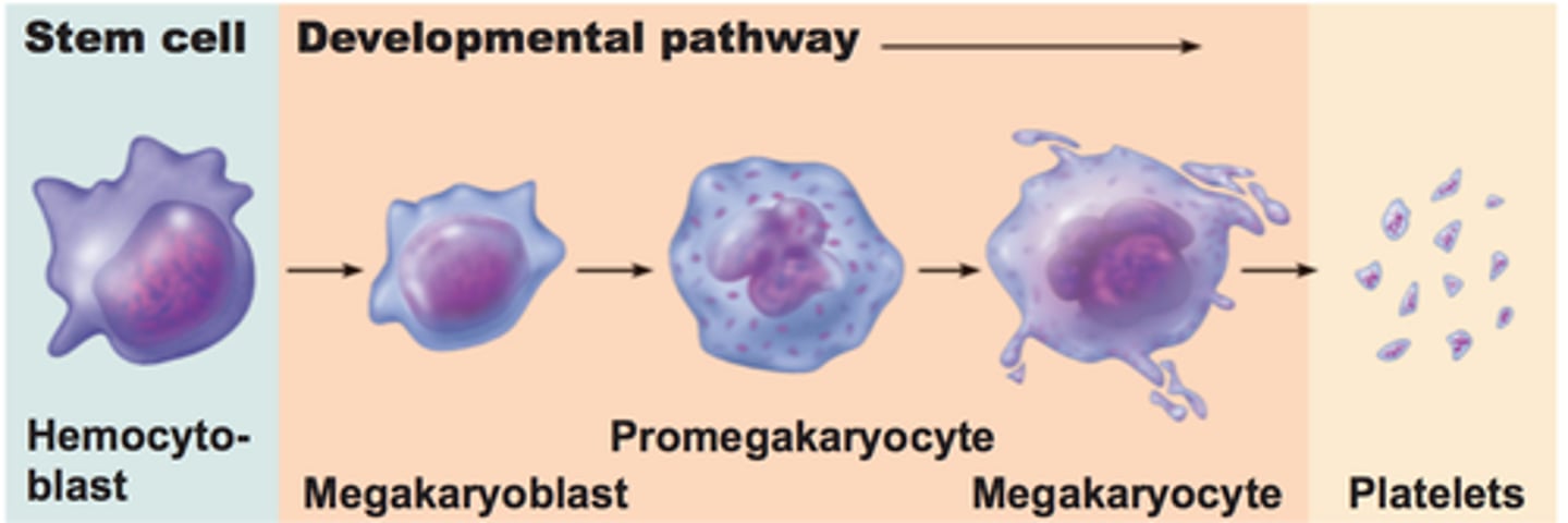
Platelets (thrombocytes)
- Cytoplasmic fragments of megakaryocytes
- Function: form temporary platelet plug that helps seal breaks in blood vessels
- Platelet formation is regulated by thrombopoietin
- Platelets age quickly and degenerate in about 10 days
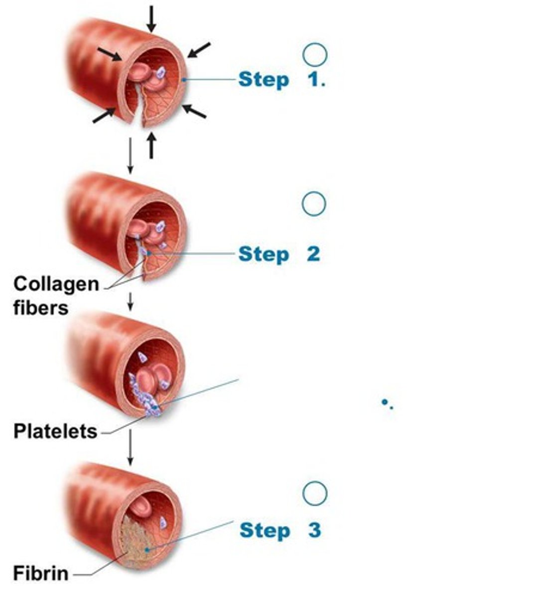
Hemostasis
Fast series of reactions for stoppage of bleeding
- Requires clotting factors and substances released by platelets and injured tissues
Three steps involved in hemostasis
Step 1: Vascular spasm
Step 2: Platelet plug formation
Step 3: Coagulation (blood clotting)
Vascular spams are triggered by:
- Direct injury to vascular smooth muscle
- Chemicals released by endothelial cells and platelets
- Pain reflexes
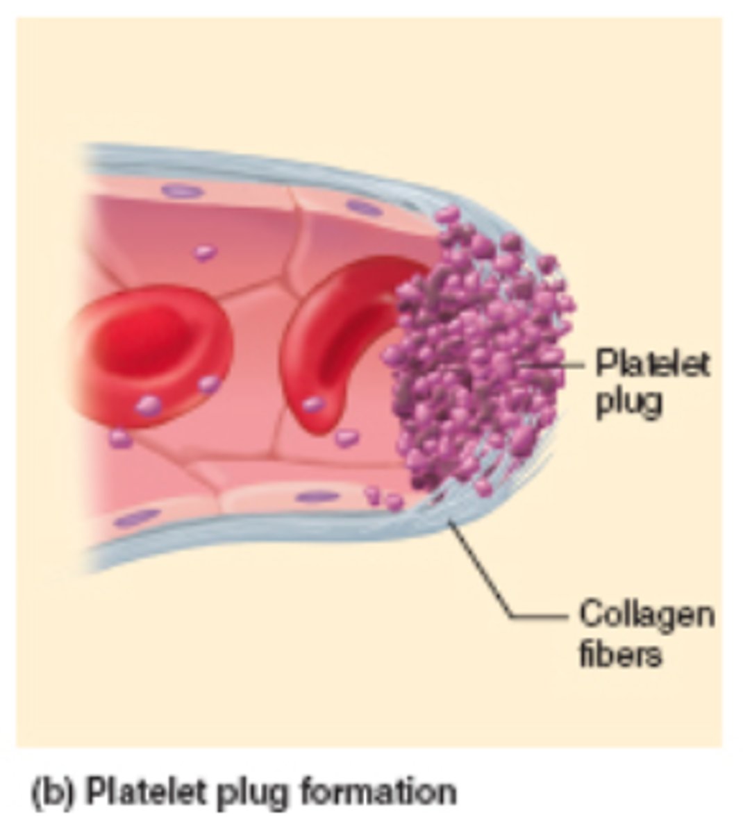
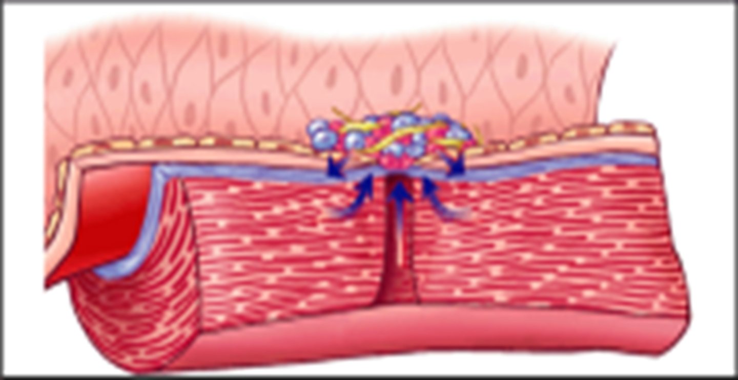
Platelet Plug Formation
- Platelets stick to collagen fibers that are exposed when vessel is damaged
- Von Willebrand factor helps to stabilize platelet-collagen adhesion
- When activated, platelets swell, become spiked and sticky, and release chemical messengers:
---> ADP causes more platelets to stick and release their contents
---> Serotonin and thromboxane A2 enhance vascular spasm and platelet aggregation
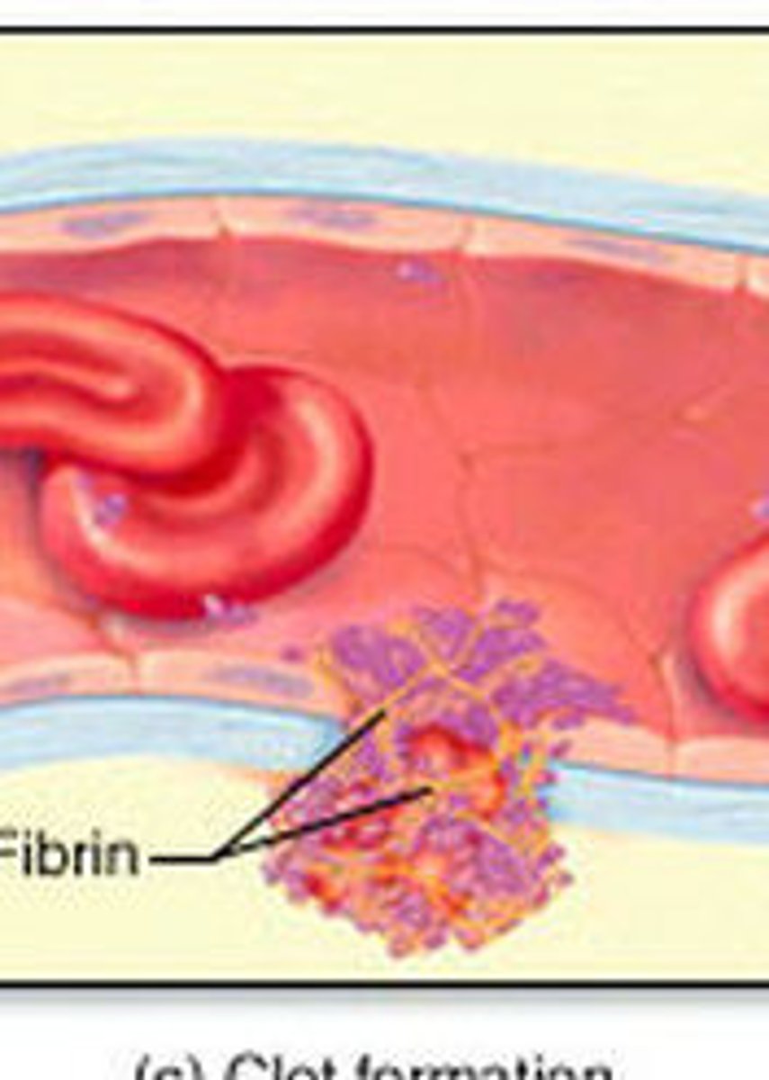
Coagulation (blood clotting)
Reinforces platelet plug with fibrin threads
- Series of reactions use clotting factors (procoagulants), mostly plasma proteins
---> Numbered I to XIII in order of discovery
---> Vitamin K needed to synthesize four factors
- Coagulation occurs in three phases
1. Two pathways to prothrombin activator
2. Pathway to thrombin
3. Common pathway to the fibrin mesh
Two pathways to prothrombin activator (Coagulation Phase 1)
– Initiated by either or both pathways:
• Intrinsic
• Extrinsic pathway
– Involves Tissue Factor, TF
–Faster than intrinsic pathway) (usually both)
– Each pathway cascades toward and ends with the activation of factor X
– Factor X then forms prothrombin activator

Pathway to Thrombin (Coagulation Phase 2)
– Prothrombin activator catalyzes transformationof prothrombin to active enzyme thrombin
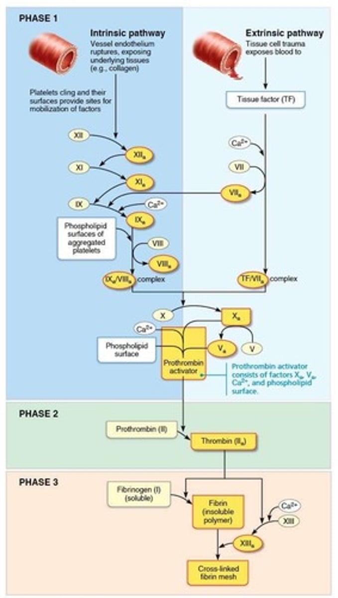
Common pathway to the fibrin mesh (Coagulation Phase 3)
– Thrombin converts soluble fibrinogen to insoluble fibrin, which forms structural basis of clot
– Fibrin causes plasma to become a gel-like trap catching formed elements
– Thrombin (along with Ca2+) activates factor XIII (fibrin stabilizing factor), which:
• Cross-links fibrin
• Strengthens and stabilizes clot

Clot retraction
– Actin and myosin in platelets contract within 30–60 minutes
---> Contraction pulls on fibrin strands, squeezing serum from clot
• Serum is plasma minus the clotting proteins
– Draws ruptured blood vessel edges together
Platelet-derived growth factor (PDGF)
- Released by platelets
-Stimulates division of smooth muscle cells and fibroblasts to rebuild blood vessel wall
Vascular endothelial growth factor (VEGF)
Stimulates endothelial cells to multiply and restore endothelial lining
Fibrinolysis
- Process whereby clots are removed after repair is completed
– Begins within 2 days and continues for several days until clot is dissolved
– Plasminogen (inactive), plasma protein that is trapped in clot, is converted to plasmin (active), a fibrin-digesting enzyme
• Tissue plasminogen activator (tPA), factor XII, and thrombin all play a role in conversion process
Factors limiting normal clot growth
–Two mechanisms limit clot size
• Swift removal and dilution of clotting factors
• Inhibition of activated clotting factors
– Limited amount of thrombin is restricted to clotby fibrin threads, preventing clot from gettingtoo big or escaping into bloodstream
• Antithrombin III inactivates any unbound thrombinthat escapes into bloodstream
• Heparin in basophil and mast cells inhibits thrombinby enhancing antithrombin III
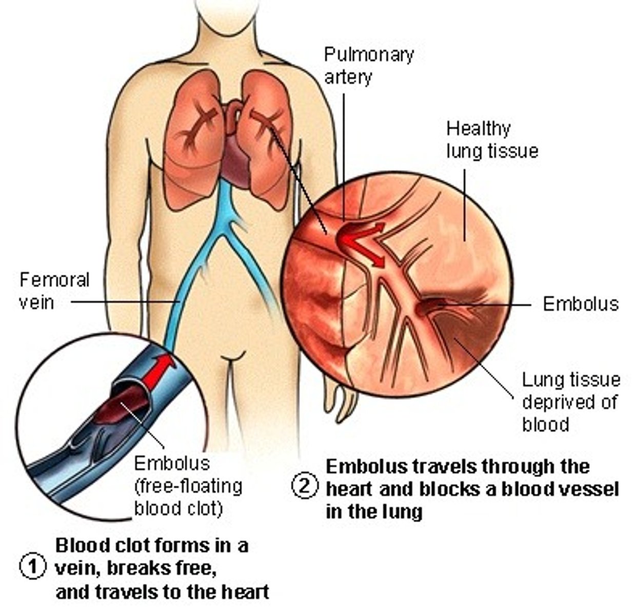
Thrombus
Clot that develops and persists in unbroken blood vessel
- May block circulation, leading to tissue death
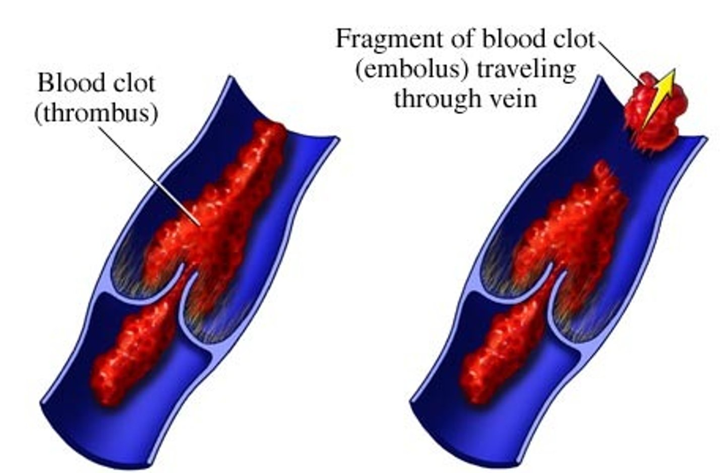
Embolus
Thrombus freely floating in bloodstream
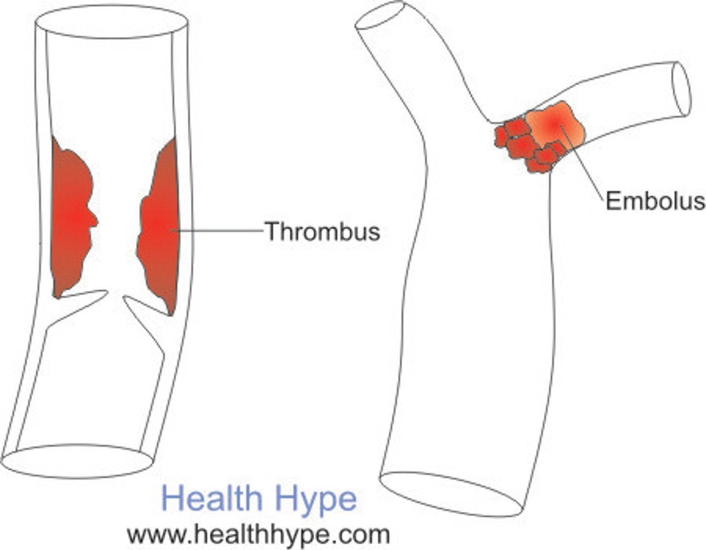
Embolism
Embolus obstructing a vessel
---> Example: pulmonary or cerebral emboli
Anticoagulant drugs
used to prevent undesirable clotting
-Aspirin
-Heparin
-Warfarin
-Dabigatran
Aspirin (anticoagulants)
Antiprostaglandin that inhibits thromboxane A2 (released by activated platelets for spasm and clotting)
Heparin (anticoagulant)
Used clinically for pre- and postoperative cardiac care
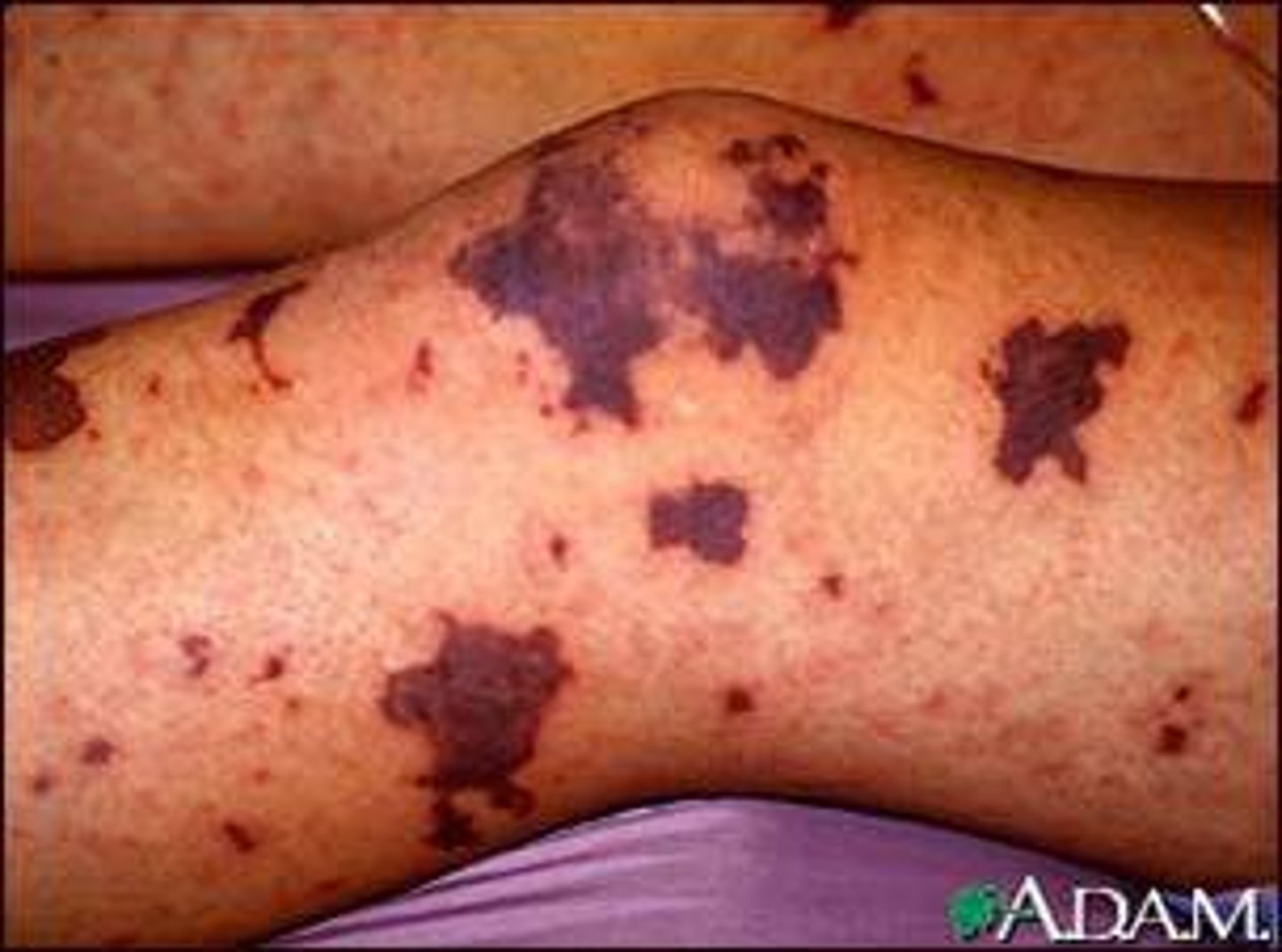
Thrombocytopenia
Deficient number of circulating platelets
– Petechiae appear as a result of spontaneous, widespread hemorrhage
– Due to suppression or destruction of red bone marrow (examples: malignancy, radiation, or drugs)
– Platelet count <50,000/ml is diagnostic
–Treatment: transfusion of concentrated platelets
Impaired liver function (bleeding disorder)
– Inability to synthesize procoagulants (clotting factors)
– Causes include vitamin K deficiency, hepatitis, or cirrhosis
– Liver disease can also prevent liver from producing bile, which is needed to absorb fat and vitamin K
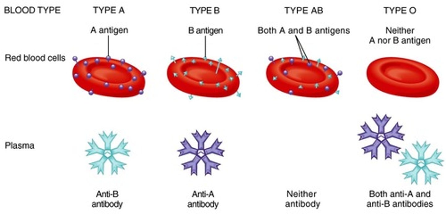
Antigen
Anything perceived as foreign that can generate an immune response
---> RBC antigens are referred to as agglutinogens because they promote agglutination
ABO blood groups
Based on presence or absence of two agglutinogens (A and B) on surface of RBCs
• Type A has only A agglutinogen
• Type B has only B agglutinogen
• Type AB has both A and B agglutinogens
• Type O has neither A nor B agglutinogens
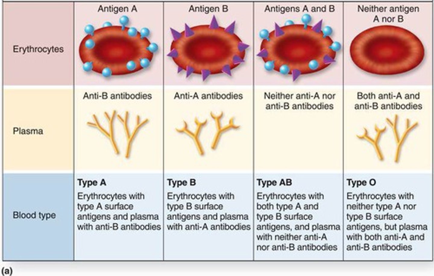
Antibodies (agglutinins)
Blood may contain preformed anti-A or anti-B
- Act against transfused RBCs with ABO antigens not present on recipient's RBCs
- Anti-A or anti-B form in blood at about 2 months of age, reaching adult levels by 8-10 years of age
Rh blood groups
– 52 named Rh agglutinogens (Rh factors)
– C, D, and E are most common
– Rh+ indicates presence of D antigen
• 85% Americans are Rh+
- Anti-Rh antibodies are not spontaneously formed in Rh– individuals
--> Anti-Rh antibodies form if Rh– individual receives Rh+ blood, or Rh– mom is carrying Rh+ fetus
Hemolytic disease of newborn (erythroblastosis fetalis)
Only occurs in Rh– mom with Rh+ fetus
- First pregnancy: Rh– mom exposed to Rh+ blood of fetus during delivery; first baby born healthy, but mother synthesizes anti-Rh antibodies
Second pregnancy: Mom’s anti-Rh antibodies cross placenta and destroy RBCs of Rh+ baby
- Baby treated with prebirth transfusions and exchange transfusions after birth
- RhoGAM serum containing anti-Rh can prevent Rh– mother from becoming sensitized
Transfusion reactions
– Occur if mismatched blood is infused
– Donor’s cells are attacked by recipient’s plasma agglutinins (antibodies)
• Agglutinate and clog small vessels
• Rupture and release Hb into bloodstream
– Result in:
• Diminished oxygen-carrying capacity
• Decreased blood flow beyond blocked vessel
• Hemoglobin in kidney tubules can lead to renal failure
Type O universal donor
no A or B antigens
Type AB universal recipient
no anti-A or anti-B antibodies
Autologous transfusions
Patient predonates own blood that is stored and available if needed
CMP (comprehensive medical panel)
Blood chemistry profile that checks various blood chemical levels
- Prothrombin time and platelet counts assess hemostasis
Fetal blood cells form in yolk sac, liver, and spleen
How do fetal blood form?
The fetus forms hemoglobin F, which has higher affinity for O2 than hemoglobin A formed after birth
What does the fetal form?
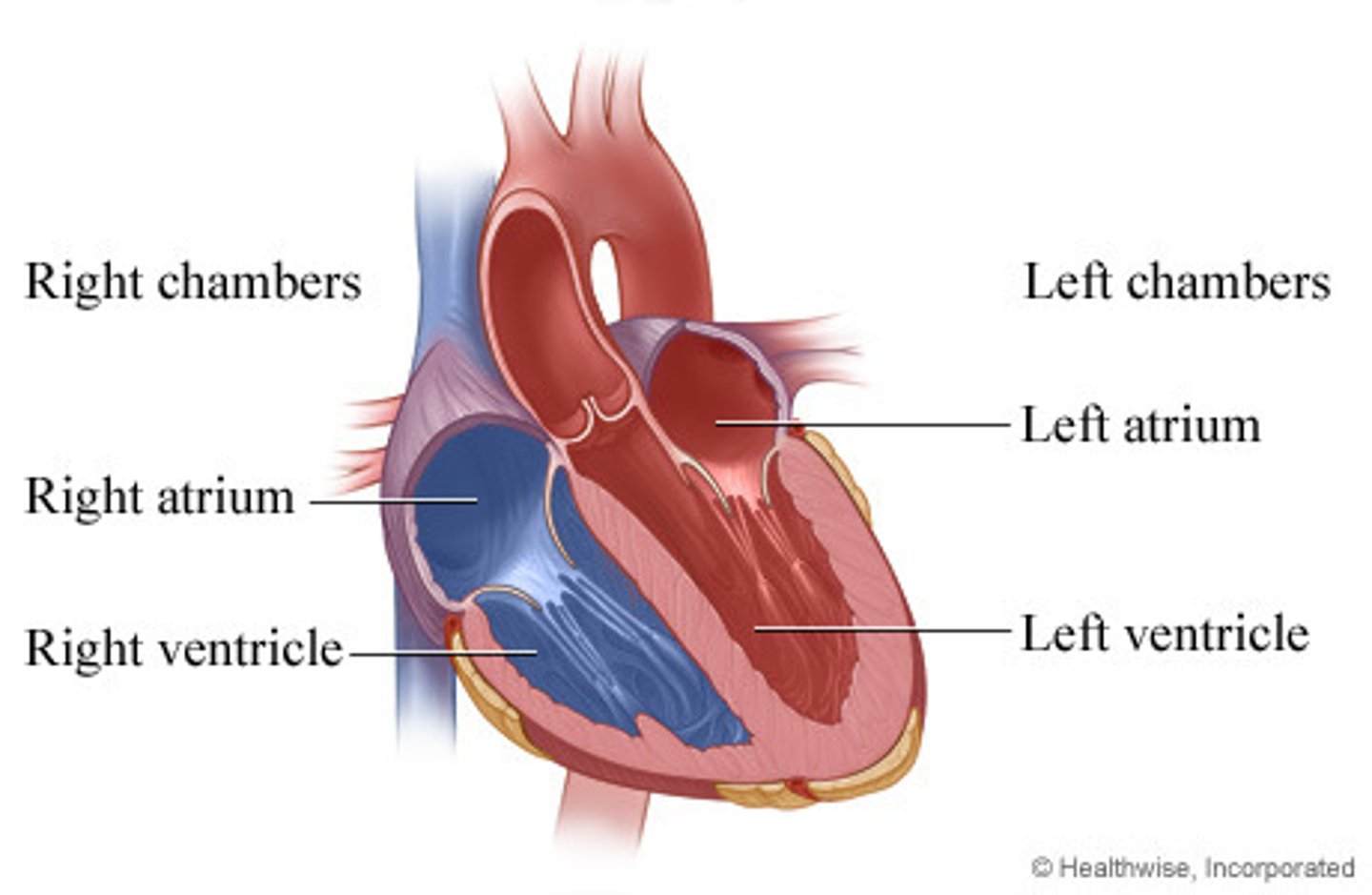
Right side of heart
Receives oxygen-poor blood from tissues
- Pumps blood to lungs to get rid of CO2, pick up O2, via pulmonary circuit
Left side of heart
Receives oxygenated blood from lungs
- Pumps blood to body tissues via systemic circuit

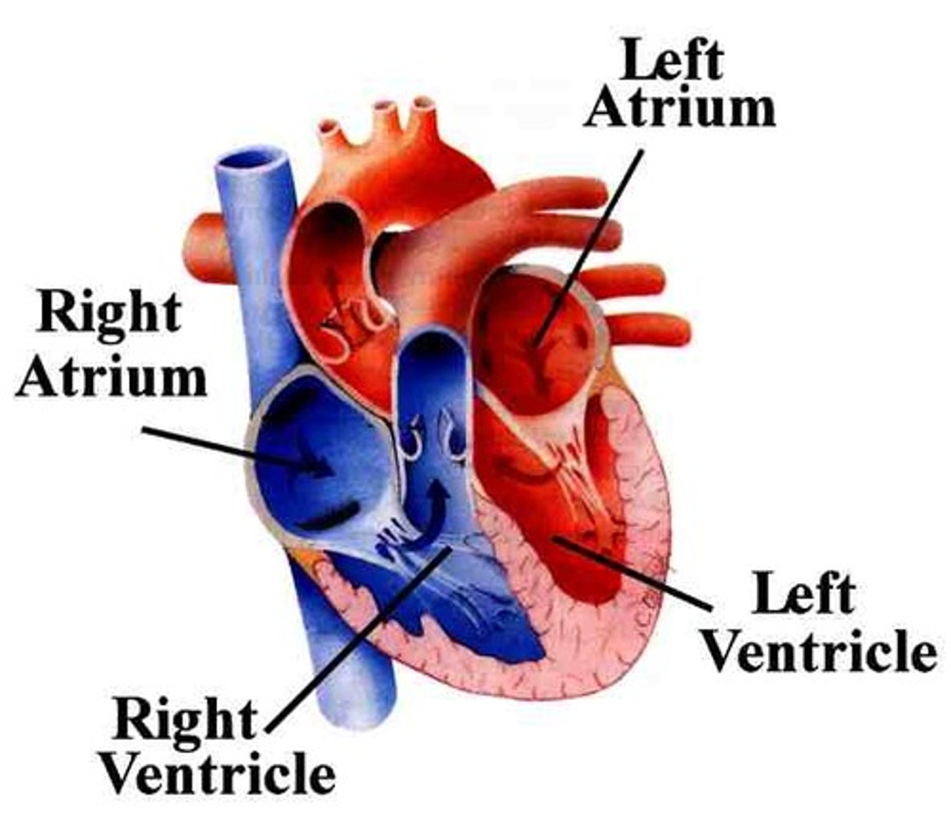
Right atrium (Receiving chambers of heart)
Receives blood returning from systemic circuit
- Receives deoxygenated blood from body
- Contains ridges formed by pectinate muscles
Left atrium (Receiving chambers of heart)
Receives blood returning from pulmonary circuit
- Receives oxygenated blood from lungs
- Pectinate muscles found only in auricle
- Four pulmonary veins return blood from lungs

Right ventricle (Pumping chambers of heart)
Pumps blood through pulmonary circuit (pulmonary trunk)

Left ventricle (Pumping chambers of heart)
Pumps blood through systemic circuit (aorta - largest artery in body)

Pericardium
Double-walled sac that surrounds heart; made up of two layers
1. Superficial fibrous pericardium
2. Deep two-layered serous pericardium
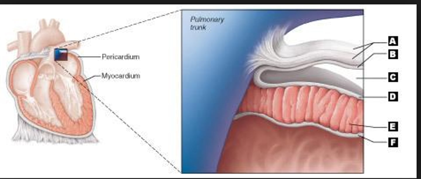
1. Superficial fibrous pericardium
Functions to protect, anchor heart to surrounding structures, and prevent overfilling
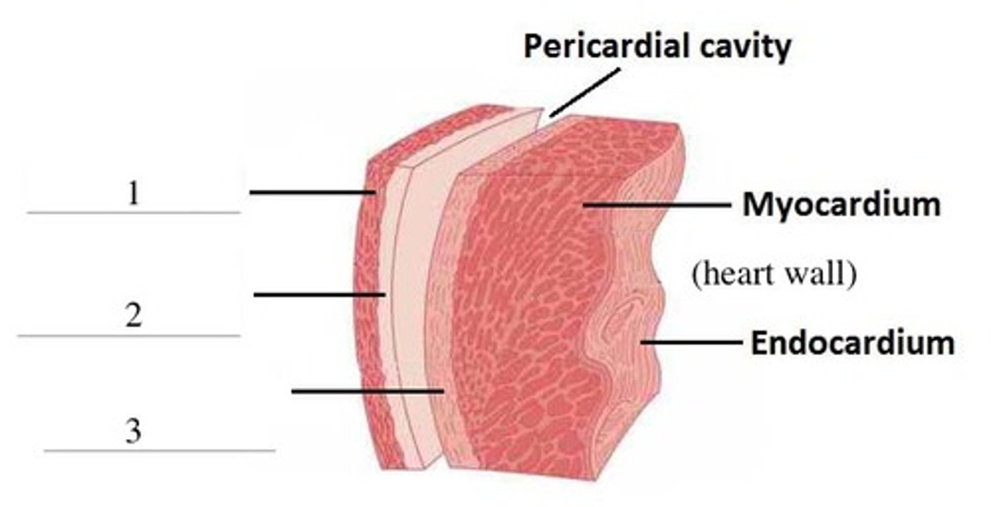
2. Deep two-layered serous pericardium
• Parietal layer lines internal surface of fibrous pericardium
• Visceral layer (epicardium) on external surface of heart
• Two layers separated by fluid-filled pericardial cavity (decreases friction)
Pericarditis
– Inflammation of pericardium
– Roughens membrane surfaces, causing pericardial friction rub (creaking sound) heard with stethoscope
– Cardiac tamponade
• Excess fluid that leaks into pericardial space
• Can compress heart’s pumping ability
• Treatment: fluid is drawn out of cavity (usually with syringe)
Four chambers of the heart
Two superior atria
Two inferior ventricles

Interatrial septum
Separates atria
---> Fossa ovalis: remnant of foramen ovale of fetal heart
Interventricular septum
Separates ventricles
Three veins empty into right atrium:
1. Superior vena cava
---> Returns blood from body regions above the diaphragm
2. Inferior vena cava
---> Returns blood from body regions below the diaphragm
3. Coronary sinus
---> Returns blood from coronary veins
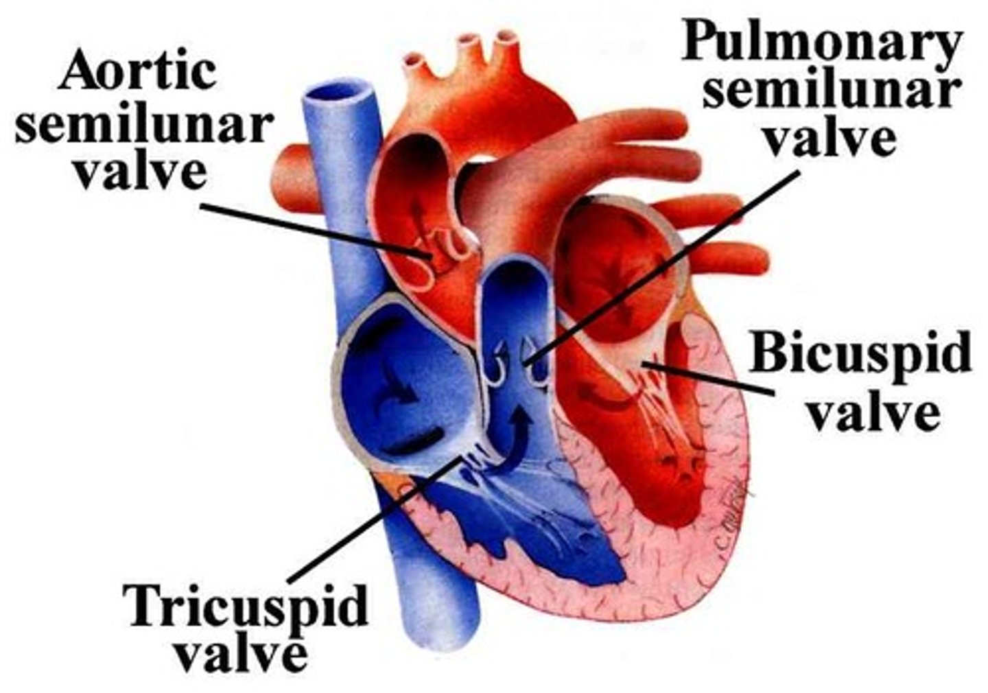
Heart Valves
Ensure unidirectional blood flow through heart
- Open and close in response to pressure changes
- Two major types of valves
---> Atrioventricular valves
---> Semilunar valves
Atrioventricular valves
Located between atria and ventricles (white spider web)
- Prevent backflow into atria when ventricles contract