Cytology Infection/Inflammation
1/29
There's no tags or description
Looks like no tags are added yet.
Name | Mastery | Learn | Test | Matching | Spaced |
|---|
No study sessions yet.
30 Terms
Cytology
the medical and scientific study of cells
Different types of cytology
ears
skin
fluid
masses
Inflammation
-characterized by the type of inflammatory cells present
-can have infectious or noninfectious cause
Infection
-agents may be visualized in cytologic specimens
-some are morphologically distinct allowing for specific identification
-other agents may require additional diagnostics
Neutrophils that undergo normal aging and cell death may be ....
pyknotic or hypersegmented
Types of inflammation
-suppurative
-purulent
-neutrophilic
Suppurative inflammation
-infectious predominant cause often seen in intracellular bacteria
-noninfectious seen in neoplasia, necrosis, trauma, or in immune medicated disease
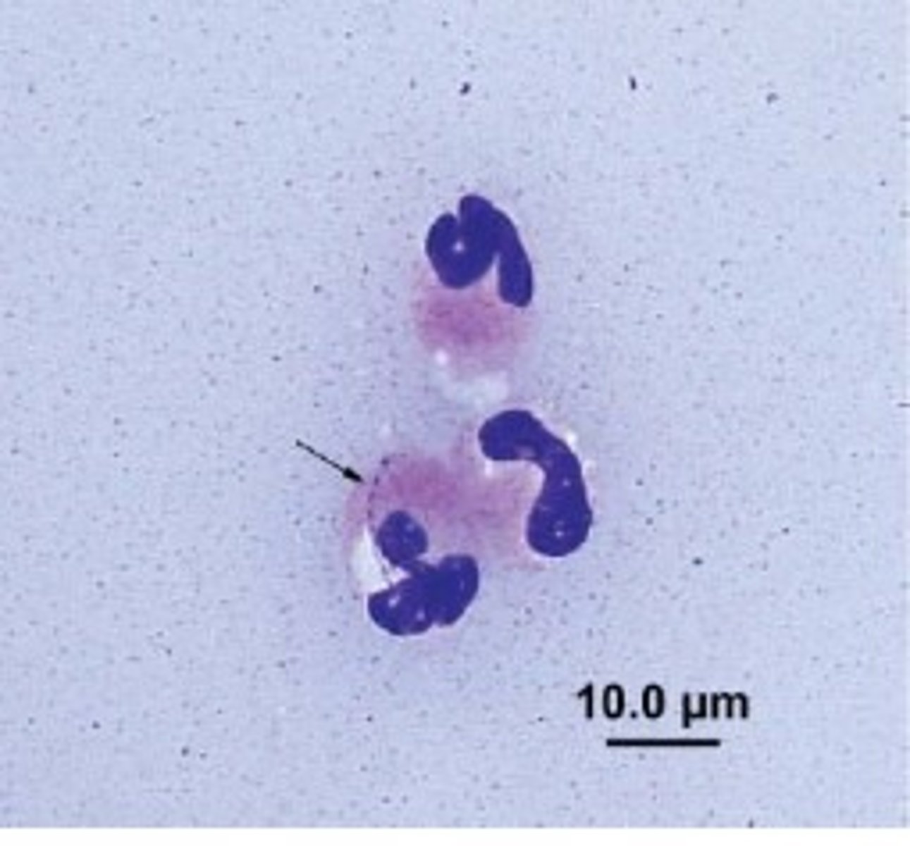
Nondegenerate neutrophils
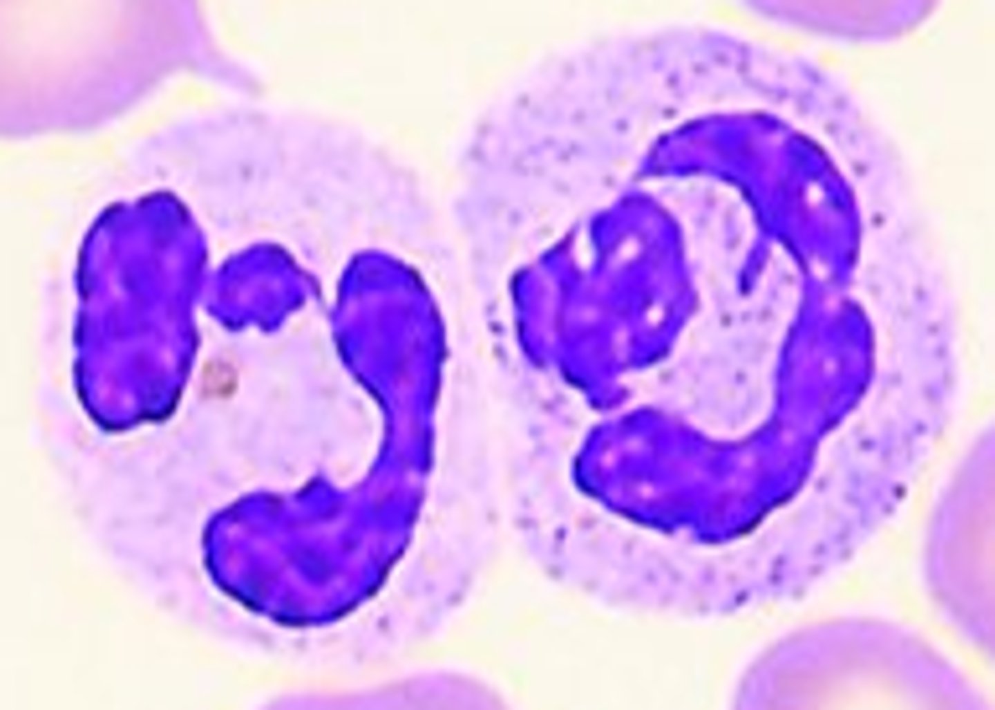
degenerate neutrophils
-nuclear changes seen in reflective of a toxic environment
-associated most with bacterial infection

nuclear changes
-swollen nuclei
-swelling of cytoplasm
-vacuolization
Macrophages in inflammation
-seen in histiocytic, granulomatous, or macrophagic inflammation
-large round cell with oval to indented nuclei and abundant basophilic cytoplasm
How do activated macrophages appear?
vacuolated
samples that are composed predominantly by neutrophils and macrophages are considered...
Mixed or pyogranulomatous
What can cause macrophages inflammation?
chronic inflammation caused by foreign body reactions, fungal infections, infectious caused by filamentous bacteria, and vaccine reactions
Lymphocytic inflammation
small to intermediate sized lymphocytes often mixed with plasma cells
-morphology similar to those seen in hyperplastic lymph node aspirates
When do we see lymphocyte/plasma cell inflammation?
occurs with immune reactions, vaccine reactions, chronic inflammation
Mast cells
round cells containing round nuclei and moderately abundant cytoplasm that contains abundant dark purple granules
what should be considered if numerous or criteria of malignancy mast cells are seen?
MCT
Eosinophils in inflammation
-inflammation process considered to have eosinophilic component if eosinophils comprise great > 10% of the inflammatory cells
-appear similar to those peripheral blood film with abundant eosinophilic cytoplasmic granules
When would we seen an abundant of eosinophils or mast cells?
occurs with hypersensitivity reactions, parasitic migrations, some fungal infections, certain neoplasia, and pythiosis
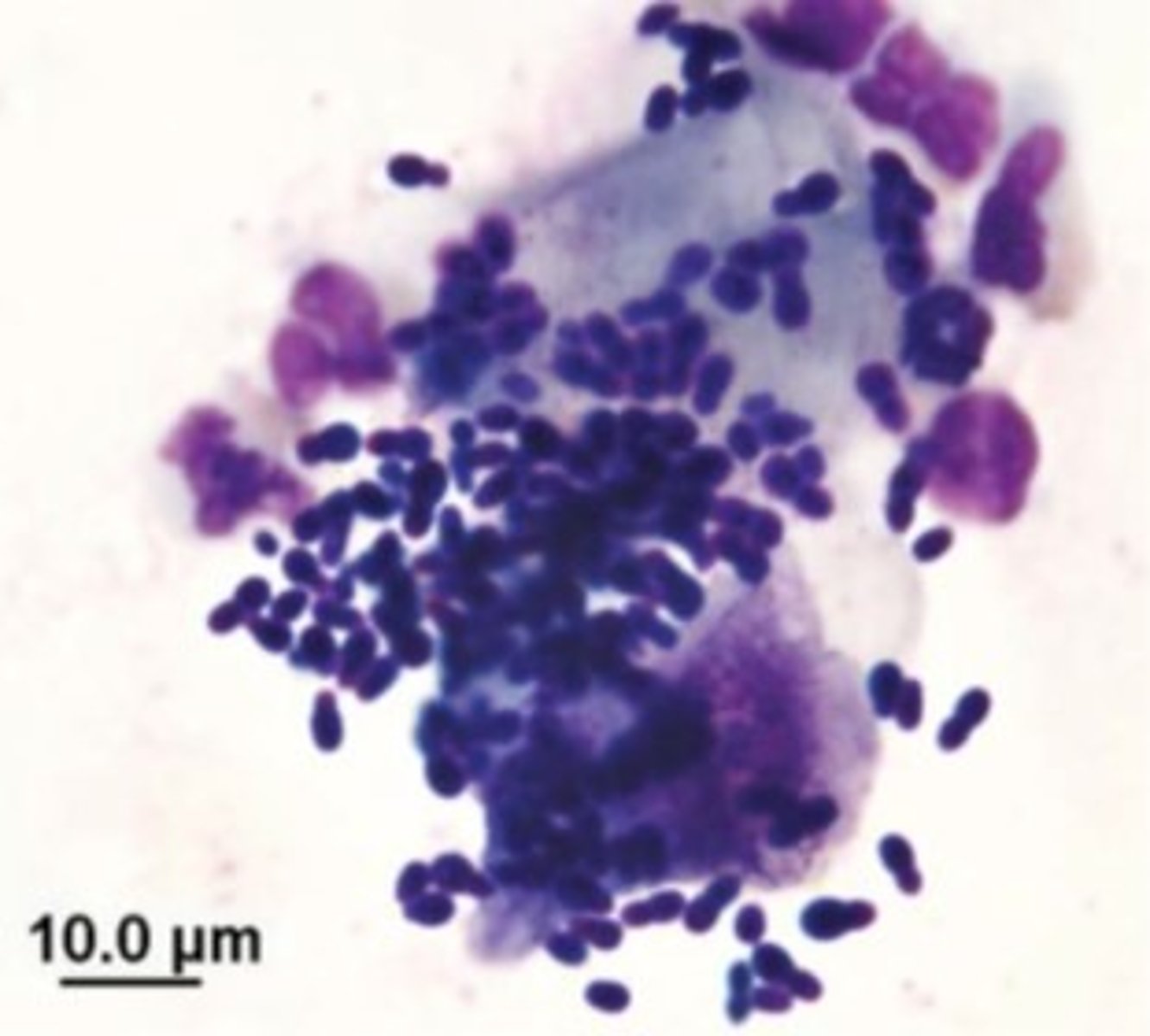
Cocci
-appear round, in chains, or clusters
-usually gram positive (streptococcus/staphylococcus)
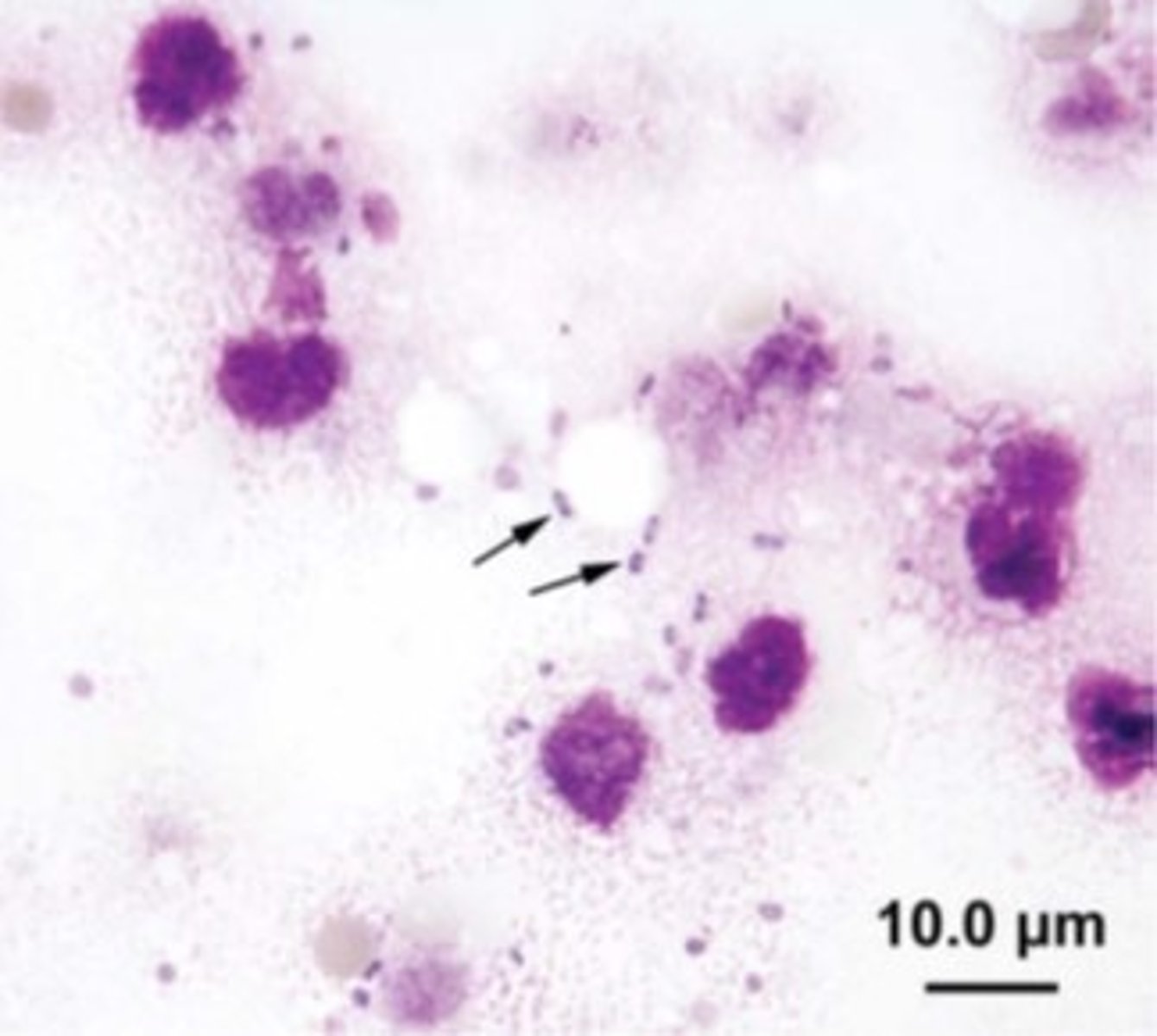
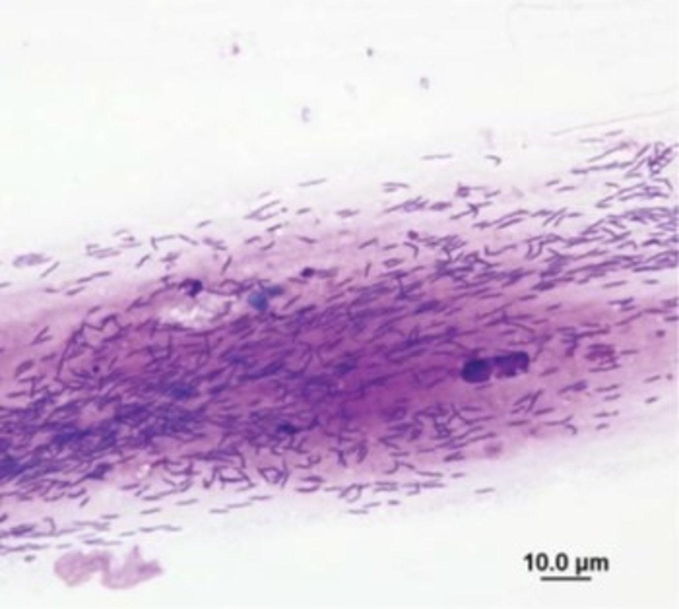
Rods
-seen individually or in chains (E.coli/clostridium)
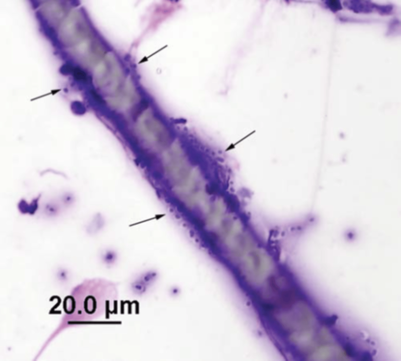
-filamentous rods grow in long chains (actinomyces)
Spirochetes
normal in oral and GI samples (Helicobacter)
Hyphae forming fungi
-candida, aspergillus
-often present within thick aggregates of macrophages, and can have variable shapes and sizes
Dermatophytes
-microsporum, thrichophyton
-cause scaly hairless skin lesions
-small brick shaped asthrospores
Malasezzia spp
-small budding yeast
-usually associated with dermatitis and otitis externa
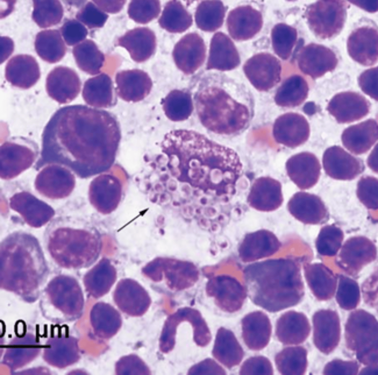
Histoplasma Capsulatum (dimorphic fungi)
-round to oval yeast that replicated by narrow based budding
-yeast in the body, hyphae in the environment
-gi signs in dogs, respiratory in cats
-thrive in nitrogen rich organic matter
Considerations to histoplasma capsulatum
-can be found in any tissue but once in peripheral blood, prognosis is poor
-Second most common fungal infection
-MIRA VISTA LAB (reference lab)
-Treated with itraconazole/fluconazole
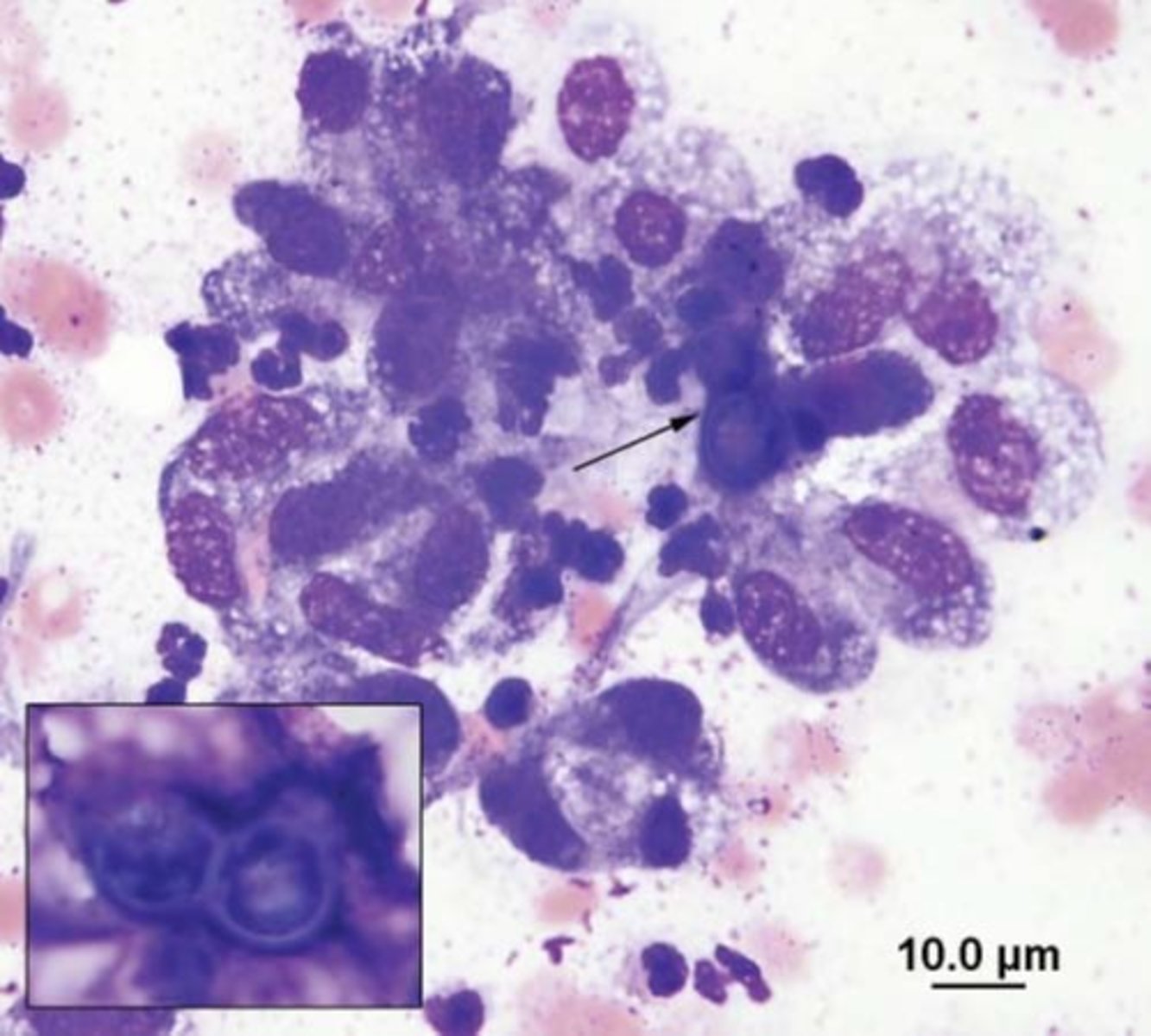
Blastomyces
-medium sized round yeast that replicates by broad based budding
-thick walled and deeply basophilic
-canine infectious are more common than feline
Considerations to Blastomyces