integumentary system 1
1/20
There's no tags or description
Looks like no tags are added yet.
Name | Mastery | Learn | Test | Matching | Spaced | Call with Kai |
|---|
No analytics yet
Send a link to your students to track their progress
21 Terms
macule
flatness
distinguished by color
papule
elevated solid area (< 5mm)
nodule
a papule > 5mm
plaques
elevated flat topped
> 5mm
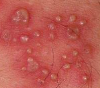
vesicle
fluid filled raised area
± 5mm

pustule
pus filled area
bullae
fluid filled area
> 5mm
scale
cornification of skin
plate-like
associated to inflammatory disease
lichenification
thickened and rough
excoriation
traumatic lesion
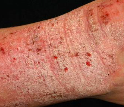
ezcematous dermatitis
atopic eczema
skin is itchy, dry and cracked
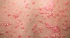
uticaria
allergic rx
IgE
wheals
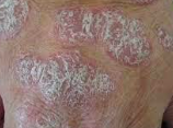
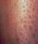
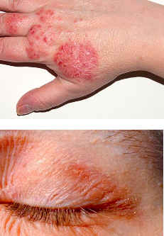
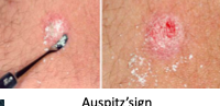
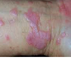
psoriasis
2% pop
Auspitz’s sign
pinpoint bleeding in areas where the plaque of a scaling rash has been removed (bleeding where you scratch)
lichen planus
self limiting 1-2 years after onset
band like lymphocyte infiltrate in dermo-epidermal junction
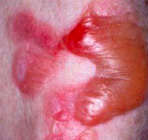
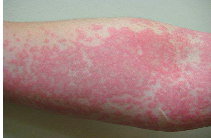
pemphigious
loss of integrity of normal intracellular attachments within the epidermis and mucosal epithelium
triggered by chemicals or drugs, OB, Stress, vaccines, viral infections
life threatening if not treated
40-60ys
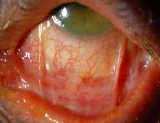
causes symblepharon (abnormal lid and eye ball adhesion)
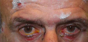
symblepharon
abnormal adhesion bn eyeball and lids
caused by pempigous
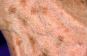
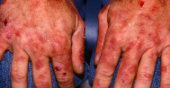
dermatitis herpetiformis
urticaria and vesicles
associated with Celiac disease
epidermolysis bullosa
blisters develop at site of pressure, rubbing or trauma
porphyria
uncommon inborn error of porphyrin synthesis
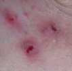
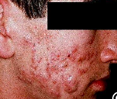
acne vulgaris
middle to late teenagers
triggered by FSH, LH, corticosteroids, heavy clothing, tropical climate
comedone : central black keratin plug
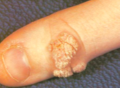
verrucae (wart)
common lesion in children and teens
associated with HPV
direct contact with dev of squamous cell carcinoma of uterine cervix
different appearances:
-vulgaris
-flat
-palmaris
-condyloma acuminatum (venereal)