Chapter 11- Membrane Structure
1/51
There's no tags or description
Looks like no tags are added yet.
Name | Mastery | Learn | Test | Matching | Spaced |
|---|
No study sessions yet.
52 Terms
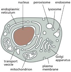
Plasma membrane
Is a selective barrier around the cell that compartmentalizes the area around the cell. Prevent molecules on one side from freely mixing wiht those on the other side
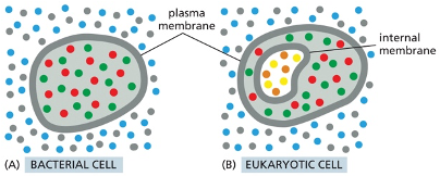
in some bacteria the plasma membrane is the only membrane that they have
eukaryotic cells: also have internal membranes that enclose individual organelle
Proteins associated w/ plasma membrane
allow cell to receive information. Help with import and export, move
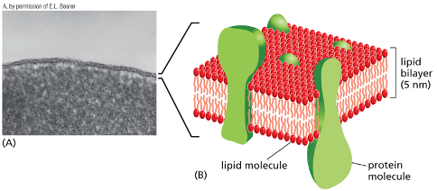
Cell membrane is made up of Lipid bilayer
embedded and associated with proteins
the proteins embedded carry out functions that give the membrane individual characteristics
lipids are arranged in 2 closely apposed sheets
permeability barrier to most water soluble molecules
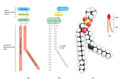
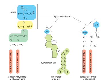
Lipid structure
Lipid head
Hydrophilic carrying a phosphate group is always NEG charged
Tail
hydrophobic
Amphiphatic!
What is the most abundant lipid in the cell membrane
phospholipid
Phosphatidylcholine
one of the most abundant phospholipids in membranes of plants and animals
has a small molecule called “choline” that is attached to the phosphate group and its hydrophilic head
Amphiphatic
molecules that have hydrophobic + hydrophilic regions
examples
phospholipid
detergent
cholesterol (found in plants + animals)
glycolipids- lipids that have sugars as their head
lipids being amphiphatic impact how the bilayers assemble in aqeous environments
Solubility + hydrophobic/ hydrophilic molecules
Hydrophilic molecules
dissolve readily in water
why? bc they contain either charged or uncharged polar groups that can form electrostatic reactions or HBs with water molecules
Hydrophobic
insoluble in water
why? bc most are uncharged + non polar so they cannot form fav reactions with water
force adjacent water molecules to reorganize into cage like structures around them. Being cage like is more highly ordered thatn the rest of the water. When you create the cage it requires energy. But the energy cost are minimized when the hydrophobic molecules cluster together which limits their contact with surrounding water molecules.
Can the head group carry the negative charge?
Yes, typically the phosphate is the one that always carries the negative charge but there are cases like, serine where it carries the negative charge as well.
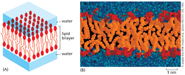
Lipid bilayer confirmation
Hydrophilic heads face the water on both surfaces of the bilayer
hydrophobic tails are shielded from the water within the interior of the bilayer
This arrangement helps with the conflicting attractions and is energetically favorable
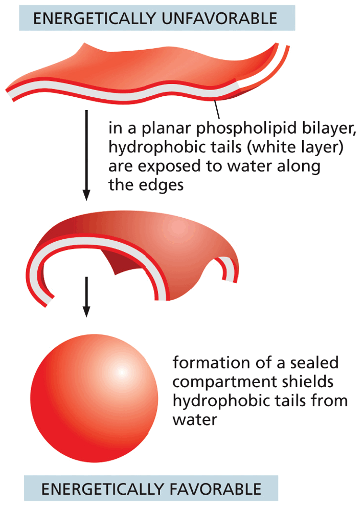
How is the lipid bilayer self sealing?
discuss if it was teared
spontaneity
Tear—> exposed to water—> this is energetically unfavorable—> the molecules in the bilayer spontaneously rearrange to eliminate the tear
if the tear is to big the sheet can fold on itself and break up into closed vesicles
Lipid bilayer forms spontaneously when the membrane lipids are placed in water
Lipid bilayer
fluidity
What does it mean for the membrane to be “fluid”
why is it important
fluidity of lipid bilayer depends on?
How does temperature affect the fluidity
does the length and double vs single bond affect fluidity
unsaturated/ saturated
flexibility
Fluidity
ease of lipid molecules moving within the plane of the bilayer—is important for membrane function and has to be maintained within certain limits.
helps proteins diffuse rapidly in the plane of the bilayer and interact with another (signaling) and allows membranes to fuse with one another + mix molecules
The fluidity of a Lipid bilayer depends on its composition
How fluid the bilayer @ certain temp depends on the phospholipid composition + the hydrocarbon tails
Closer packing of tails= more viscous + less fluid
the length of the double bonds to affect fluidity it's a major property of hydrocarbon tails that effect how tightly they pack. For example a short chain can reduce the tendency to interact between hydrocarbon tails
Unsaturated (contains double bond)
double bond- creates kink in the tail- which makes it more difficult for the tails to pack closely together
doesn’t contain the max # of H atoms
Saturated (no double bonds)
has all of its H atoms
Flexibility (able to bend)
- important for membrane function
sets a lower limit of about 25nm to the vesicle diameter that cell membranes can form
Membrane phospholipid movement within the lipid bilayer
name some ways that the lipids molecules can move
Flip flop: rare
later diffusion is rapid
is the result of thermal motion
the lipids are continously exchanging places with their neighbors within the same monolayer and this can lead to lateral diffusion within the plane of each monolayer
Rotate: rapidly occurs
Flexion
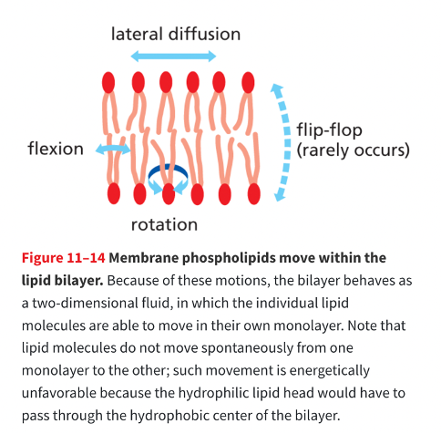
The membrane components can ______ ______ and rotationally but wont switch monolayer spontaneously
The membrane components can diffuse laterally and rotationally but wont switch monolayer spontaneously
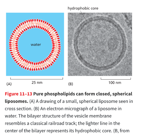
liposomes
pure phospholipids can form closed spherical liposomes
Cholesterol
how does it affect the fluidity of the membrane
is there a lot of cholesterol in the membrane
structure
role
impact on flexibility + permeability
@ high temp vs @ low temp
in animal cells the fluidity of the membrane is adjusted by including cholesterol (Steriod), which is present in large amounts in the plasma membrane
structure: short, rigid planar steroid ring a the top what has a polar head + a non polar tail
role: fill in the spaces between the neighboring phospholipid molecules that were left by the unsaturated hydrocarbon tails.
Cholesterol can stiffen the bilayer, making it less flexible and less permeable
@ Hight temp
cholesterol restricts the movement of fatty acids - this maintains the stability
@ Low temp
cholesterol prevents tight packing of fatty acids - maintains fluidity
Where does membrane assembly begin?
what enzyme transfers the phospholipids
result?
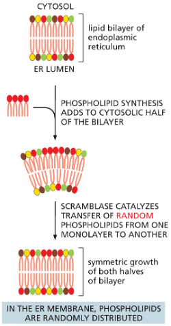
In the endoplasmic recticulum
new phospholipids are made by enzymes bound to the cytosolic surface of the ER.
Scrambalase
type of transporter protein that removes randomly selected phospholipids from 1 half of the bilayer and then inserts them to the other half
the enzyme that transfers phospholipids
result: symmetric growth of both halves of the bilayer
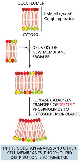
When membranes leave the ER what happens next, where are they incorporated?
Describe the importance of flipases:
When membranes leave the ER and are incorporated in the Golgi apparatus, they encounter a different set of transporters called flippases, which selectively remove phosphatidylserine (light green) and phosphatidylethanolamine (yellow) from the noncytosolic monolayer and flip them to the cytosolic side. This transfer leaves phosphatidylcholine (red) and sphingomyelin (brown) concentrated in the noncytosolic monolayer. The asymmetric addition and distribution of membrane lipids can help curve or bend a membrane bilayer, which is necessary for essential processes such as the budding of membrane vesicles
Flipases:
help to establish and maintain the asymmetric distribution of phospholipids characteristic of animal cell membranes
is a phospholipid handling transporter that uses the NRG from ATP hydrolysis to transfer specific phospholipids from the monolayer facing the noncytoplasmic side to face the cytosol
all cell membranes have distinct “inside” and “outside” faces
explain what the cytosolic monolayer is vs non cytosolic monolayer.
Cytosolic monolayer: always faces the cytoplasmic side
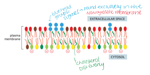
the negatively charged phospholipids are maintained on the cytoplasmic
Non cytosolic monolayer: is exposed to the cell exterior (in the case of the plasma membrane) or the interior space LUMEN or an organelle

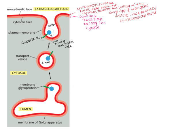
Where are lipids glycosylated, and where do they remain?
lipids- glycosylated in the golgi app and they remain on the non cytoplasmic side of the membrane
once a glycolipid molecule has been created it remains trapped in the monolayer, why?
how is the glycolipid oriented in the plasma membrane
because there are no flipasses that transfer glycolipids to the cytosolic side
When the glycolipid is delivered to the plasma membrane it displays the sugar to the EXTERIOR of the cell on the non cytosolic membrane
SUGARS FACE THE CELL EXTERIOR
Question: All of the carbohydrates in the plasma membrane face the cell exterior. Which direction do the carbohydrates on the internal cell membranes face?
A. The cytosol
B. The cell exterior
C. the lumen of the vesicle or organelle
D. the glycocalyx
C. the lumen of the vesicle or organelle
Glycolipids are located only in the noncytosolic half of the bilayer; the same orientation holds true for glycoproteins. For the plasma membrane, this means that sugars face the cell exterior. For internal membranes, any sugars will face the lumen of the vesicle or organelle .
What are membrane proteins?
these are proteins associated with the lipid bilayer of a cell
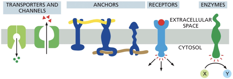
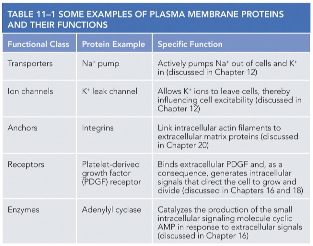
examples of plasma membrane proteins and their functions:
Transmembrane Proteins
Monolayer Associated
Lipid linked
Protein Attached
Transmembrane
extend through the bilayer and is amphiphatic
as a single α helix, as multiple α helices, or as a rolled-up β sheet (called a β barrel).
some can put most of their mass on just one side while others will put their mass on both sides
Monolayer associated
anchored to the cytosolic half of the lipid bilayer by an amphiphatic alpha helix
Lipid linked
lie outside of the bilayer (either side)
attached to the membrane by one or more covalently attached lipid groups
Protein Attached
bound indirectly to one side of the membrane or the other
held in place by their weak non covalent interactions with other membrane proteins
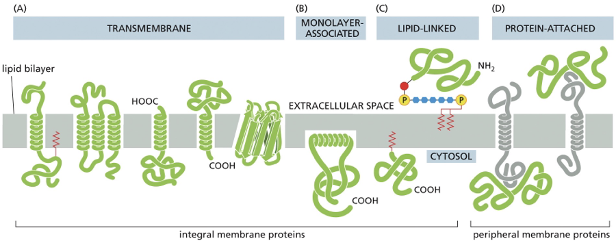
What are Integral membrane proteins vs peripheral membrane proteins?
Integral membrane proteins
are physically integrated into the membrane in some way
ex: transmembrane, lipid linked, and monolayer associated
can be removed by disrupting the bilayer with detergents
Peripheral membrane protein
indirectly associated with the membrane
ex: protein attached
can be released from the membrane by more gentle extraction procedures that interfere with protein-protien interactions, but they leave the lipid bilayer intact

The C-N backbone of polypeptides is
A. Hydrophilic
B. hydrophobic
Hydrophilic
peptide bonds that join the successive amino acids in a protein are normally polar, making the polypeptide backbone itself hydrophilic
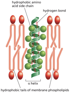
The atoms on either side of a peptide bond (red line) are polar and carry partial positive or negative charges (δ+ or δ–). These charges allow these atoms to hydrogen-bond with one another when the polypeptide folds into an α helix that spans the lipid bilayer
Are the side chains hydrophobic or hydrophilic
hydrophobic
they cannot form favorbale reactions with water molecules
hydrogen bonding + polypeptide chain
hydrogen bonding is maximized if the polypeptide chain forms a regular alpha helix
the alpha helix structure allows hydrophobic R-groups to be exposed while the backbone is shielded from the membrane
the hydrophobic side chains of amino acids forming the alpha helix make contact with the hydrophobic hydrocarbon tails of the phospholipid molecules
many single pass transmembrane proteins are ______
receptors for extracellular signals
multiple alpha helices can be used to produces ?
hydrophilic channels or pores across the membrane
What is the most common from that a polypeptide chain can cross a lipid bilayer
what is another way?
The alpha helix is the most common
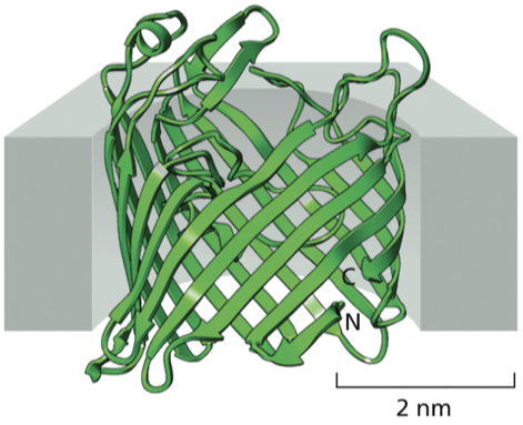
but the polypepetide chain of some transmembrane proteins crosses the lipid bilayer as a BETA sheet that is rolled into a cylinder like a keg, sometimes called a β barrel
β barrel
rolled into a cylinder like a keg
amino acid side chains - face inside of the barrel- line the aqueous channel- mostly hydrophilic
outside of barrel = hydrophobic
contact the hydrophobic core of the lipid bilayer
Give an example of a beta barrel structure
Porin proteins
large, water filled pores in mitochondrial and bacterial outer membranes
porins allow the passage of small nutrients, metabolites, and inorganic ions across their outer membranes, while preventing unwanted larger molecules from crossing
What is the primary function of porin proteins?
A. Act as enzymes for ATP synthesis
B. Allow passive transport of small molecules across the outer membrane
C. Pump ions against a gradient
D. Anchor membranes to the cytoskeleton
B. Allow passive transport of small molecules across the outer membrane
Porins are typically found in which of the following locations?
A. Cytosol
B. Inner mitochondrial membrane only
C. Plasma membrane of all eukaryotic cells
D. Outer membranes of mitochondria and some bacteria
D. Outer membranes of mitochondria and some bacteria
How do porins prevent large unwanted molecules from passing through?
A. They use ATP to block entry
B. They are gated by hormones
C. Their pore size selectively allows only small molecules
D. They break down large molecules enzymatically
C. Their pore size selectively allows only small molecules
How can lipids be solubilized?
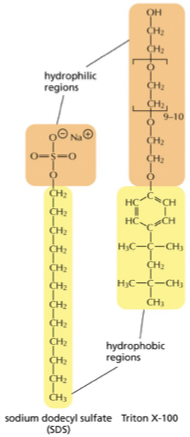
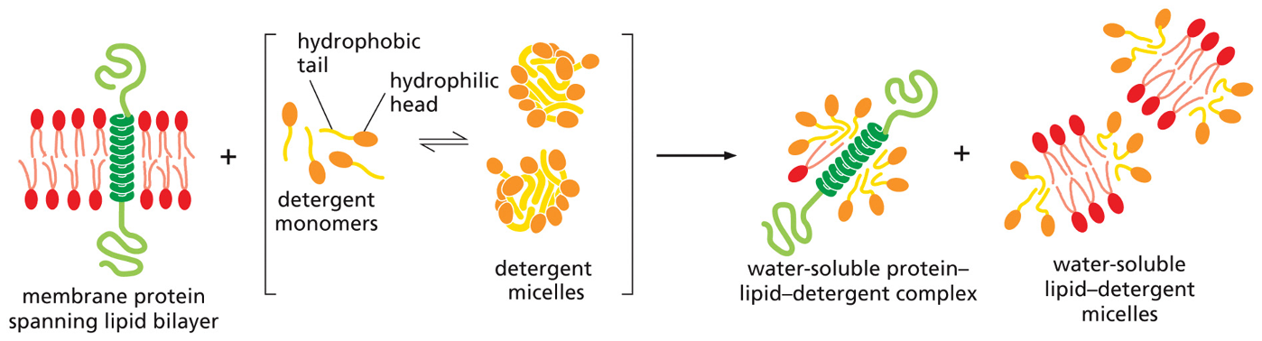
by detergents (small, amphiphatic, lipidlike molecules, interact with membrane proteins and membrane lipids)
1st step in the tipurification process
involves solubilizing the membrane with agents that destroy the lipid bilayer by disrupting hydrophobic associaitons
How do detergents differ from membrane phospholipids
How does this difference affect their shape
the detergents only have a single hydrophobic tail
the detergent molecules are shaped like cones
detergent micelles
in water, these conical molecules tend to aggregate into small, irregularly shaped clusters called micelles
What advancements have been made to determine membrane proteins in high resolution
advancements in x-ray chrystallography + new approaches like cyro- electron microscopy
Bacteriorhodopsin
Why is it important
What is it
Does it do anything with protons
Structure
important because it was the structure that 1st revealed exactly how alpha helices cross the lipid bilayer
Bacteriorhodopsin: small protein, found in large amounts in the plasma membrane of Halobacterium salinarum, an archaeon that lives in salt marshes. Bacteriorhodopsin is a pump, a class of transmembrane protein that actively moves small organic molecules and inorganic ions into and out of cells.
acts as a membrane transport protein that pumps H+ (protons) out of the cell
Each bacteriorhodopsin contains
1 chromophore (light absorbing, non protein, gives the organism a purple color) called RETINAL
Retinal is covalently attached to one of the bacteriorhodopsins transmembrane alpha helices
absorbs a photon of light and it changes shape
How does retinal cause a shape change and what is the affect
Retinal is light absorbing and when it absorbs a photon of light it changes shape
the shape change causes the surrounding helices to go through small conformational changes
which pump 1 proton from retinal to the the outside of the organism
In the presence of sunlight, the bacteriorhodopsin molecules pump?
pump H+ out of the cell
generates a concentration gradient of protons across the plasma membrane
cell uses proton gradeint to store NRG and convert it into ATP
what is the first step of examining protein function and determining the 3D shape of the protein
protein isolation
the plasma membrane of animals is stabilized by a meshwork of filamentous proteins called?
cell cortex that is attached to the underside of the membrane
True or false
a membrane is a 2 dimensional fluid
true
and many of its proteins and lipids can move freely within the plane of the bilayer
Most proteins have what type of diffusion
lateral diffusion
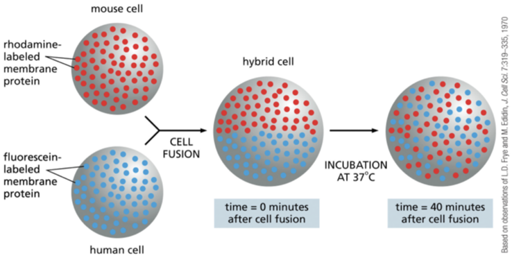
how was lateral diffusion initially demonstrated
fusing a mouse cell with a human cell
formed a double sized hybrid cell
@first the mouse and human proteins were confined to their own halves but within a hour the proteins evenly mixed over the entire cell surface
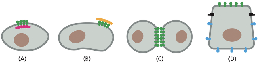
what does this figure demonstrate?
proteins:
A. attached to the cytoplasmic proteins
B.attached to the extracellular matrix molecules outside the cell
C. attached to transmembrane proteins on the neighboring cell
D. diffusion barrier (black) can restrict the proteins to a particular membrane domain
restriction via tight junctions to other protein barrier
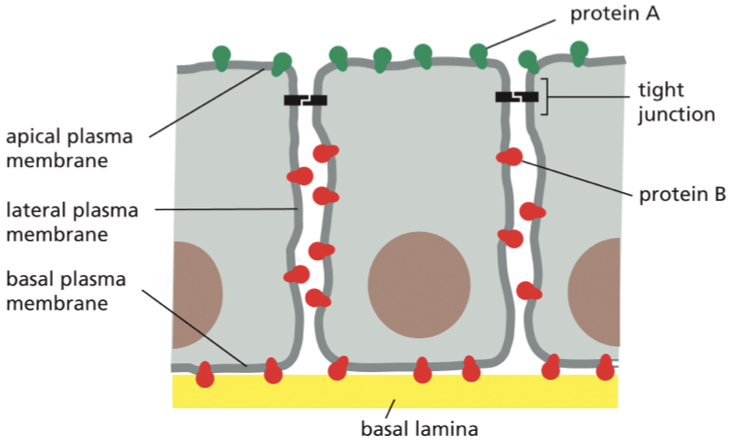
membranes proteins are restricted to particular domains of the plasma membrane of epithelial cells in the gut explain this figure
Protein A (green) and protein B ( red) can diffuse laterally within their own domains but they are prevented from entering each others domains by a specialized tight junction
The restriction of protein movement allows for?
directionality and sideness of cells
By preventing membrane proteins from moving all around the cell, the cell can keep certain proteins on one side and others on a different side. This creates functionally different regions, giving the cell directionality or polarity.