Lab 1: Microscopes, Experimental Design, and Data Presentation (Celine)
1/66
There's no tags or description
Looks like no tags are added yet.
Name | Mastery | Learn | Test | Matching | Spaced | Call with Kai |
|---|
No study sessions yet.
67 Terms
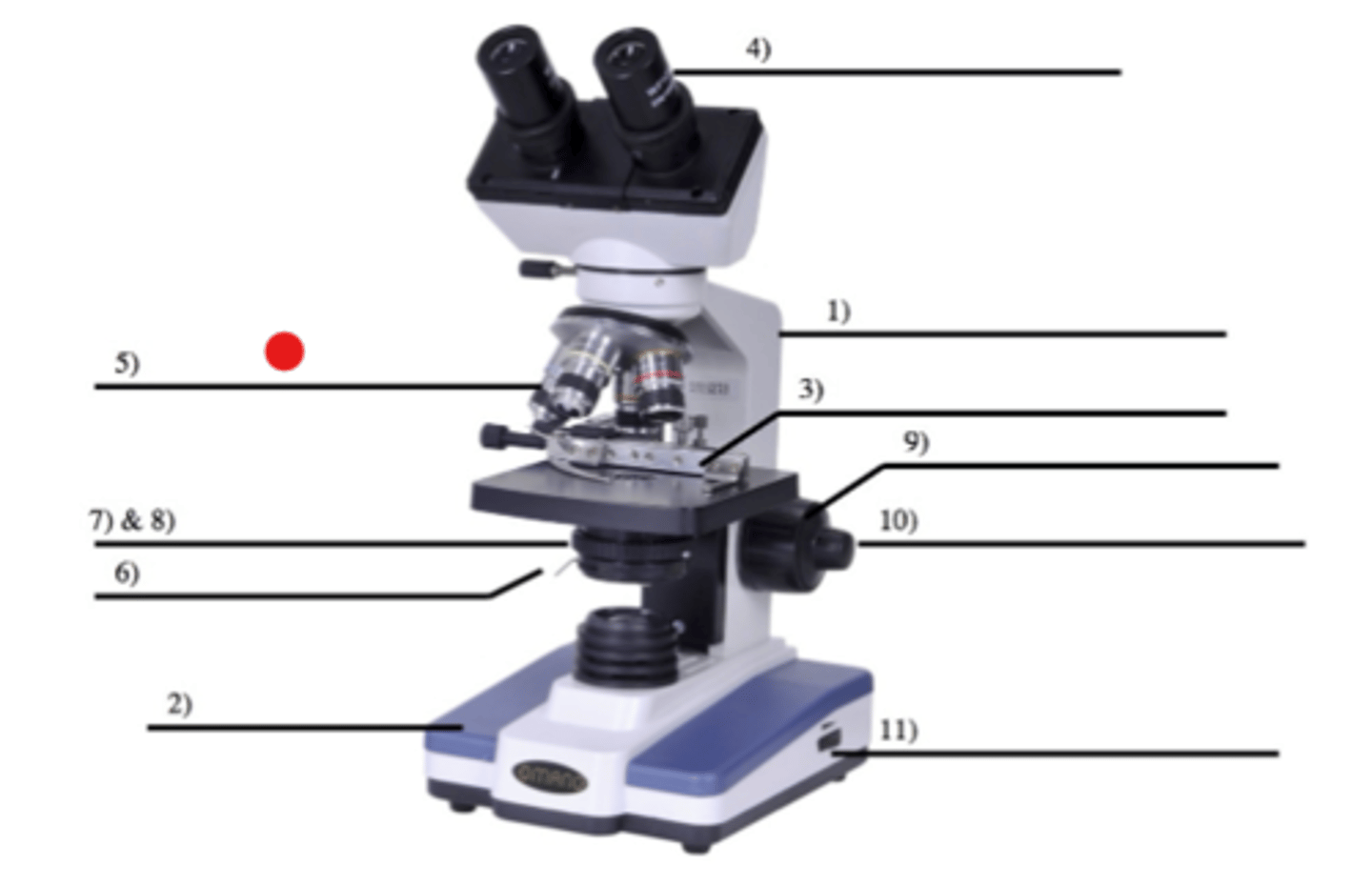
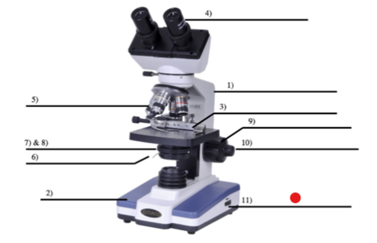
Microscope: Arm
Usage?
1
- Used to carry the microscope (grasping)
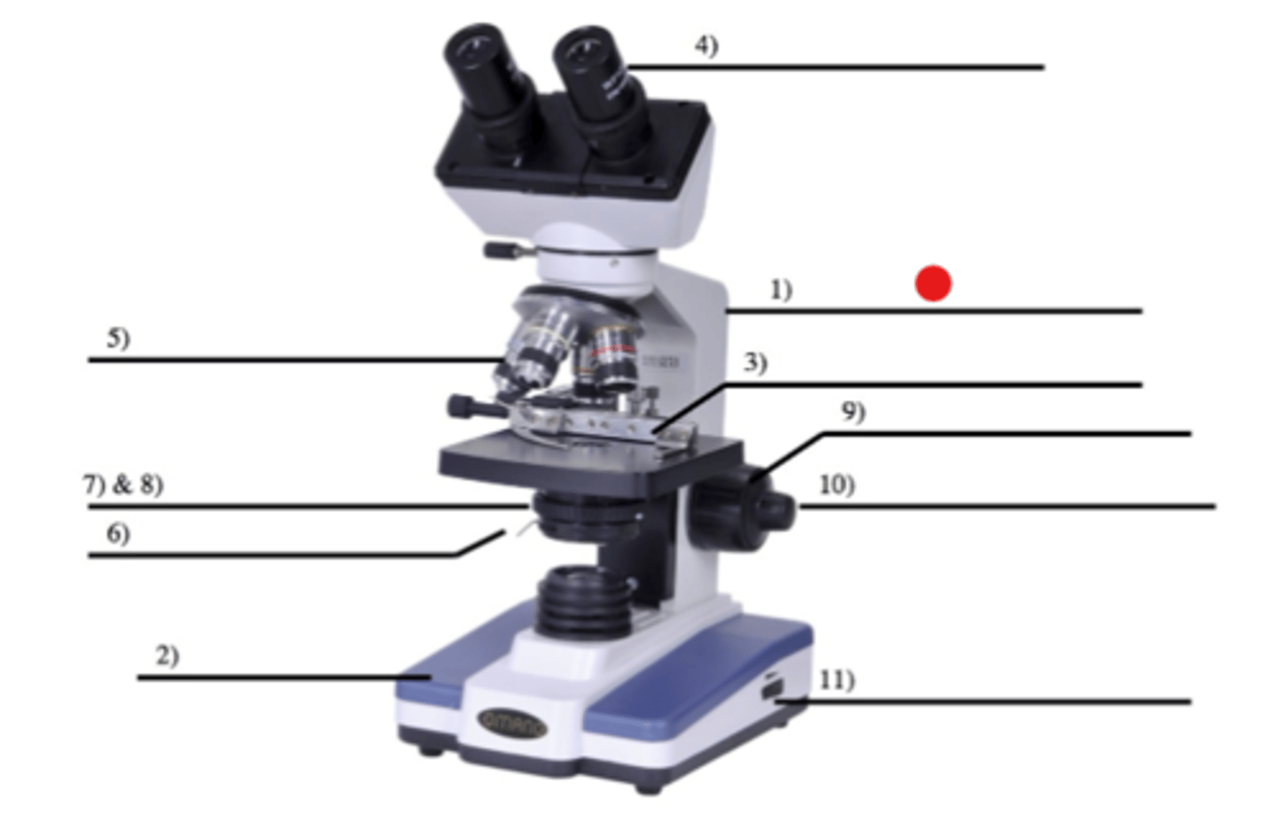
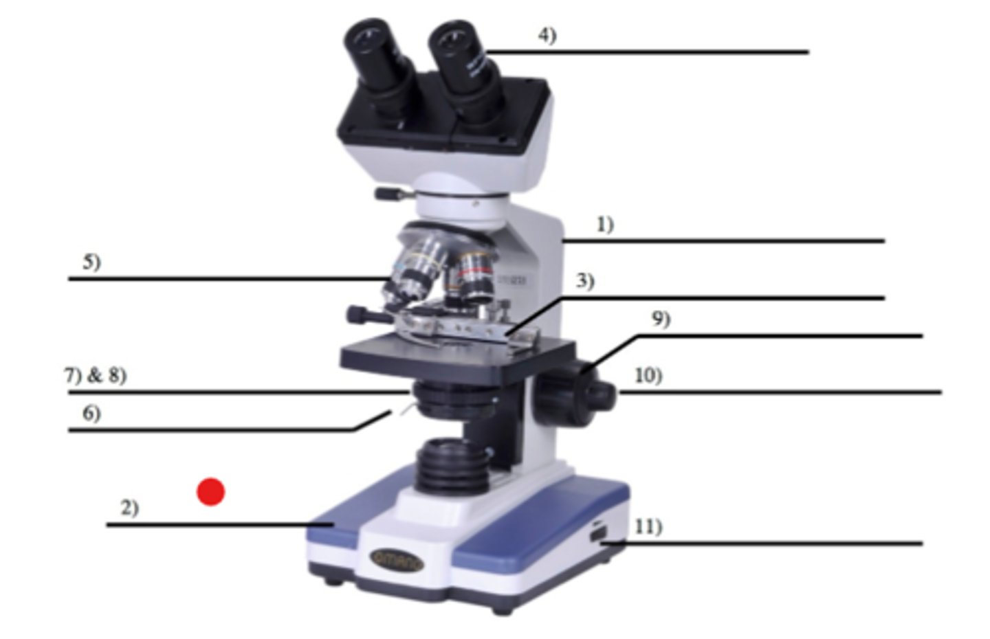
Microscope: Base
Usage?
2
- Used to carry the microscope (supporting)
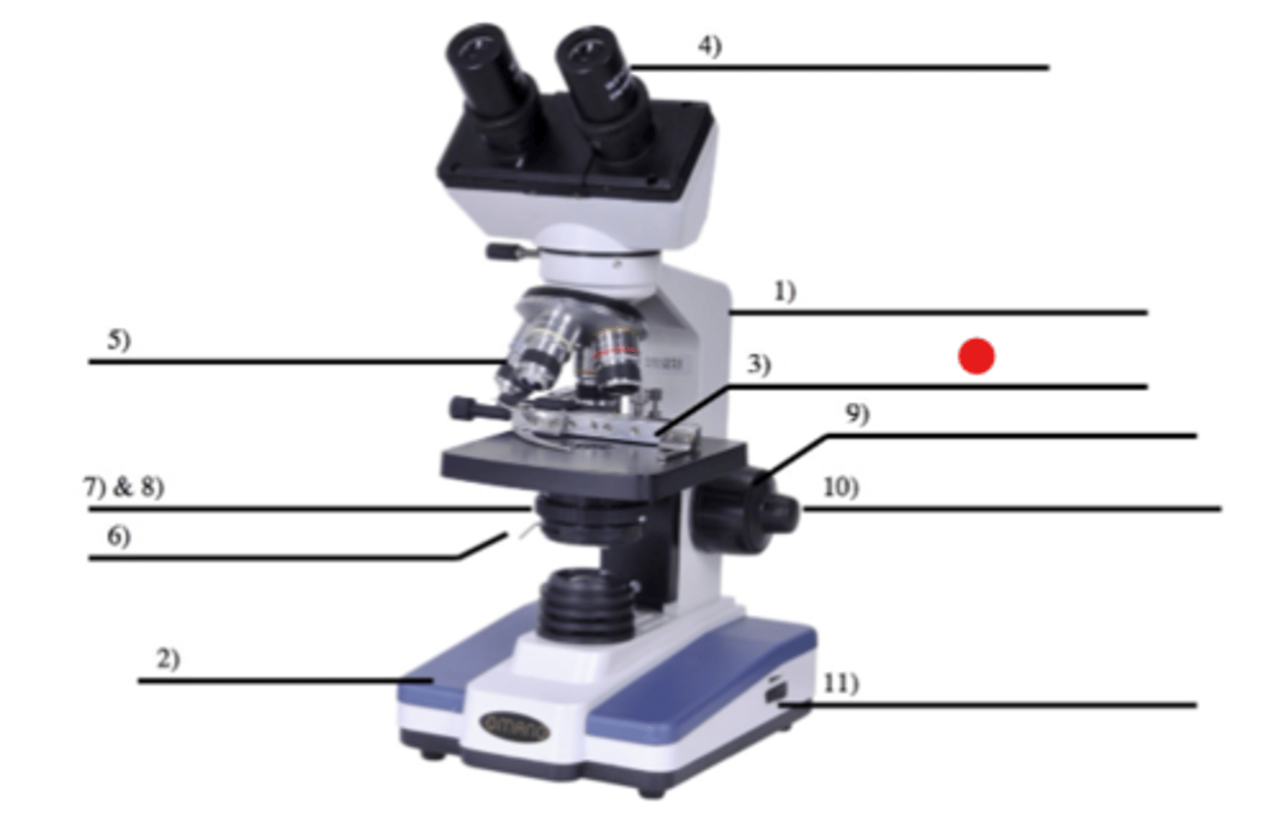
Microscope: Mechanical Stage
Usage?
3
- It's the platform on which microscope slides are placed
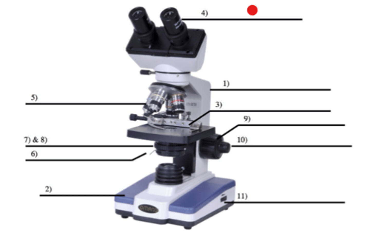
Microscope: Ocular
Usage?
4
- Otherwise known as the "eyepiece"
- Objects are viewed by looking through
- The magnification of our microscopes are 10x
Microscope: Objective
Usage?
5
- The line of vision passes from the ocular through the lens of the objectives
In Bio51, what kind of "ocular" are our microscopes?
Binocular (having two eyepieces)
How many objectives do our microscopes have, and what are they?
3 objectives: 4x, 10x, 40x
What is the 4x objective called?
Scanning
What is the 10x objective called?
Low power
What is the 40x objective called?
High power
Microscope: Condenser
Usage?
7
- Located beneath the stage
- Focuses light rays on the surface of the slide
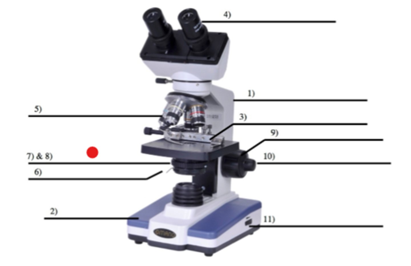
Microscope: Iris diaphragm
Usage?
8
- Regulates the amount of light passing through the condenser

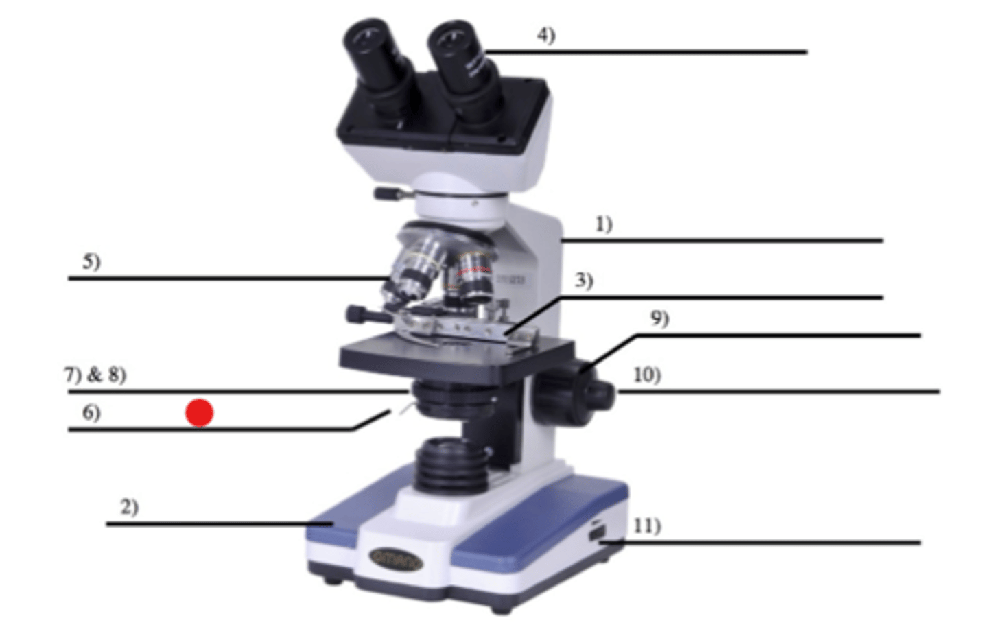
Microscope: Diaphragm lever
Usage?
6
- Controls the iris diaphragm
What are the concentric knobs on the side of the microscope used for?
Focusing the instrument
Microscope: Coarse adjustment
Usage?
9
- Large knob
- Used for large-scale adjustment
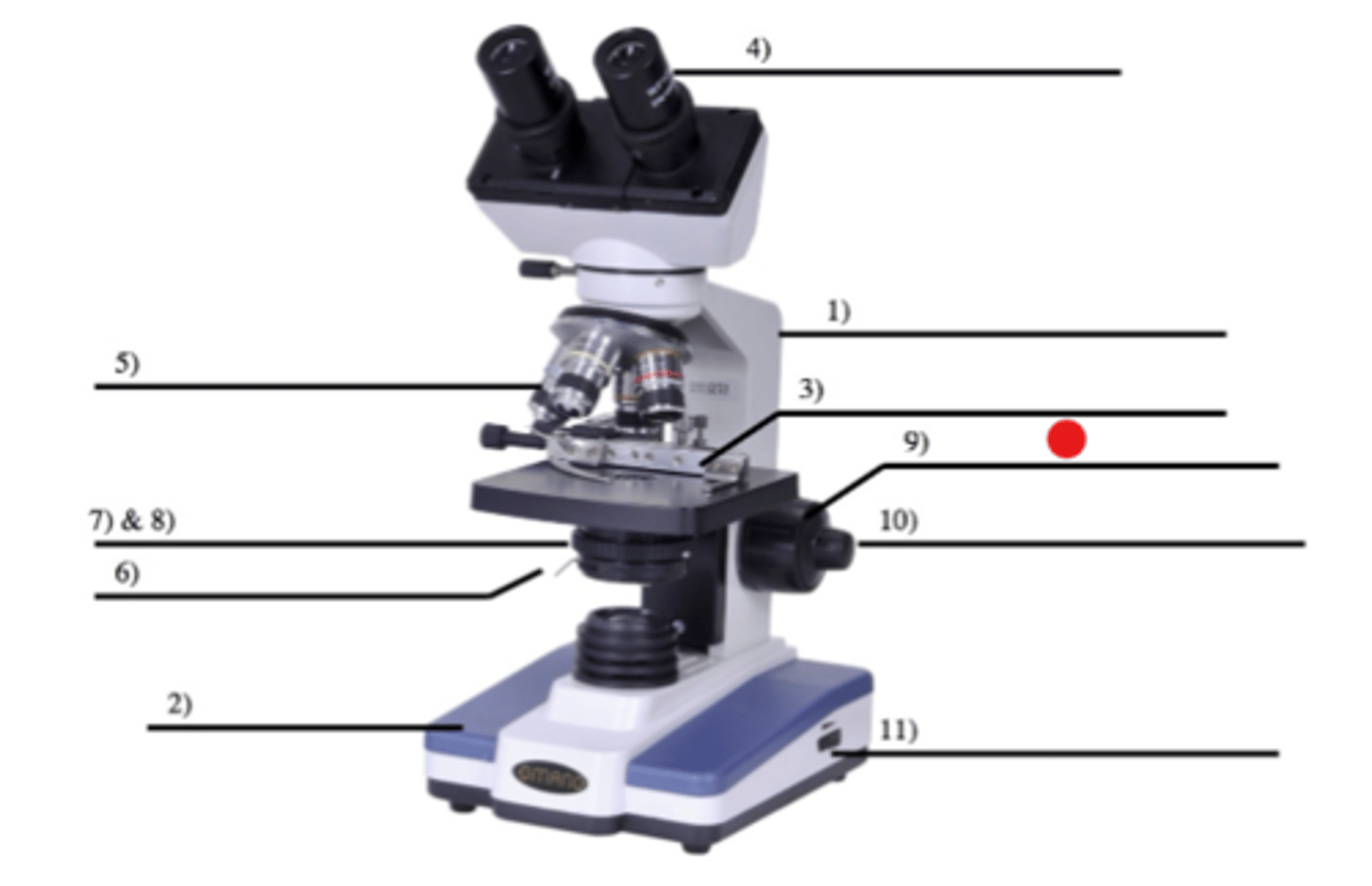
Microscope: Fine adjustment
Usage?
10
- Small knob
- Used for small-scale adjustment
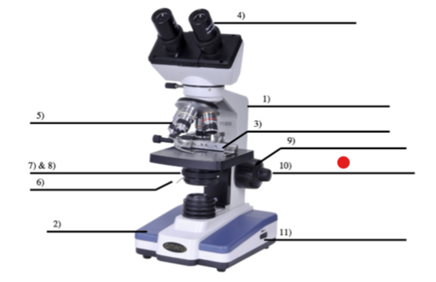
Microscope: Rheostat
Usage?
11
- Adjusts light intensity (brighter, darker)
- You can follow this up by adjusting the iris diaphragm
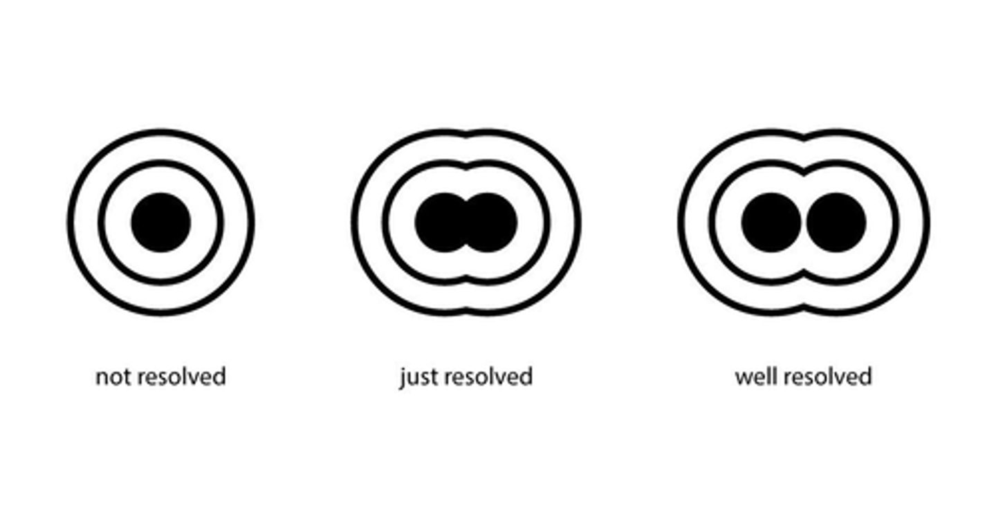
What is resolving power/resolving limit?
A measure of the microscope's ability to distinguish between adjacent points on a slide
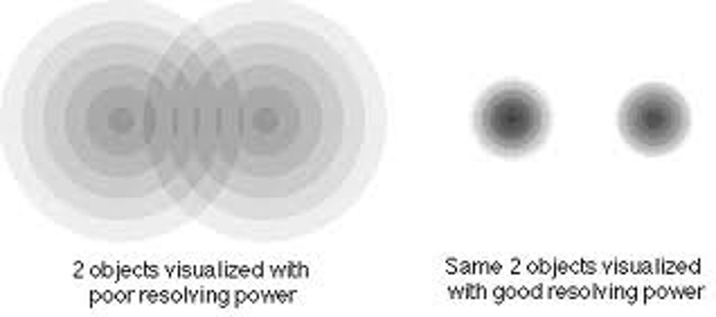
What does having "high resolving power" imply?
That you can distinguish between points that are very close together and this property will give you a very sharp, precise image
What resolving power does the human eye have?
0.1 mm
What is resolving power the function of? Essentially, how does it work?
It's a function of the wavelength of light used to visualize the object and the design of the condenser
(Bonus knowledge: Oil immersion is also used with a 100x objective, but oil immersion will not be likely to be used in this class)
What wavelength provides maximum resolution?
Shortest wavelength of visible light (blue)
- That's why a blue filter is normally used over the light source

The best compound microscope lenses have a resolving power of about 0.2 µm—what does this mean?
Two objects 0.2 µm apart will be seen as separate entities, but if they're closer than this they will be seen as a single object because their images will fuse together
What is the simplest way to increase resolution?
Opening the iris diaphragm, letting in more light
When resolution increases, what will happen to the image and contrast?
Resolution will increase, image will tend to wash out, contrast will decrease
Magnification
How many times larger the object appears in the microscope as compared to its size as seen with the unaided eye
How is magnification calculated?
Power of oculars x Power of objectives
What is the magnification of a 10x ocular and 10x objective?
100x
What is the magnification of a 10x ocular and a 100x objective?
1000x
The more you magnify the object, what happens to the clarity
It will not decrease, and the fuzziness will just get bigger
Parfocal
Microscopes are parfocal.
When the image is in focus in low power, it will be nearly in focus when a higher power objective is rotated into position
If your image isn't clear in a low power, don't even bother trying to make it clear in a high power!
When you raise the magnification, the object will get bigger, but what will happen to its brightness?
The image will get darker
Field
The area of observation or the area you can see under the microscope
When magnification gets bigger, what happens to your field?
It gets smaller
(This is why the image gets darker: The concentration of light on that portion is lower than before, so you'll need to increase light)
How do you increase the light?
1) Adjusting the rheostat
2) Opening the iris diaphragm
Why should you only use fine focus at high powers?
You may smash the slide!
The working distance (distance between slide and objective lens) is decreased when you increase magnification

Depth of field
The amount of the object that is in focus at one time
What happens to the depth of field of power increases?
It will get smaller
Large depth of field
You will clearly see through the object with most parts in focus
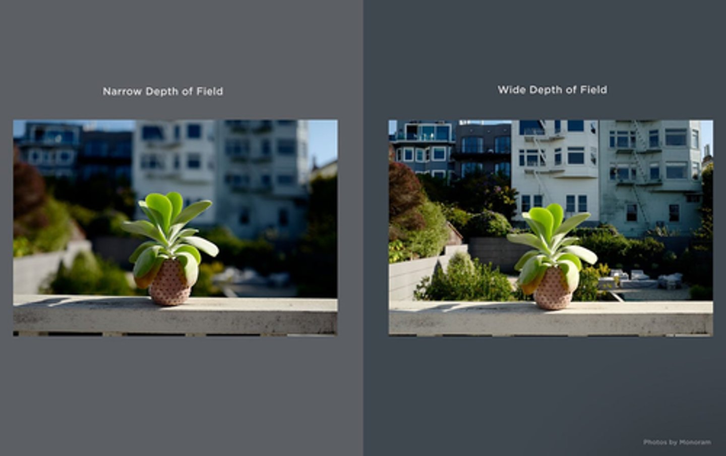
Small depth of field
Only a tiny portion of the object (optical section) will be in focus, and the areas above and below the plane of focus will be fuzzy
Experimental design
Describes the manner in which a hypothesis is tested through experimentation
What do scientists use an experiment for?
To search for cause and effect relationships in nature
- They design an experiment so that changes to one variable causes another variable to change a predictable way
Independent variable
Changed by the researcher and observations are made to see if the change of the independent variable has an impact on the dependent variables
- CONTROLLED by researchers
Dependent variable
Change as a result of the independent variable
- AFFECTED by independent ("depends" on the independent variable)
Where does experimental data come from?
Observations on the dependent variable to see how it responds to the change made to the independent variable
Controlled variables
Remain constant in an experiment regardless of the experimental treatment
Ex. If Celine wants to see which jelly tastes better (dependent variable) depending on what flavor she uses (independent variable), she'll keep the bread type, peanut butter type, and toast level all the same, and these will NOT be affected by the independent variable (controlled variables)
Ex. (From lab) If a scientist wanted to quantify the change in plant growth (i.e., dependent variable) due to nitrogen supplementation, he or she would manipulate the independent variable (soil nitrogen level) of plants while maintaining controlled variables (e.g., soil water content, sunlight exposure, etc.) at a constant level because water and sunlight affect plant growth.
Controlled group
Group is used to compare as "normal" or "baseline"
Treatment group
The groups that were changed/affected
Where is the independent variable plotted on?
The x-axis (horizontal)
Where is the dependent variable plotted on?
The y-axis (vertical)
What graph should you use if the independent variable exhibits continuous variation?
A scatter plot
Continuous variation example: mg of nitrogen per m^3 of soil
What graph should you use if the dependent variable exhibits discrete variation?
A column chart
Discrete variation example: low nitrogen vs. high nitrogen
How do plants perform photosynthesis?
Plants must receive sunlight and exchange respiratory gases (CO2 and O2) with the ambient environment
Where is gas exchange largely mediated through the plant?
Stomata
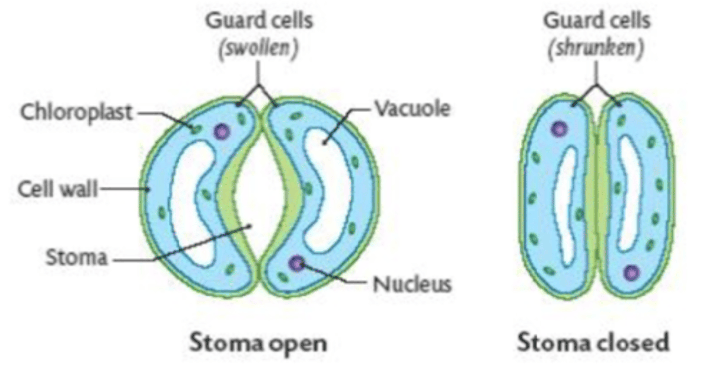
What happens when a plant has sufficient water?
The vacuoles in its guard cells that surround each stomata fill with water --> Causes guard cells to be TURGID or swollen, which OPENS stomata and allows gas exchange
What happens if a plant is dehydrated?
Guard cells become flaccid or shrunken, which reduces water loss and also reduces gas exchange

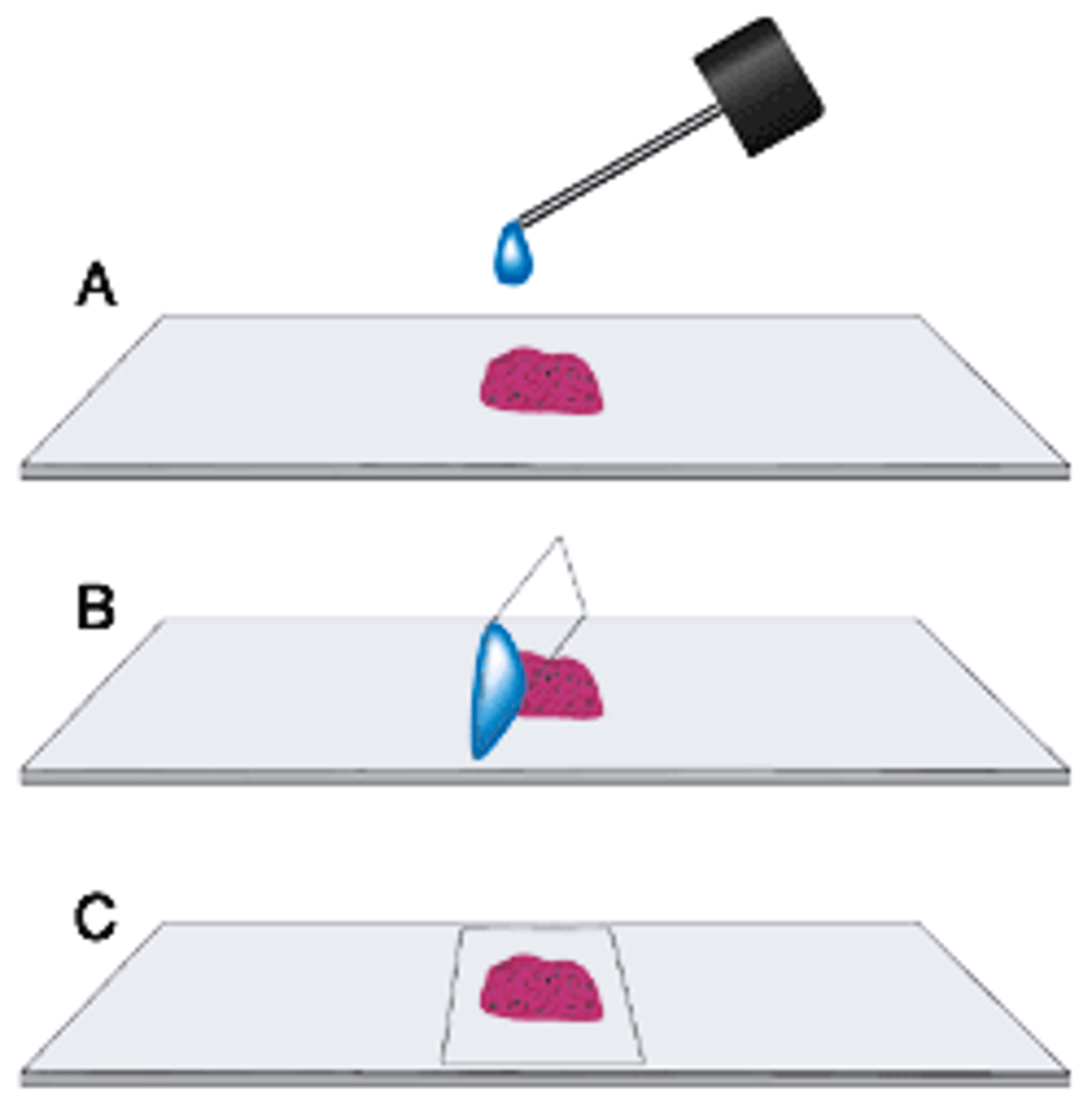
Wet mount
A standard laboratory procedure and is prepared by
1) Placing a drop of liquid on the slide
2) If the material is dry, place it directly on the slide and add a drop of water or stain
3) The mount is covered with a coverslip (at a 45* angle to reduce bubble formation)
In the 3 different-colored yarn fibers, how can you instantly tell which one is on top and which one is on the bottom?
Moving the stage upwards, whichever one comes into focus first is the top.
Moving the stage downwards, whichever one comes into focus first is the bottom
As a last resort... lwky just look at the slide lol
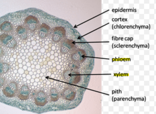
1D. Preparation of a cross-section plant vascular tissue
GOAL: Explore how plants exchange gases (O2, CO2, and water vapor) with their environments
1) Prepare a cross section of a stem by making a thin (~1mm) slice of the waxed stem tissue with a razor blade
2) Submerge the slice in the test plate well with 50% ethanol for 5 minutes
3) Use forceps to remove the cross section from the ethanol
4) Submerge in the test plate well with 0.2% Toluidine Blue for 5-10 seconds
5) Immediately place in a test plate well with water for at least 2 minutes
6) Make a wet mount slide and observe
CLEAN-UP: Gently wipe off the plant tissue and water from the slide with Kim Wipe, and throw wipe into regular trash
Toluidine blue O stain
- A metachromatic stain
- Vascular tissue (ground tissue and cells from phloem, which transports sugars, and lignified tissue of xylem, which transports water and minerals) stain bluish and greenish
- Non-vascular structures take on purplish hue
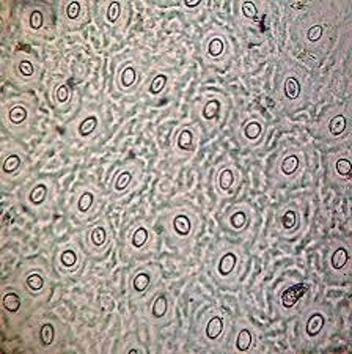
2A. Leaf casts
GOAL: Observe the differences in stomatal activity of leaves subjected in light and dark treatments
0) Dark treatment plants have been placed in darkness for >24 hours.
1) Remove a leaf from the plant. Prepare a cast of the underside of a leaf surface by painting a thin layer of clear fingernail polish
2) Let dry for 10 mins
3) Take a clear piece of tape and firmly press it on the nail polish. When you peel the tape, it should remove the cloudy leaf cast
4) Tape the peeled impression to the DARK labeled slide, trip any excess
5) Examine the leaf cast under the light microscope, focusing on low power, then 100x power
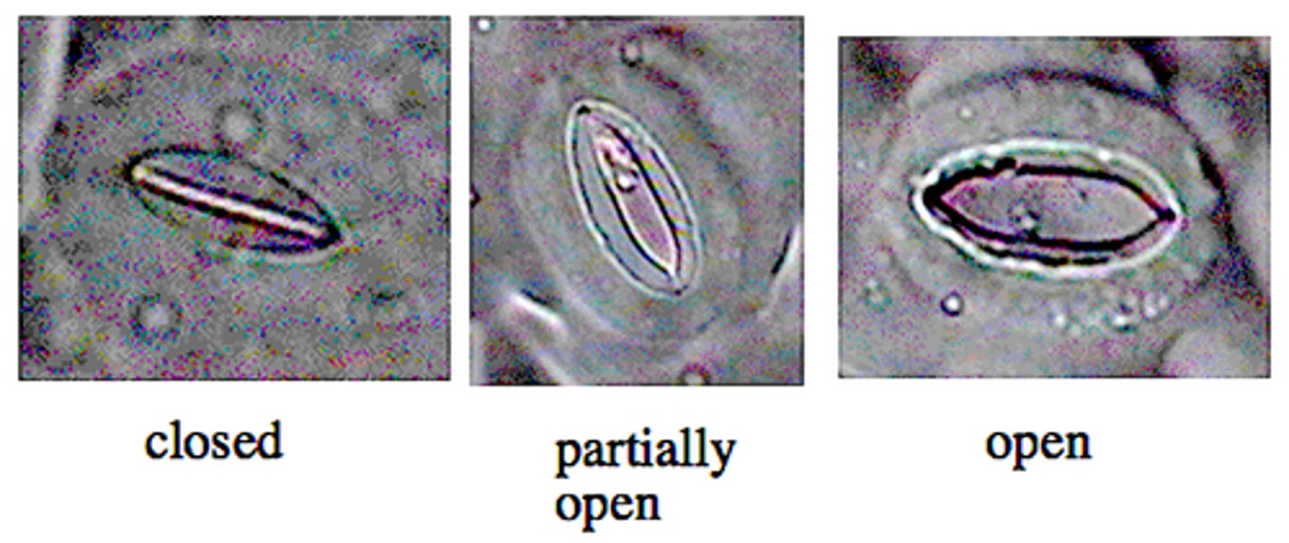
6) Search for areas with concentrated stomata, move to 400x power
7) Record data and count number of stomata
8) Use the fine focus to determine if stomata are open and closed. Open stomata will appear rounder, while closed stomata resemble flat footballs
9) Repeat for other microscope fields
10) Follow the same procedure to test the plants that have been in the light for at least 30 minutes
Dissecting (stereoscopic) microscope
The image is formed from reflected light and has a considerable depth of field because the magnification powers are generally low
- The image is not as altered as with a compound microscope
What happens when you put an "e" slide under a compound microscope? How will it alter the appearance of the image?
1) Amplifies the image
2) Flips the image upside down
3) Darkens the image
Be able to locate the phloem and xylem. What are they?
Phloem: the vascular tissue in plants that conducts sugars and other metabolic products downward from the leaves
Xylem: the vascular tissue in plants that conducts water and dissolved nutrients upward from the root and also helps to form the woody element in the stem.
What happens when you put an "e" slide under a dissecting microscope? How will it alter the appearance of the image?
It amplifies the image, but unlike the compound microscope, it doesn't flip it upside down. The image should still be darkened.
Compound microscope
What is the LOWEST magnification?
What is the HIGHEST magnification?
Lowest: 40x
Highest: 400x
Dissecting microscope
What is the LOWEST magnification?
What is the HIGHEST magnification?
Lowest: 10x
Highest: 30x