Lab Chapter 16
1/31
There's no tags or description
Looks like no tags are added yet.
Name | Mastery | Learn | Test | Matching | Spaced | Call with Kai |
|---|
No analytics yet
Send a link to your students to track their progress
32 Terms
How does air enter the respiratory tract?
Through a Naris. Before air passes through the nasal cavity, air enters the respiratory tract through this. This is the primary entryway for air.
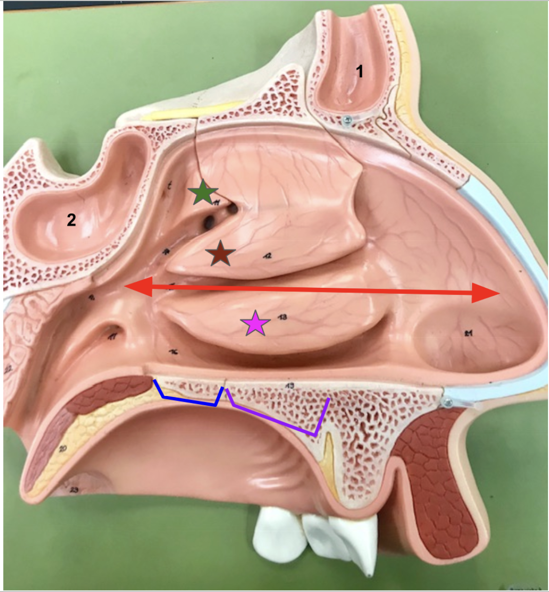
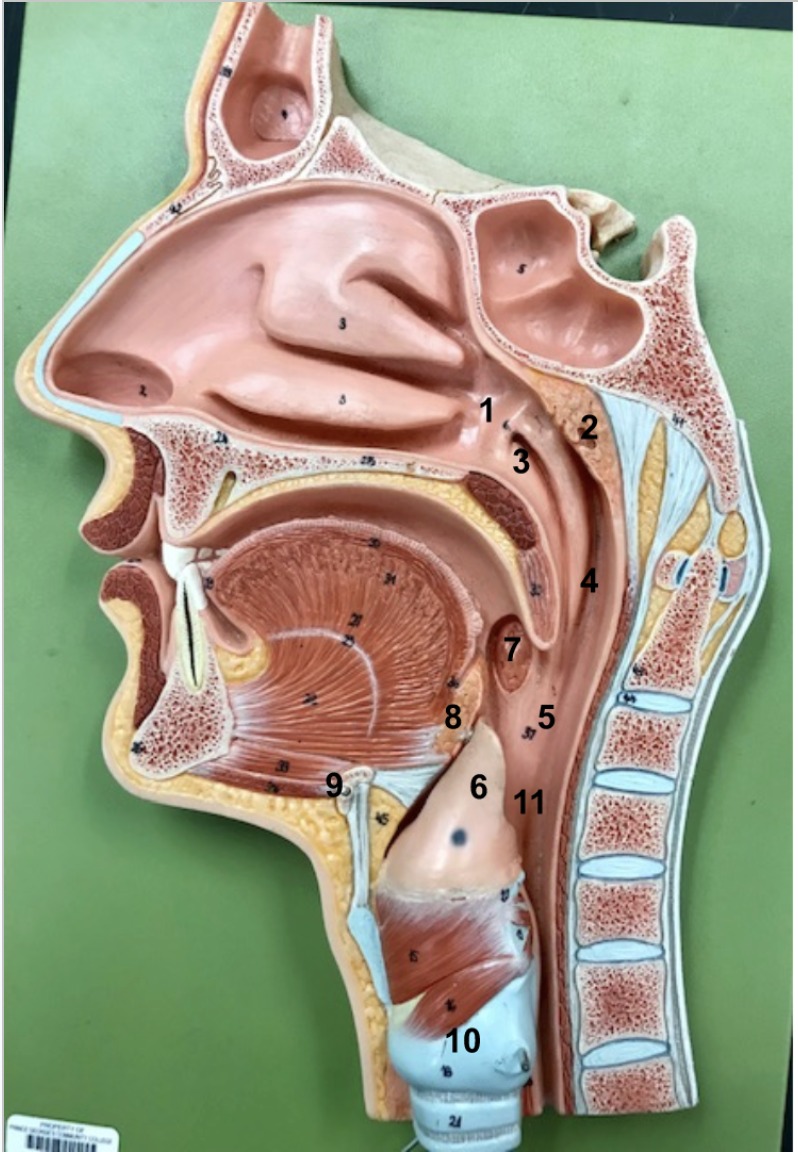
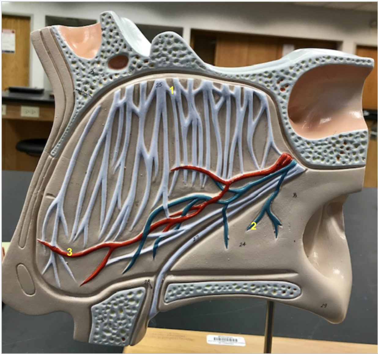
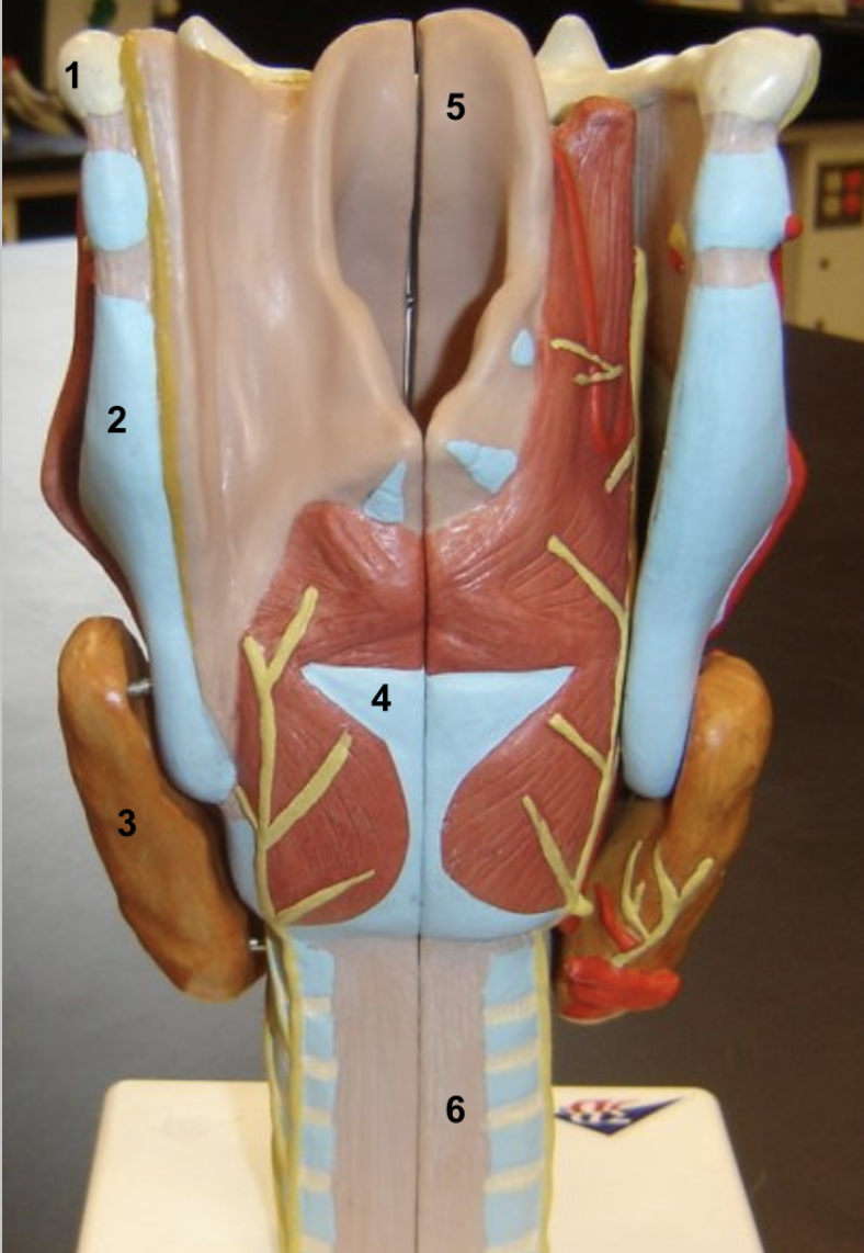
Identify. Function of stars & numbers?
Red: nasal cavity (notice how it stretches from the nostril to the nasal pharynx)
3 stars are the nasal conchae. Create turbulence (swirling air around) and help filter humidify, and warm air through nasal hairs and nasal mucus, which comes from the sinuses (#1&2).
Green: Superior nasal conchae of ethmoid bone
Red: Middle nasal conchae of ethmoid bone
Purple: Inferior nasal conchae
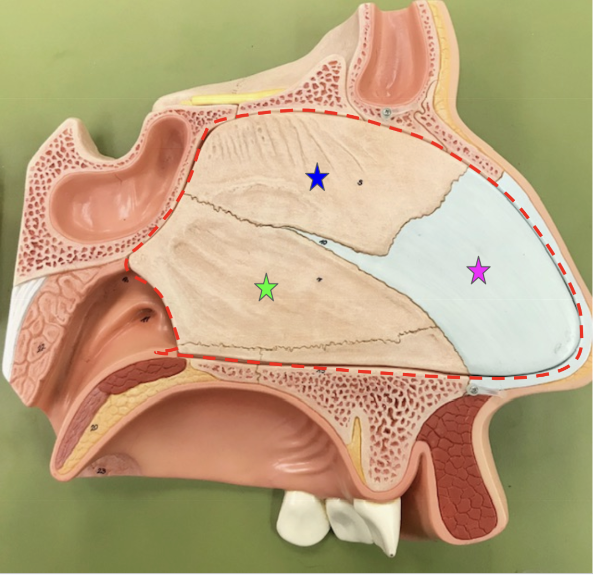
Branches represent the hard palate (bony area) composed of:
Palatine process of the maxilla (Blue)
Horizontal process of the palatine bone (Purple )
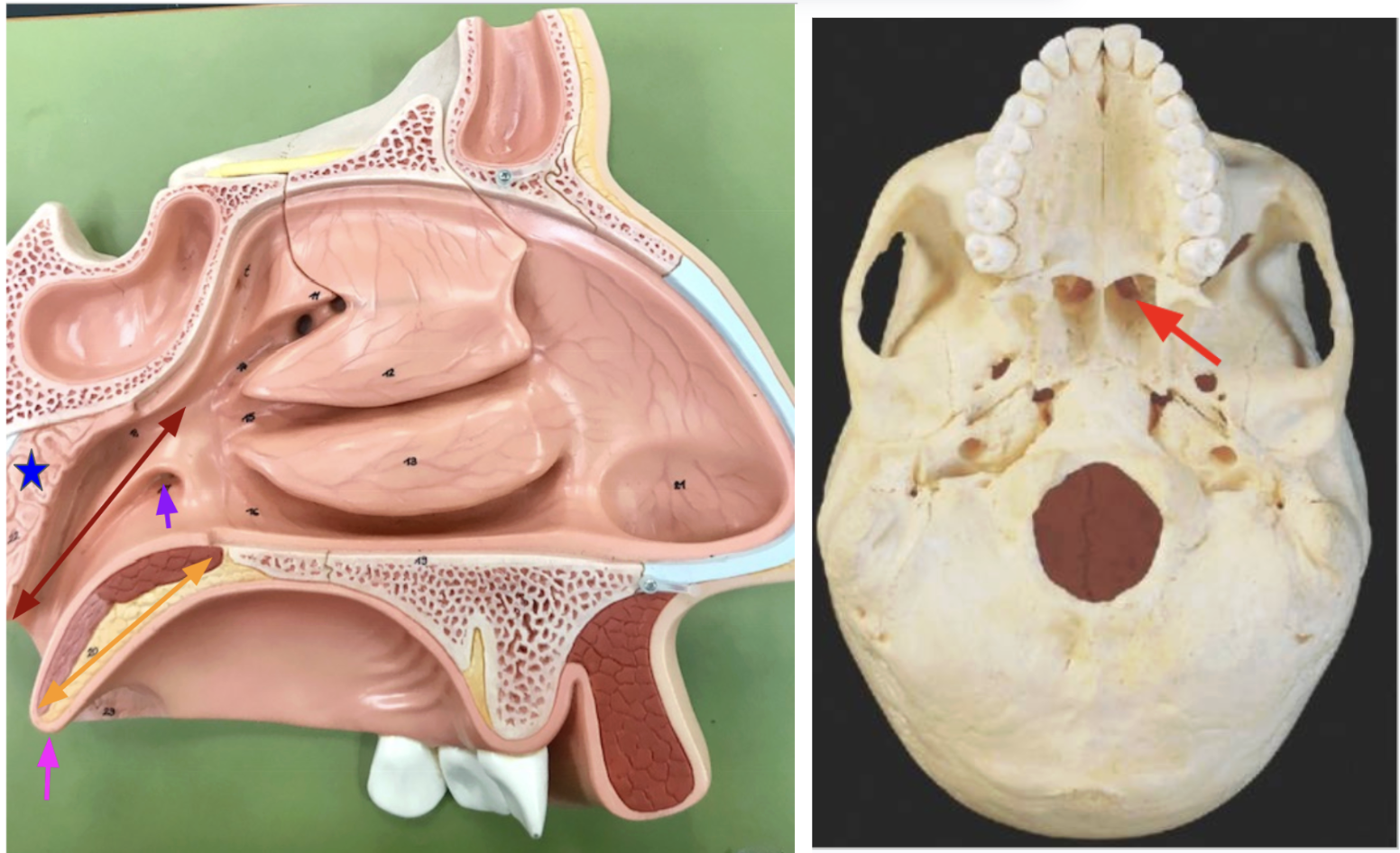
Sinuses: contain nasal mucus
#1: Frontal sinus
#2: Sphenoid sinus
Ethmoid and maxillary sinuses (not visible)


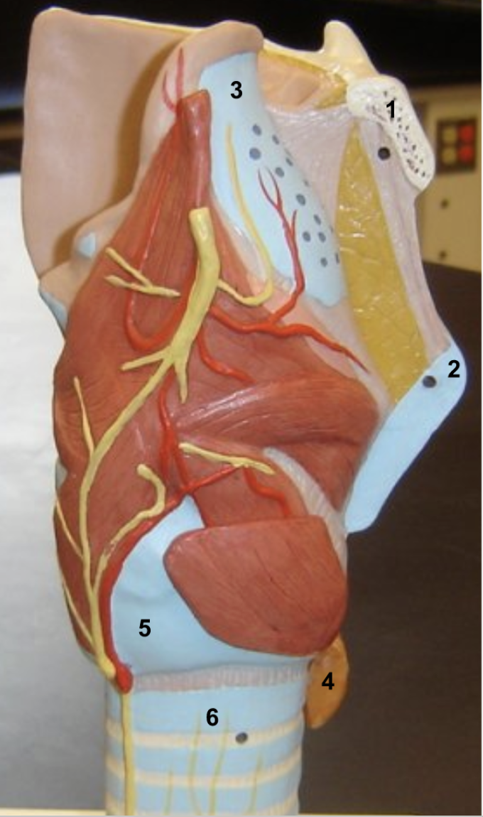
Identify. Function of pink and purple
Dark Red: Nasopharynx
Bright red: posterior nasal apertures
Yellow: Soft palate (tissue area)
Pink: Uvula - Prevents food from entering the nasal cavity when eating (bc it raises upward).
Blue: Pharyngeal tonsils
Purple: Auditory tube - Connect the ear, nose, and throat

Identify. Function of red.
Red area: Nasal septum - divides the nasal cavity into left & right
Broken into 3 parts: pink, blue, & green stars
- Anterior portion of nasal septum:
Pink: Septal cartilage
Posterior portion of nasal septum: (bony portion)
Blue: Perpendicular plate of ethmoid bone
Green: Vomer
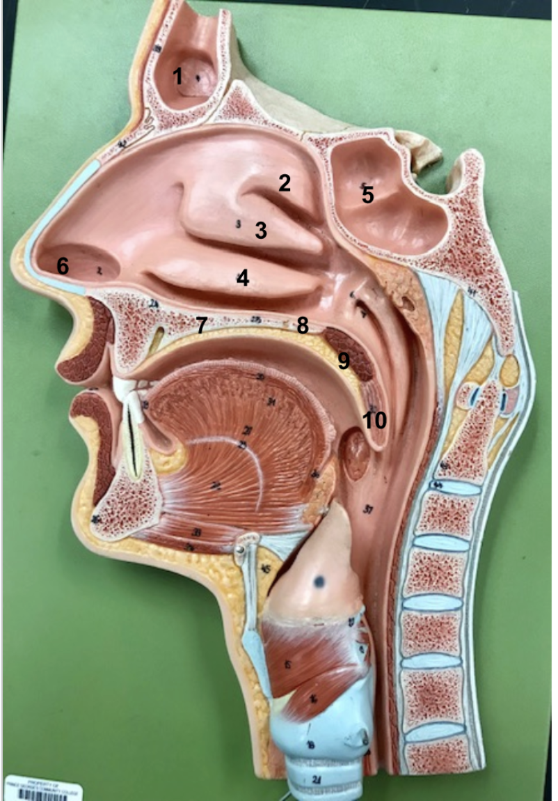
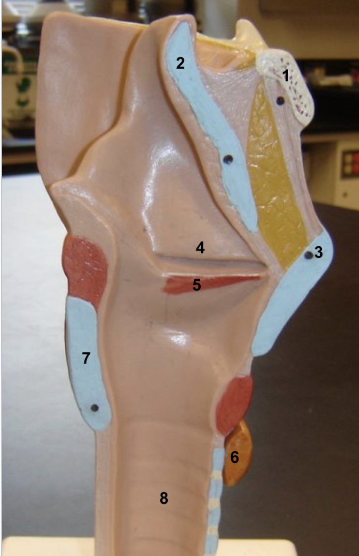
Identify
Frontal sinus
Superior nasal concha of ethmoid bone
Middle nasal concha of ethmoid bone
Inferior nasal concha
Sphenoid sinus
Vestibule (just within naris)
Palatine process of maxillary bone
Horizontal plate of palatine bone
Soft palate
Uvula


Identify. Function of 9.
Posterior nasal aperture
Pharyngeal tonsil
Opening to the auditory tube
Nasopharynx
Oropharynx
Epiglottis (covers the opening to the larynx when swallowing)
Palatine tonsil
lingual tonsil
Hyoid bone (anchors many laryngeal structures)
Cricoid cartilage (major laryngeal cartilage)
Laryngopharynx
- Notice the trachea and larynx are infront (anterior), esophagus in the back (posterior, it looks flat but when you eat it expands). From the pharynx, air travels down the front, food travels down the back.
Where does sound production come from?
Identify. Function of 2, 4 & 5.
- Sound production comes from the larynx (it is our voicebox), which this image depicts. As air moves in and out sound is created.
Hyoid bone
Epiglottis: Helps prevent choking. Opens when breathing, closes when swallowing food.
Thyroid cartilage (largest laryngeal cartilage)
True vocal cords - vibrate when air rushes past to produce sound. It is called true bc it has muscle.
False vocal cords - protect the vocal folds (true vocal chords). It is called false bc it lacks muscle.
Cricoid cartilage (visible in both the anterior and posterior larynx)
Esophagus

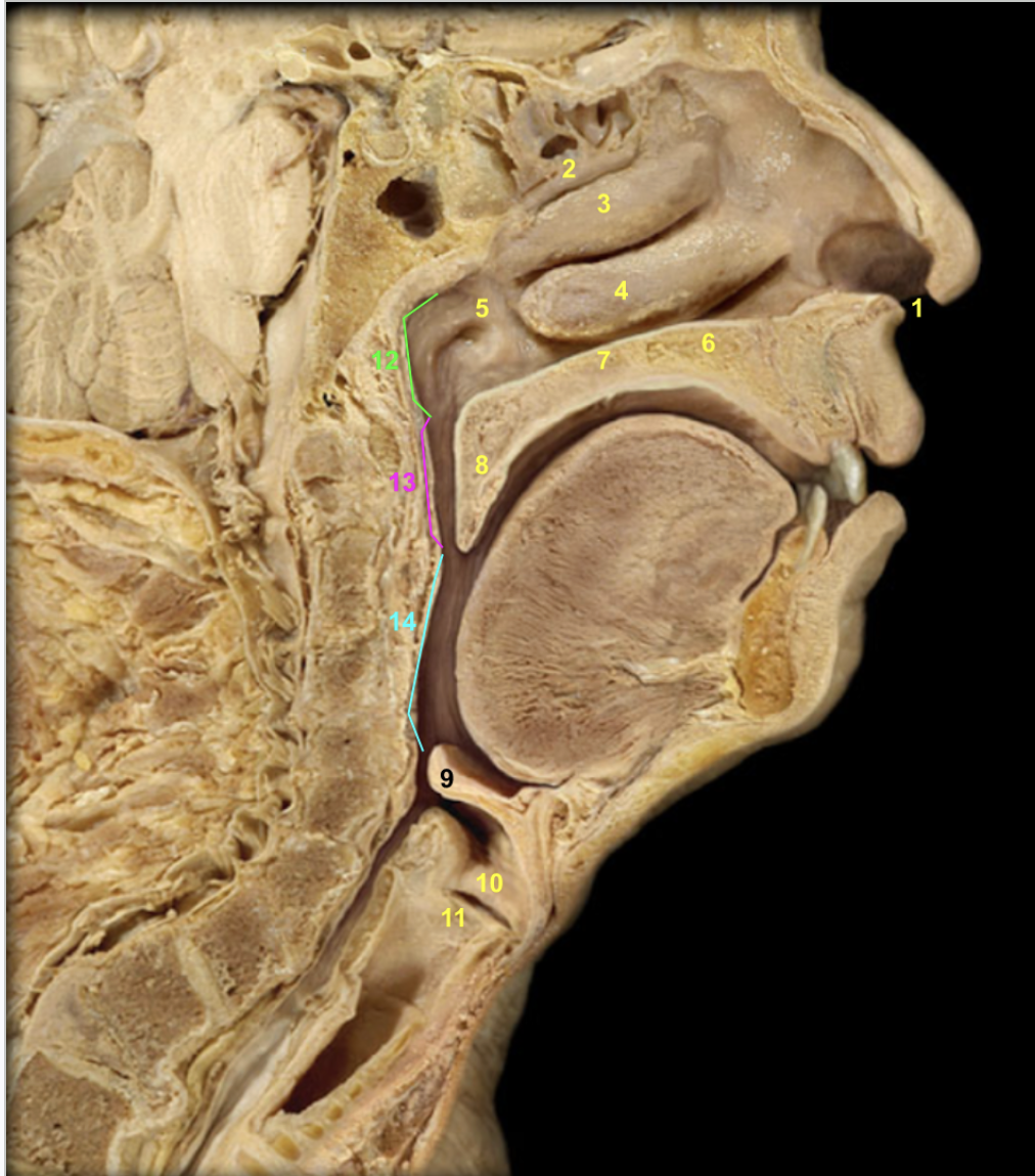
This image depicts what? Identify
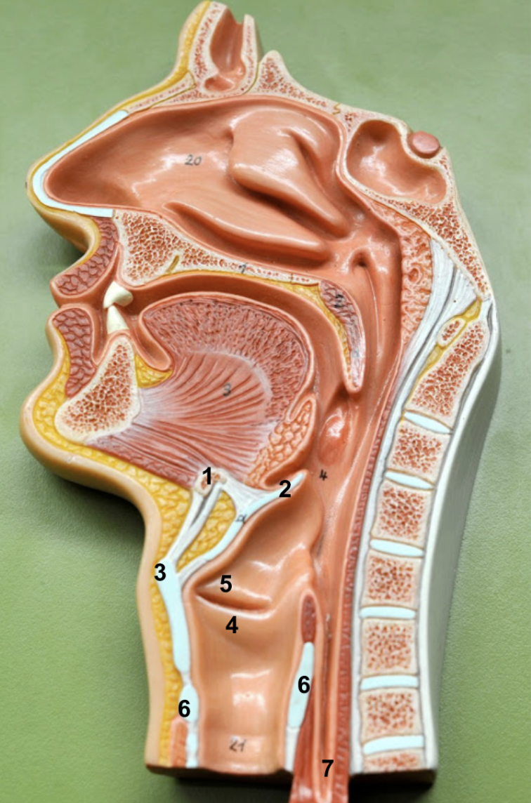
Depicts the nasal septum
Perpendicular plate of ethmoid bone
Vomer
Septal cartilage

Identify 1-13. Function of 7, 8, & 9
Hyoid bone
Thyroid cartilage
Trachea
Superior lobe of right lung
Middle lobe of right lung
Inferior lobe of right lung
Horizontal fissure of right lung - separates the right superior and right middle lobes of the lung
Oblique fissure of right lung - separates the right middle and right inferior lobes
Superior lobe of left lung
Inferior lobe of left lung
Oblique fissure of left lung - separates the left superior and inferior lobes
Cardiac notch
Diaphragm
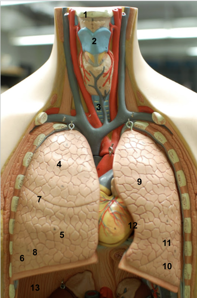
Compare the lobes & fissures of the left and right lung. Why are the lobes like this?
Right lung
3 lobes (superior, middle, and inferior)
2 fissures: Horizontal fissure (separates superior and middle lobe), Oblique fissure (separates the middle and inferior lobe)
Left lung
2 lobes (superior and inferior)
One fissure: Oblique fissure which separates the superior and inferior lobes
- There are more lobes on the right than left bc the heart is situated more towards the left side of the heart. Nd this creates an indentation in the left lung (cardiac notch).
What is the bronchial tree?
Trachea > main bronchus > lobar bronchi > segmental bronchi
Trunk: Trachea.
This branches into two parts left & right main bronchus, which carry air to each lung.
These split into left & right lobar bronchi which carry each to each lobe of the lung. The left only has superior & inferior lobar bronchi, while the right has superior, middle, and inferior lobar bronchi.
These split into left & right segmental bronchi which carry air deep within the lung.
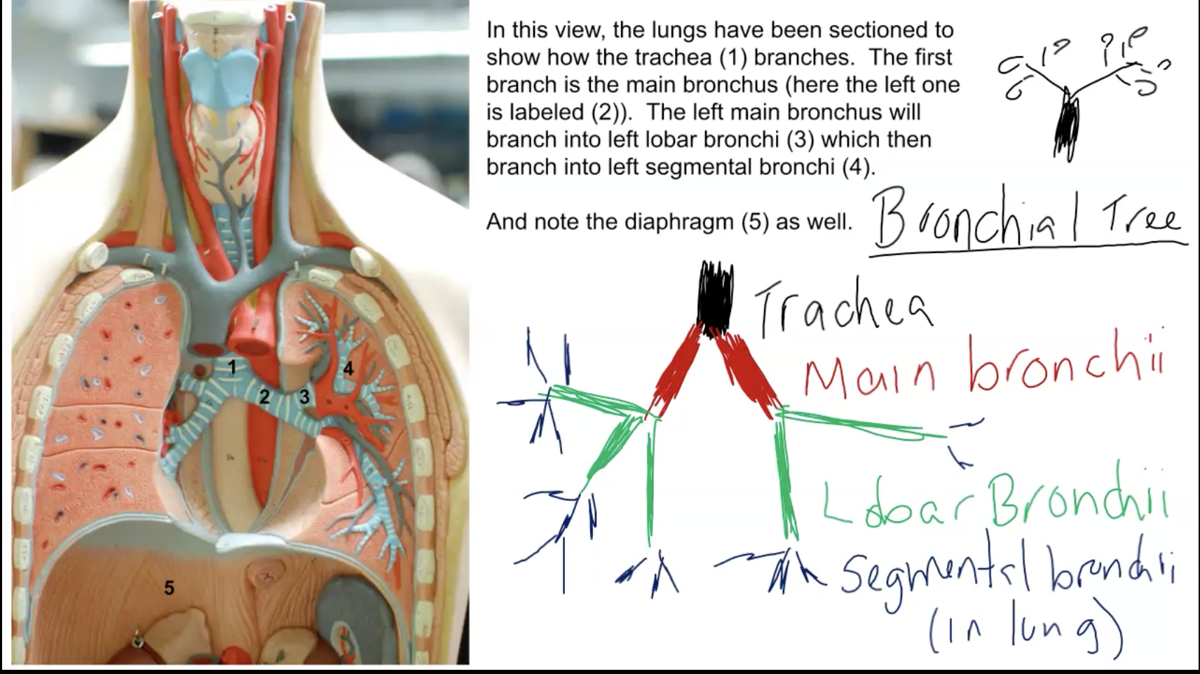

Identify
Trachea
Left main bronchus.
Left superior lobar bronchi
Left segmental bronchi
Diaphragm
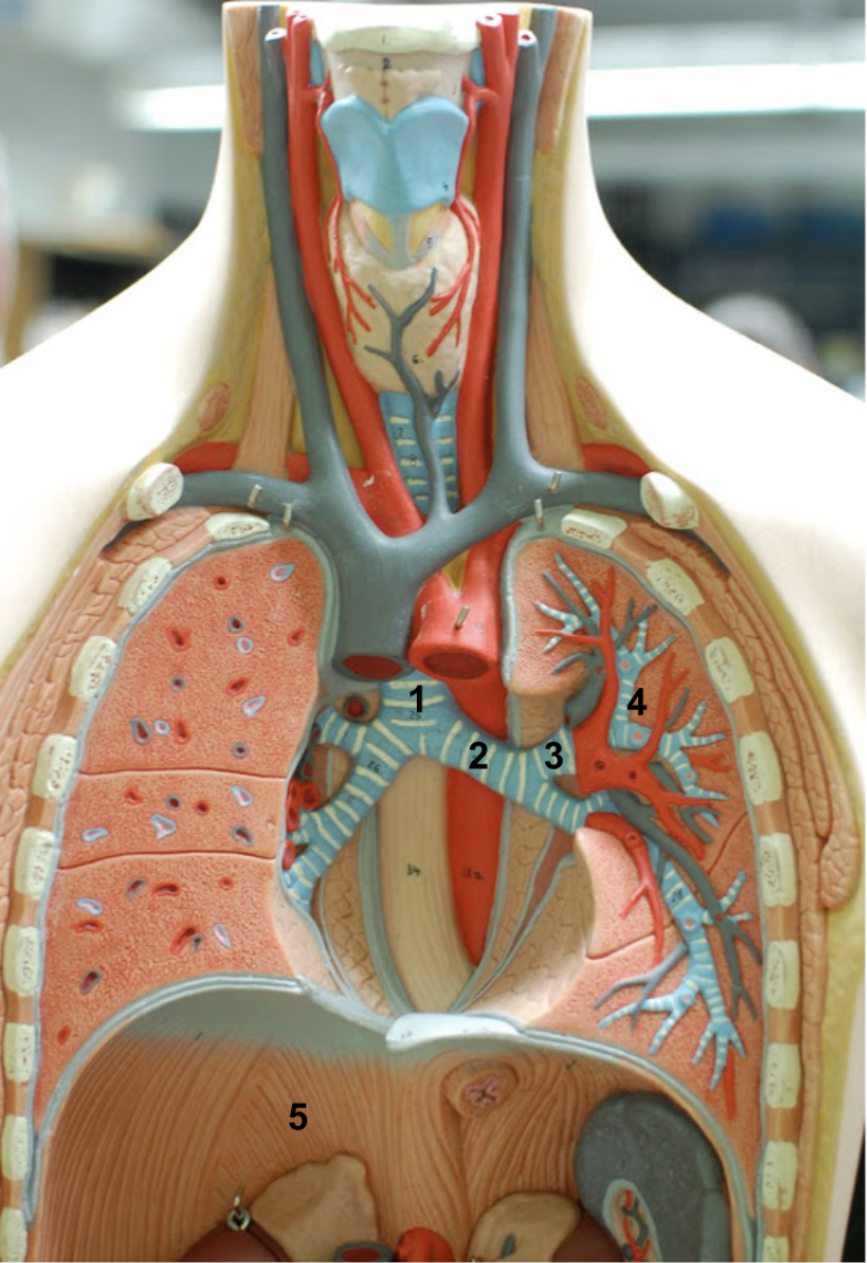

This is an image of the left lung. Identify
Recall that the colors switch in the lungs. So blue is an artery and red is a vein.
Pulmonary artery
Pulmonary vein
Segmental bronchus
Diaphragm
Mediastinal surface

Identify
Hyoid bone
Thyroid cartilage (2).
Laryngeal prominence
Thyroid gland (covering the cricoid cartilage)
Trachea
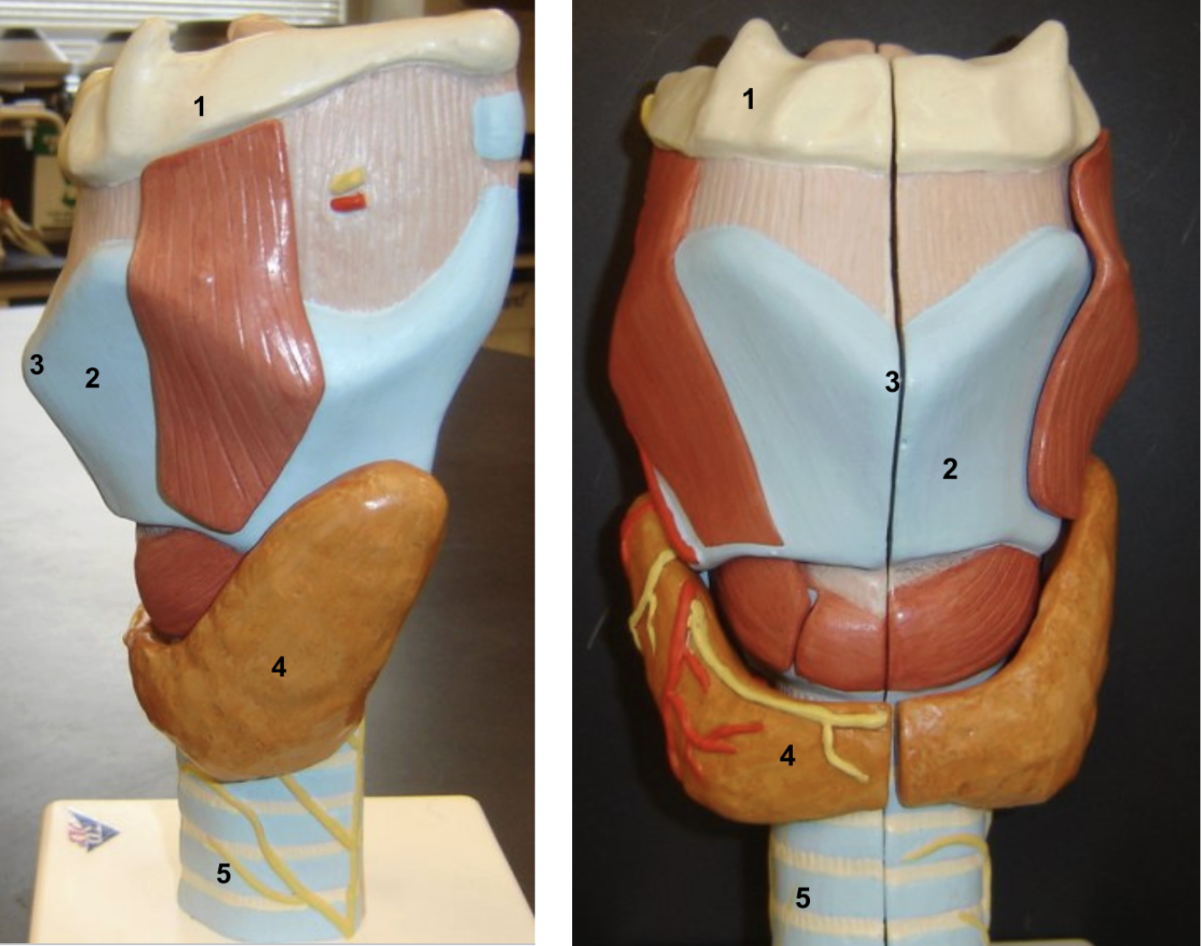
True or false: Only males have a laryngeal prominence.
False. Both male and females have one, but males typically have a larger one.
Identify. How do number 2 & 4 differ, how are they similar? How does #5 differ from the two?
How does the arrangement of cartilage on #6 aid in function?
Hyoid bone
Thyroid cartilage
Thyroid gland
Cricoid cartilage
Epiglottis
Trachea
- Thyroid cartilage does not fully encircle the larynx (The word "thyroid" refers to its shield-shaped structure), while the cricoid cartilage does encircle the larynx entirely. Both are composed of hyaline cartilage and help keep the airway open. The epiglottis is composed of elastic cartilage and covers the larynx during swallowing.
- The cartilage of the trachea is C-shaped and does not fully encircle the tube. The lack of cartilage in the posterior trachea (replaced primarily by muscle) makes the trachea flexible and able to expand during swallowing.

Why is the trachea always open?
Due to the tracheal cartilage. It keeps it open at all times because we always need air.
Identify
Hyoid bone
Thyroid cartilage (notice the Laryngeal prominence)
Epiglottis
Thyroid gland
Cricoid cartilage
Trachea


Identify
Hyoid bone
Epiglottis
Thyroid cartilage (laryngeal prominence)
False vocal cord (Vestibular fold)
True vocal cord (vocal fold)
Thyroid gland
Cricoid cartilage
Trachea
Identify
Naris
Superior nasal concha of the ethmoid bone
Middle nasal concha of the ethmoid bone
Inferior nasal concha
Opening to the auditory tube
Palatine process of the maxillary bone
Horizontal plate of the palatine bone
Uvula
Epiglottis
False vocal chord/Vestibular fold
True vocal chord/Vocal fold
Nasopharynx
Oropharynx
Laryngopharynx
Identify
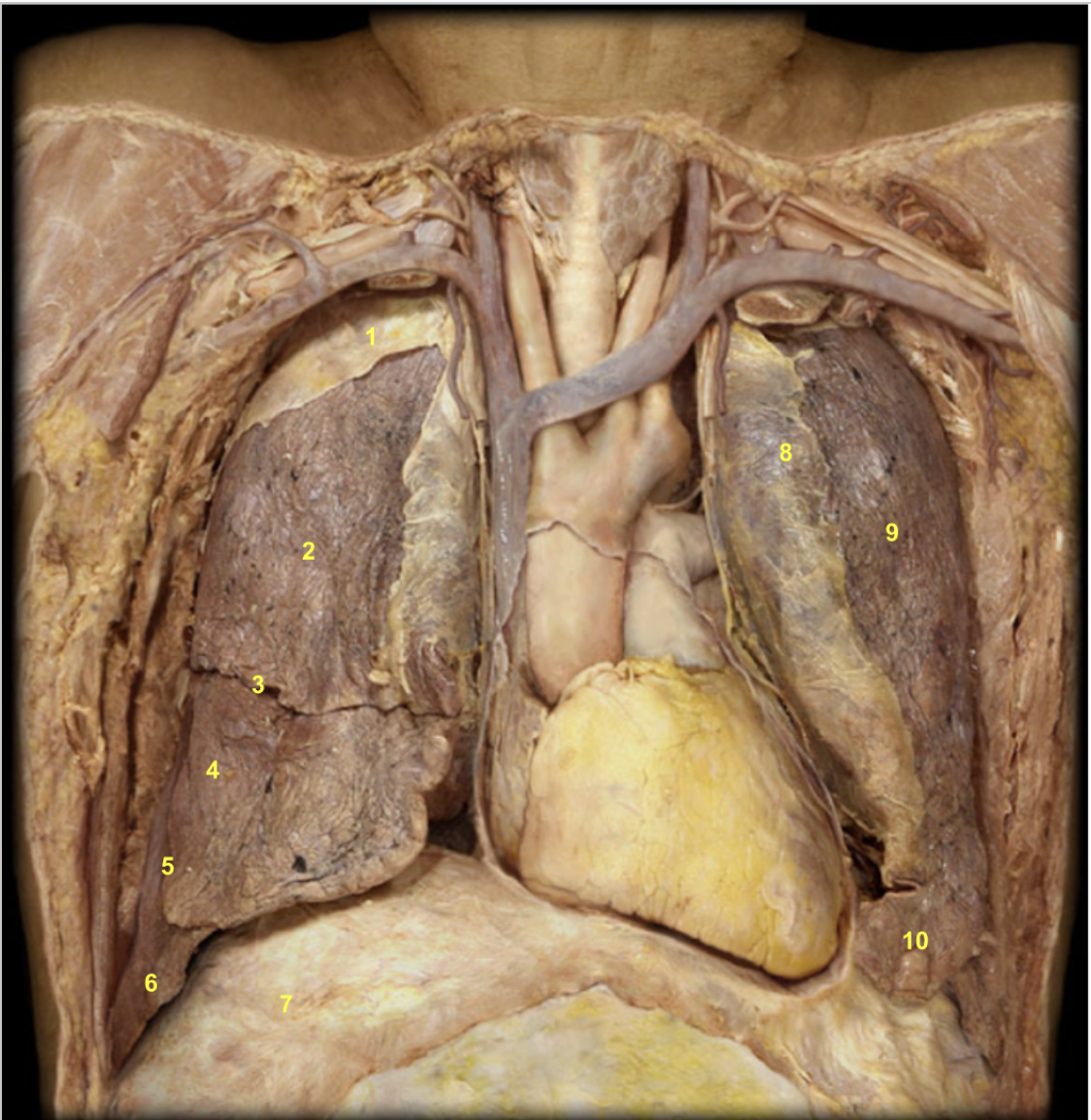
Parietal pleura of right lung
Superior lobe of right lung
Horizontal fissure of right lung
Middle lobe of right lung
Oblique fissure of right lung
Inferior lobe of right lung
Diaphragm
Parietal pleura of left lung
Superior lobe of left lung (covered by visceral pleura)
Inferior lobe of left lung


Identify
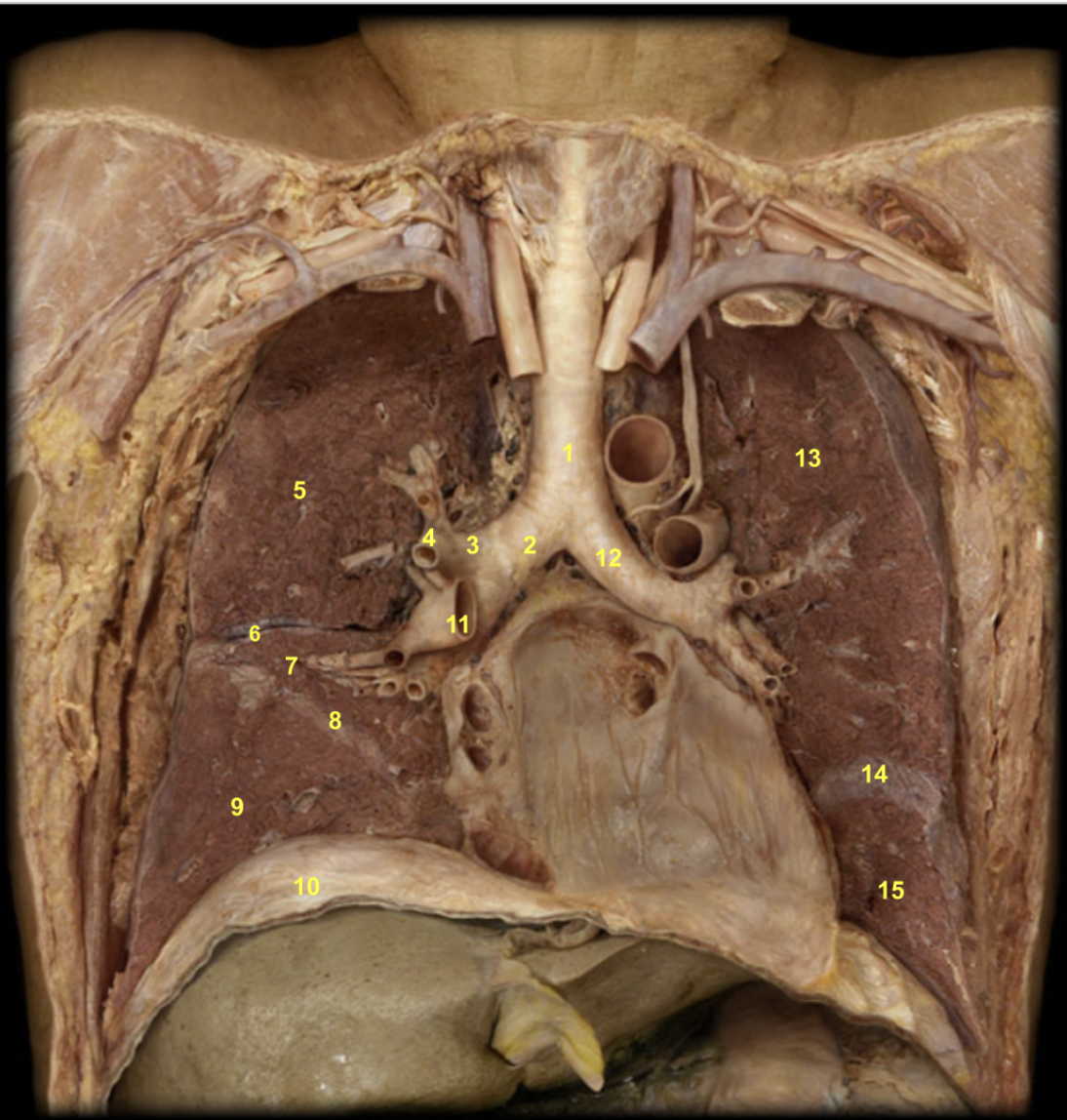
Trachea
Right main bronchus
Right lobar bronchus
Right segmental bronchus
Superior lobe of right lung
Horizontal fissure of right lung
Middle lobe of right lung
Oblique fissure of right lung
Inferior lobe of right lung
Diaphragm
Right pulmonary artery
Left main bronchus
Superior lobe of left lung
Oblique fissure of left lung
Inferior lobe of left lung

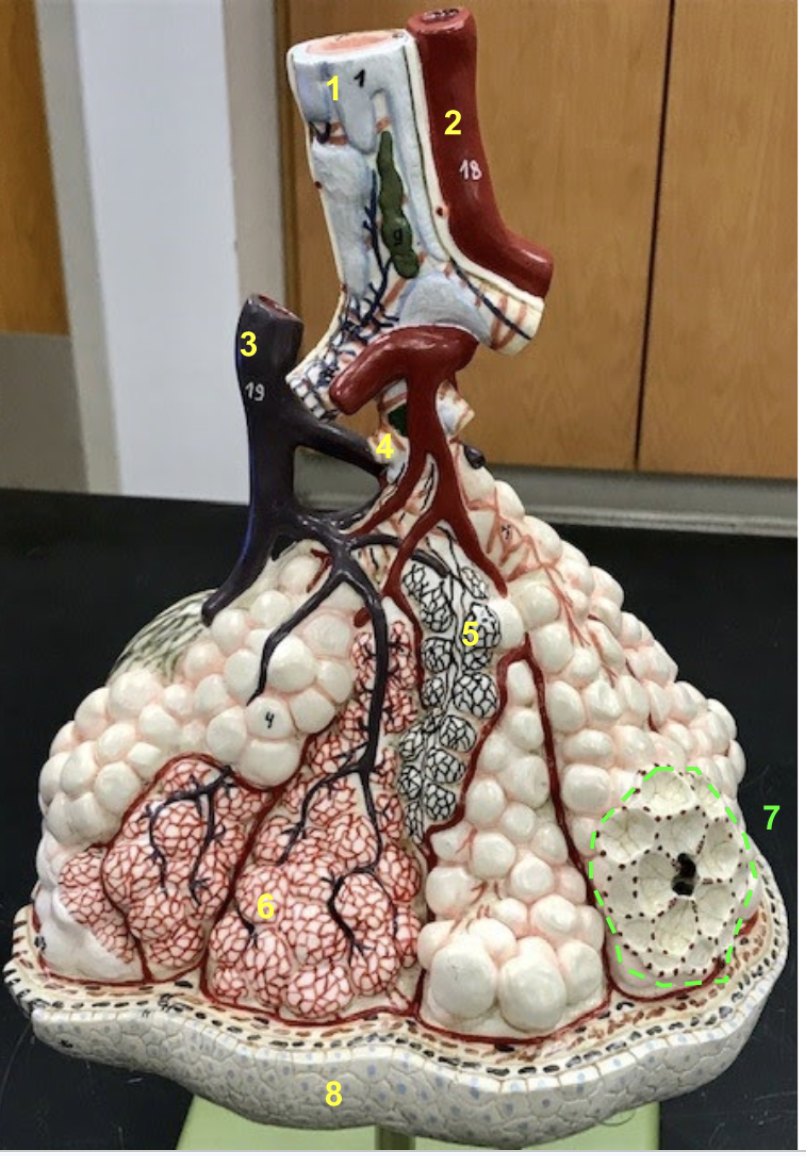
Identify. Relation between 5 & 6. Function of 5.
Remember in the lungs, the colors are switched for arteries and veins.
Small bronchus
Pulmonary vein
Pulmonary artery branch
Terminal bronchiole (the last airway structure w/o alveoli)
Alveoli (notice how it is encased by elastic fibers in an alveolar duct)
Pulmonary capillaries (notice how they sit on top of the alveoli)
Alveolar sac (cluster of alveoli).
Visceral pleura
- Alveoli is the site of diffusion. Gas exchange occurs between alveolar air and pulmonary capillary blood.

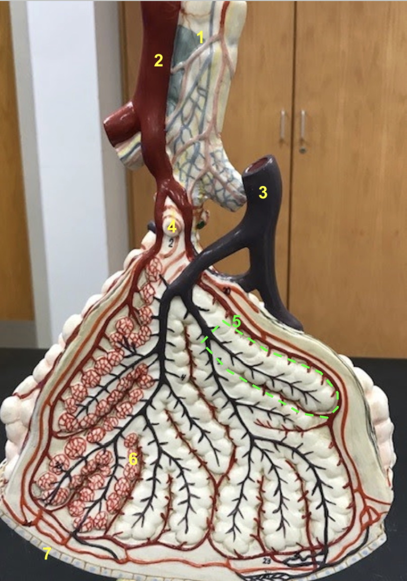
Identify
1. Intrapulmonary bronchus
2. Pulmonary vein branch
3. Pulmonary artery branch
4. Terminal bronchiole
5. Alveolar duct
6. Alveolus with pulmonary capillaries
7. Visceral pleura
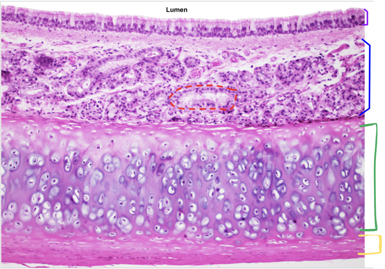
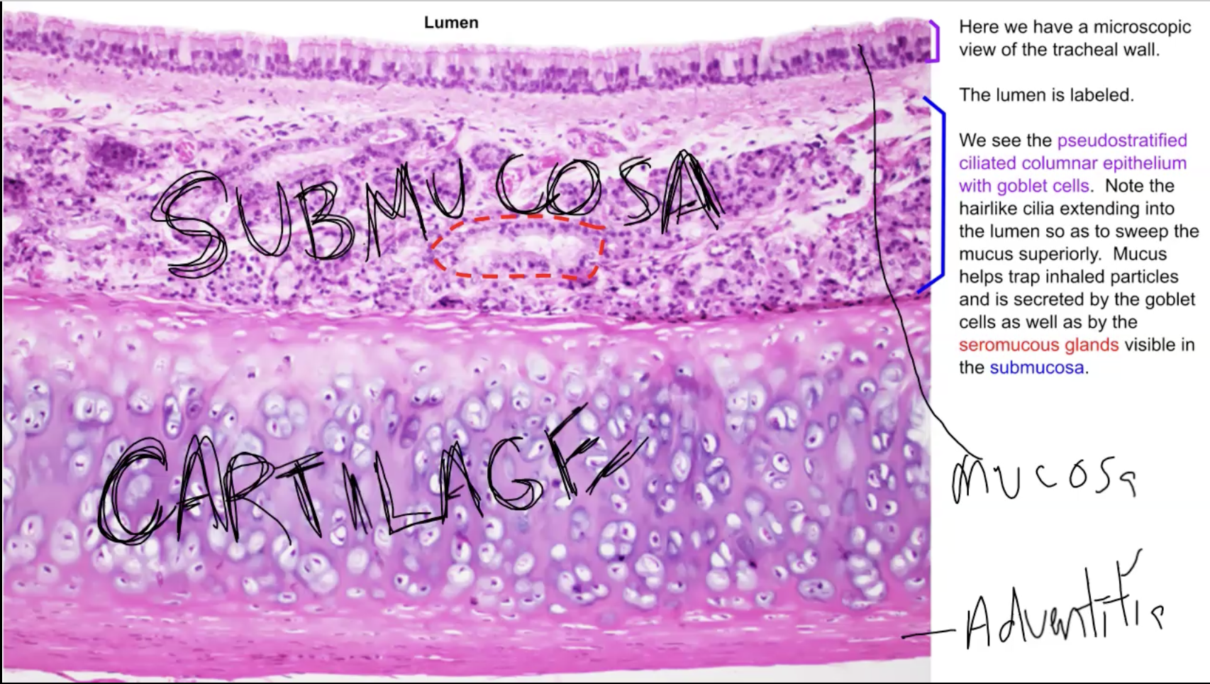
This image represents what organ? Identify each label
Function of green and yellow?
What is the
Red: Seromucous glands
Purple: Mucosa (made of Pseudostratified ciliated columnar epithelium with goblet cells)
Blue: Submucosa (contains various things like nerves, arteries, veins, glands, etc)
Green: Cartilage - strengthens the trachea and keeps it open.
Yellow: Adventitia. The adventitia is sticky which keeps the trachea in place and allows it to stick to neighboring structures.

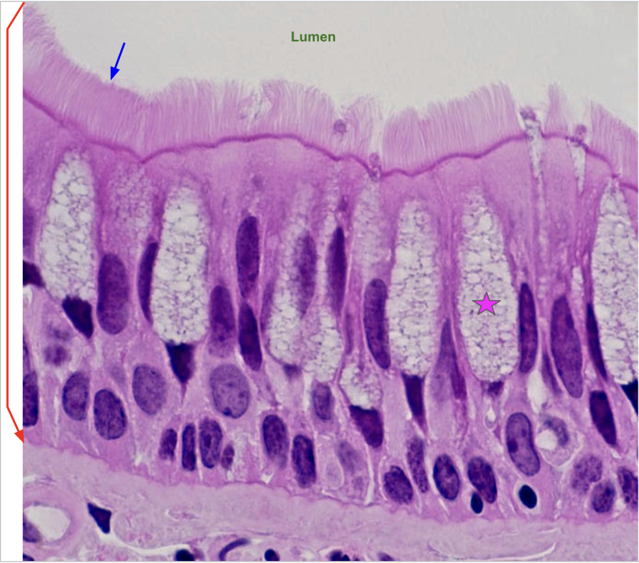
What is this image? Identify labels. Function of star?
The mucosa of the trachea
Red: Pseudostratified ciliated columnar epithelium.
Blue: Cilia
Star: Goblet cells - make mucus
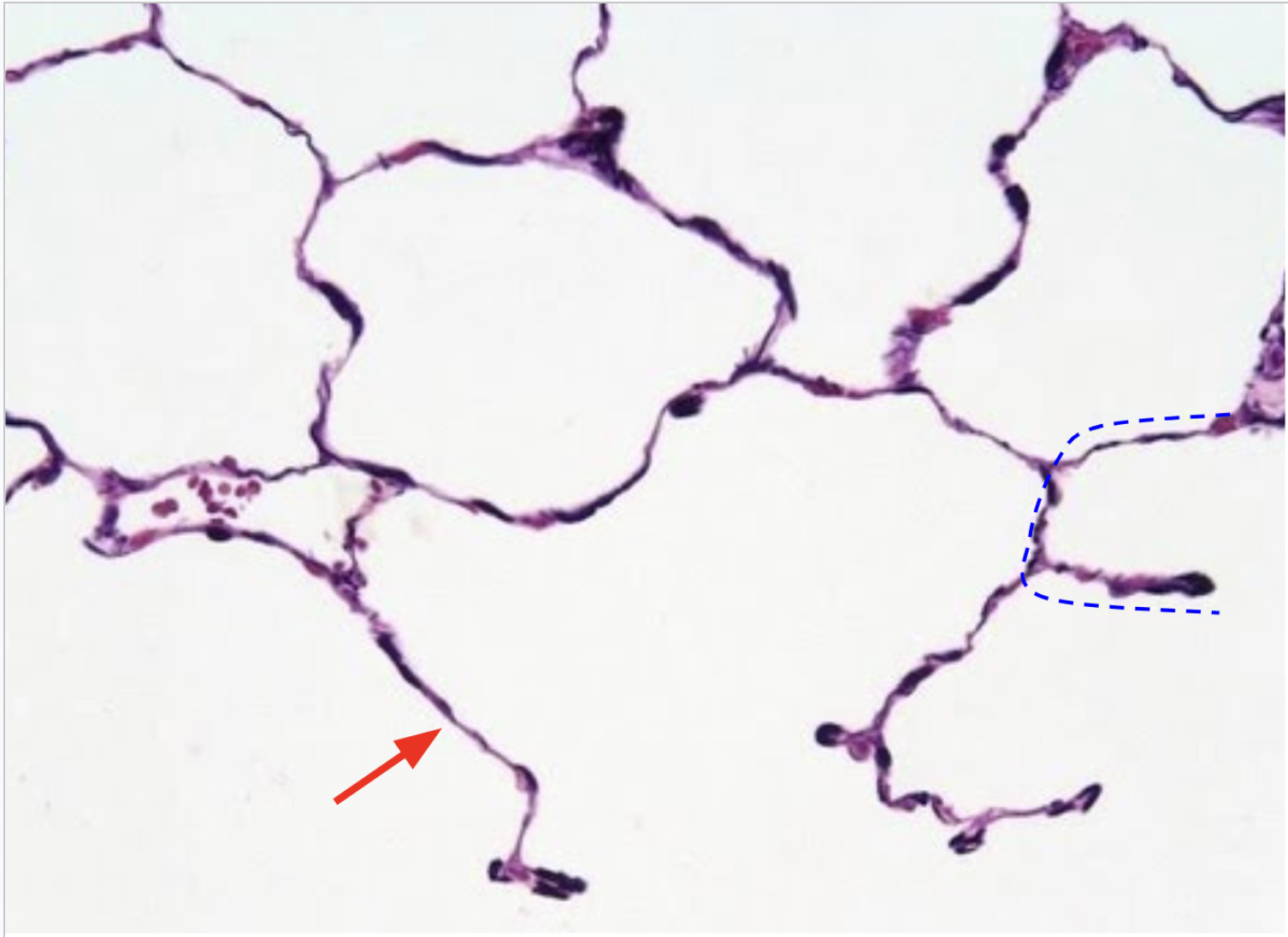
This image depicts what? Identify labels. Function?
Aveoli in the lungs
Red: Simple squamous epithelium
Blue: Aveolus - function is diffusion

Smoking can cause an increase in mucus production and a decrease in cilia function. Explain why this combination can be so detrimental to one’s health?
Without cilia, it becomes difficult to move the excess mucus superiorly to the pharynx where it can be swallowed. Pathogens can build up in the mucus and chronic infections may result.
Why is the epithelium lining the nasopharynx different from the epithelium that lines the oropharynx and laryngopharynx?
Bc the nasopharynx is not a passageway for food/drink and thus lacks the need for the protection provided by the stratified squamous epithelium.
Nasopharynx – pseudostratified ciliated columnar epithelium (only one layer, respiratory epithelium)
Oropharynx & Laryngopharynx – stratified squamous epithelium (several layers)
What two structures are involved in routing food and liquid away from the respiratory passages?
Soft palate/uvula and the epiglottis
If an individual was born with O-shaped tracheal cartilage, what activity would be difficult?
Swallowing because the esophagus would be unable to expand anteriorly when eating large food.
Lois has an obstruction of her right main bronchus. How would this affect the carbon dioxide levels in her right pulmonary veins?
It would increase. This is bc less oxygen will be able to reach the blood in that lung, so more CO2 will buildup.