Chapter 4 - Functional Anatomy of Prokaryotic & Eukaryotic cells
1/57
There's no tags or description
Looks like no tags are added yet.
Name | Mastery | Learn | Test | Matching | Spaced |
|---|
No study sessions yet.
58 Terms
Shape and arrangement of Bacterial cells
Prokaryotic & Eukaryotic Cells
Prokarote comes from the Greek words for pre-nculeus.
Eukaryote comes from the Greek words for true nucleus
Prokaryote
Pre-nucleus
One circular chromosome, not in a membrane
No organelles
Peptidoglycan cell walls
Binary fission
Eukaryote
True nucleus
Paired chroosomes, in nuclear membrane
several organnelles
Simple cell walls
Mitosis, Meiosis
It is primarily the structure of cell walls and ribosomes, and the absence of organelles (specialized cellular structures that have specific functions), that distinguish prokaryotes from eukaryotes. The chief distinguishing characteristics of prokaryotes (from the Greek words meaning prenucleus) are as follows:
1. Typically their DNA is not enclosed within a membrane and is usually a singular, circularly arranged chromosome. Gemma obscuriglobus has a double membrane around its nucleus. (Some bacteria, such as Vibrio cholerae, have two chromosomes, and some bacteria have a linearly arranged chromosome.)
2. Their DNA is not associated with histones (special chromosomal proteins found in eukaryotes); other proteins are associated with the DNA.
3. They generally lack organelles. Advances in microscopy reveal a few membrane-enclosed organelles (for example, some inclusions). However, prokaryotes lack other membrane-enclosed organelles such as nuclei, mitochondria, and chloroplasts.
4. Their cell walls almost always contain the complex polysaccharide peptidoglycan.
5. They usually divide by binary fission, where DNA is copied, and the cell splits into two cells. This involves fewer structures and processes than eukaryotic cell division
Eukaryotes (from the Greek words meaning true nucleus) have the following distinguishing characteristics:
Their DNA is found in the cell’s nucleus, which is separated from the cytoplasm by a nuclear membrane, and the DNA is found in multiple chromosomes.
. Their DNA is consistently associated with chromosomal proteins called histones and with nonhistones.
They have a number of membrane-enclosed organelles, including mitochondria, endoplasmic reticulum, Golgi complex, lysosomes, and sometimes chloroplasts.
Their cell walls, when present, are chemically simple.
. Cell division usually involves mitosis, in which chromosomes replicate and an identical set is distributed into each of two nuclei. Division of the cytoplasm and other organelles follows so that the two cells produced are identical to each other.
The Prokaryotes cell - The size, shape, and Arrangement of Bacterial Cells
Prokaryotes make up a vast group of very small unicellular organisms that include bacteria and archaea. The majority are bacteria. Although bacteria and archaea look similar, their chemical composition is different
Most bacteria range from 0.2 to 2.0 µm in diameter and from 2 to 8 µm in length. They may be spherical-shaped coccus (plural: cocci, meaning berries), rod-shaped bacillus (plural: bacilli, meaning little rods or walking sticks), and spiral
Shapes and Arrangements of Bacterial Cells
3 Basic Shapes
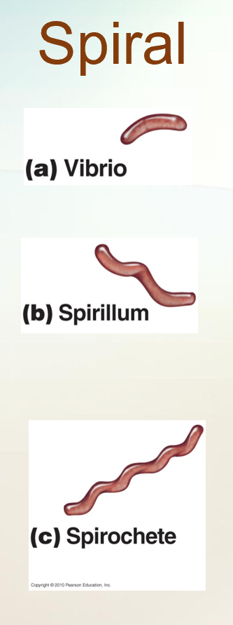
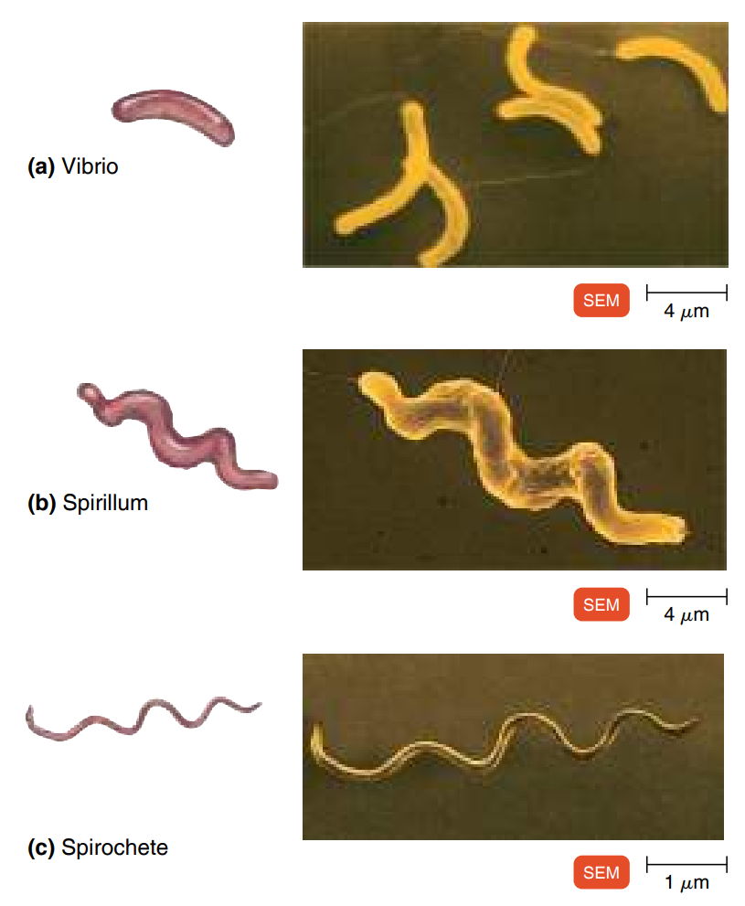
Spiral
Vibrio: Slight curve (like a kidney).
Example: Vibrio cholerae → causes cholera.Spirillum: Rigid spiral; motile via flagella.
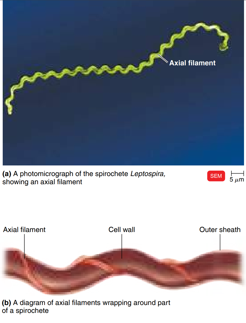
Spirochete: Flexible, corkscrew-like; motile via axial filaments (endoflagella).
Example: Treponema pallidum → causes syphilis.
Spiral bacteria have one or more twists; they are never straight. Bacteria that look like curved rods are called vibrios (Figure 4.4a). Others, called spirilla (singular: spirillum), have a helical shape, like a corkscrew, and fairly rigid bodies (Figure 4.4b). Yet another group of spirals are helical and flexible; they are called spirochetes (Figure 4.4c). Unlike the spirilla, which
use propeller-like external appendages called flagella to move, spirochetes move by means of axial filaments, which resemble flagella but are contained within a flexible external sheath. There are also star-shaped and rectangular prokaryotes
1. Vibrio 🫘
Think: a bean or a comma shape (just a little curve).
Example: Vibrio cholerae → cholera.
2. Spirillum 🌀
Think: a spring or corkscrew, but stiff and rigid.
It doesn’t bend much.
Moves with external flagella (tiny tails).
3. Spirochete 🧵
Think: a long, thin, curly hair – flexible and wiggly.
Moves by twisting its whole body using axial filaments inside (like wriggling).
Example: Treponema pallidum → syphilis.
👉 Easy memory trick:
Vibrio = a little curve.
Spirillum = stiff corkscrew.
Spirochete = flexible curly hair.
Bacillus or Bacillus (rod-shaped)
- Can be used as a scientific name or to describe a shape
Example: Bacillus anthracis discovered by Robert Koch.
Bacilli = rod-shaped bacteria (like little sticks). They divide across their short axis (side to side).
Here are the main forms:
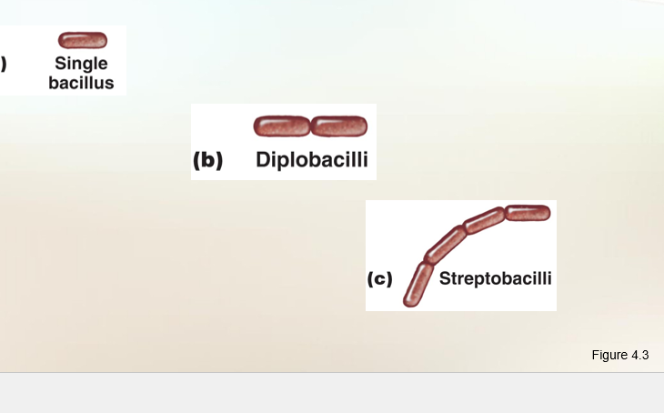
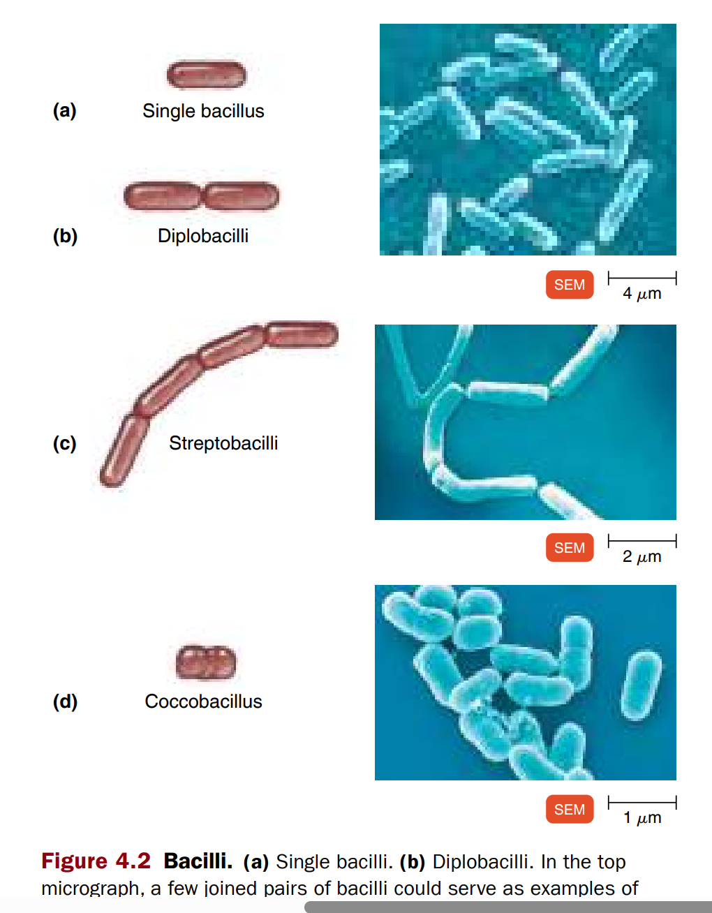
1. Single bacillus
Just one rod.
Shape: like a tiny straw.
2. Diplobacilli
After division, two rods stay stuck together → looks like a pair of straws side by side.
3. Streptobacilli
They form a chain of rods, like a string of sausages or train cars.
4. Coccobacilli
Short and fat rods → look kind of like round cocci (spheres).
Think: between a rod and a ball.
👉 Easy picture in your head:
Single = one straw
Diplo = two straws stuck
Strepto = chain of straws
Coccobacilli = oval/ball-like rod
Single bacillus → one rod.
Diplobacilli → pairs.
Streptobacilli → chains.
Coccobacillus → short, fat rods that look like cocci.
Side note: Some look like straws, some like cigars.
Only a few cause disease; others are not clinically important
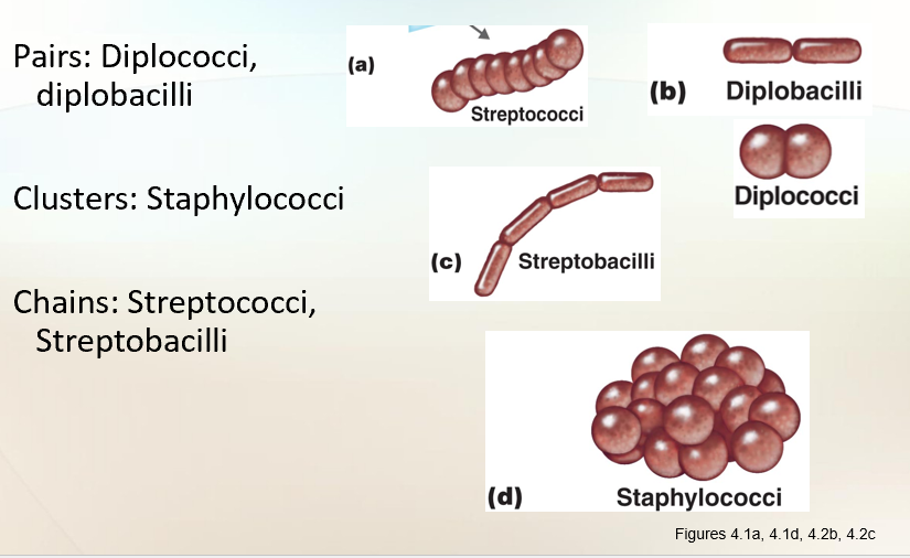
Coccus (spherical)
Coccus (spherical)
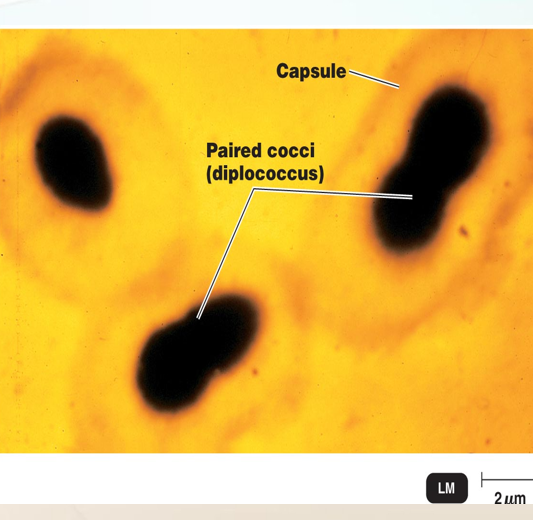
Diplococcus: pairs → Neisseria gonorrhoeae.
Streptococcus: chains → Streptococcus pyogenes → strep throat, scarlet fever, rheumatic fever.
Staphylococcus: grape-like clusters → causes skin infections, TSS, and food poisoning.
Other Shapes
Coccobacillus: between rod and sphere (short, plump).
Pleomorphic: variable shapes.
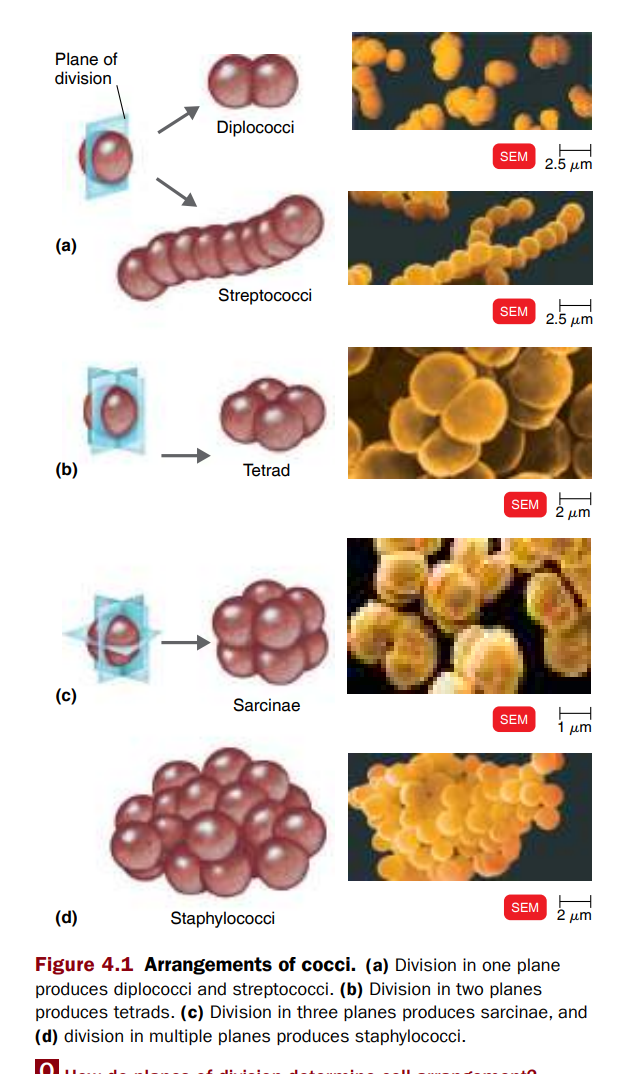
Cocci = round (ball-shaped) bacteria. After they divide, they can stay stuck together in different patterns:
1. Diplococci 🟢🟢
Pairs of two (like two marbles side by side).
2. Streptococci 🟢🟢🟢🟢🟢
Chains (like a bead necklace).
Example: Streptococcus pyogenes (strep throat).
3. Tetrads
4 cocci in a square (like four corners of a box 🎲).
4. Sarcinae
Cube of 8 cocci (like dice 🎲).
Formed when they divide in 3 planes.
5. Staphylococci
Clusters (like grapes 🍇).
Example: Staphylococcus aureus (staph infections).
👉 Easy picture in your head:
Diplo = 2 balls
Strepto = chain of balls
Tetrad = 4 balls (square)
Sarcinae = cube of 8 balls
Staphylo = bunch of grapes
c. Coccus (round, berry-like)
Diplococcus → pairs. Example: Neisseria gonorrhoeae.
Streptococcus → chains. Example: Streptococcus pyogenes.
Diseases: strep throat, scarlet fever, rheumatic fever (can affect the heart).
Staphylococcus → clusters like grapes. Example: Staphylococcus aureus.
Causes skin infections, TSS (toxic shock syndrome from tampons), and food poisoning.
Side note: Food poisoning can be immediate (toxin) or delayed (infection).
Other Forms
Coccobacillus → between rod and coccus (chubbier).
Pleomorphic → variable shapes and arrangements.
Structures External to the Cell Wall
Learning Objectives
Describe the structure and function of the glycocalyx.
Glycocalyx
Many prokaryotes secrete on their surface a substance called glycocalyx. Glycocalyx (meaning sugar coat) is the general term used for substances that surround cells
Outside cell wall
Usually sticky
Increases virulence
Allows bacterial cells to attach
Capsules prevent phagocytosis
Glycocalyx (Sugar Coat)
Many bacteria secrete a sticky, jelly-like coat outside their cell wall → glycocalyx.
It is made of sugars (polysaccharides), proteins, or both.
Built inside the cell → secreted to the outside.
Types of Glycocalyx
Capsule → organized, firmly attached.
Makes bacteria more dangerous (virulence).
Protects bacteria from being eaten (phagocytosis).
Examples:
Bacillus anthracis → capsule of D-glutamic acid (causes anthrax).
Streptococcus pneumoniae → capsule needed to cause pneumonia.
Klebsiella → capsule prevents immune attack in the lungs.
Slime layer → unorganized, loosely attached.
Helps bacteria stick to surfaces.
Functions of Glycocalyx
Virulence → makes pathogens harder to kill.
Prevents phagocytosis (immune cells can’t eat them).
Helps attachment → bacteria stick to teeth, intestines, water pipes, rocks, etc.
Forms biofilms → community of bacteria sticking together.
Biofilm glycocalyx = EPS (extracellular polymeric substance).
EPS protects, helps communication, and makes bacteria harder to remove.
Nutrition → some bacteria (e.g., Streptococcus mutans) break down their capsule for sugar when food is low.
Protection → against dehydration (drying out).
Attachment in disease:
S. mutans → sticks to teeth → dental caries (cavities).
Vibrio cholerae → attaches to intestine cells → cholera.
Quick Memory Tips
Capsule = strong shield (serious infections).
Slime layer = sticky glue (helps bacteria stick).
EPS = biofilm slime (community protection).
Examples to remember:
Anthrax (capsule = deadly).
Pneumonia (S. pneumoniae needs capsule).
Tooth decay (S. mutans).
Cholera (V. cholerae uses glycocalyx to stick to intestine).
👉 So, glycocalyx is basically a sticky sugar/protein coat that:
Protects bacteria,
Helps them stick to places,
Makes them harder to kill,
And can even feed them when food is low.
Polysaccharide, or polysaccharide + protein → forms a sticky “sugar cup.”
Teacher’s image: “like putting sugar around the cell to make it pretty.”
Functions (important for patients):
Virulence factor: makes bacteria more dangerous.
Example: Streptococcus pneumoniae → capsule needed for causing pneumonia.
Vaccines target capsule; without capsule → not virulent.
Attachment: helps bacteria stick to surfaces.
Example: Streptococcus mutans → tooth decay.
Prevent phagocytosis → capsule blocks immune cells from “eating” bacteria.
The glycocalyx itself isn’t “good” or “bad” — it’s just a coat that bacteria make.👉 But here’s the trick:
When harmless bacteria have a glycocalyx, it just helps them stick to rocks, water, or soil. That’s not bad.
When disease-causing bacteria (pathogens) have a glycocalyx, it makes them much harder for your body to fight → that is bad.
Examples:
Streptococcus mutans → glycocalyx helps it stick to teeth → cavities.
Vibrio cholerae → glycocalyx helps it stick to the intestine → cholera.
Streptococcus pneumoniae → only causes pneumonia if it has a capsule. Without it, your body kills it easily.
Bacillus anthracis → capsule makes anthrax very deadly.
⚖ So:
Not always bad, but in pathogens, glycocalyx makes them extra dangerous.
If the substance is organized and is firmly attacthed to the cell wall —→ the glycocalyx is described as a capsule. attached. The presence of a capsule can be determined by using negative staining.
If the substance is unorganized and only loosely attached to the cell wall, the glycocalyx is described as a slime layer.
In certain species, capsules are important in contributing to bacterial virulence (the degree to which a pathogen causes disease). Capsules often protect pathogenic bacteria from phagocytosis (a cellular process, often described as "cell eating," where a cell engulfs and digests large particles like bacteria, dead cells, or foreign substances, typically to protect the body from harm or to maintain tissue health) by the cells of the host.
For example,
Bacillus anthracis produces a capsule of d-glutamic acid. ) Because only encapsulated B. anthracis causes anthrax, it is speculated that the capsule may prevent it from being destroyed by phagocytosis.
Streptococcus pneumoniae which causes pneumonia I only when the cells are protected by a polysaccharide capsule. Unencapsulated S. pneumoniae cells cannot cause pneumonia and are readily phagocytized. The polysaccharide capsule of Klebsiella also prevents phagocytosis and allows the bacterium to adhere to and colonize the respiratory tract.
A glycocalyx that helps cells in a biofilm attach to their target environment and to each other is called an extracellular polymeric substance (EPS). The EPS protects the cells within it, facilitates communication among them, and enables the cells to survive by attaching to various surfaces in their natural environment.
Bacteria can stick to many surfaces using their glycocalyx (sugar coat).
They can grow on:
Rocks in rivers
Plant roots
Human teeth
Medical implants
Water pipes
Even on other bacteria
🦷 Example: Streptococcus mutans
Lives in the mouth.
Uses its glycocalyx to stick to teeth → starts forming plaque.
Plaque → leads to dental caries (cavities).
If it runs out of food, it can even eat its own glycocalyx (because it’s made of sugar!) to survive.
Vibrio cholerae, the cause of cholera, produces a I glycocalyx that helps it attach to the cells of the small intestine.
A glycocalyx also can protect a cell against dehydration, and its viscosity may inhibit the movement of nutrients out of the cell.
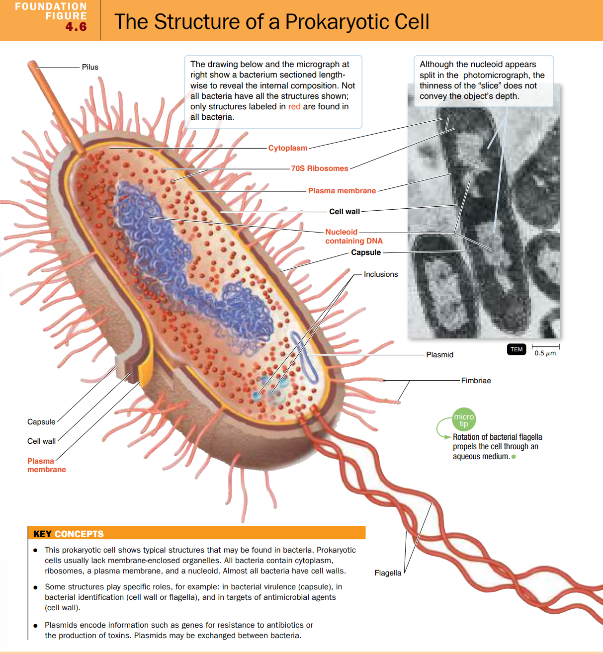
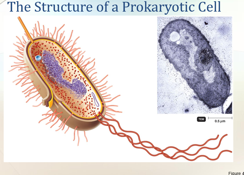
The structure of a Prokaryotic cell
KEY CONCEPTS
This prokaryotic cell shows typical structures that may be found in bacteria. Prokaryotic cells usually lack membrane-enclosed organelles. All bacteria contain cytoplasm, ribosomes, a plasma membrane, and a nucleoid. Almost all bacteria have cell walls.
Plasmids encode information such as genes for resistance to antibiotics or the production of toxins. Plasmids may be exchanged between bacteria.
Some structures play specific roles, for example: in bacterial virulence (capsule), in bacterial identification (cell wall or flagella), and in targets of antimicrobial agents (cell wall).

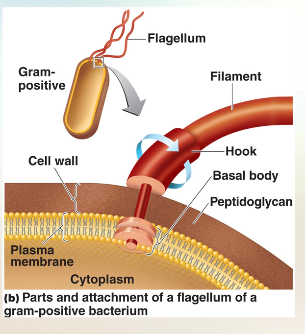
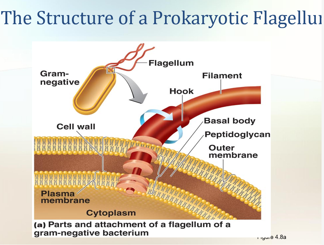
Flagella
Filament made up of chains of flagellin
Attached to a protein hook
Anchored to the wall and membrane by the basal body
Flagella = tails that help bacteria move
Some bacteria have long, whip-like tails called flagella (singular = flagellum).
Bacteria that don’t have them are called atrichous (no projections).\
Parts of a flagellum
Filament → the long tail, made of protein (flagellin).
Twisted like a spiral spring (helix).
Hollow inside.
Hook → connects the filament to the base, like a joint.
Basal body → anchors the flagellum into the cell wall & membrane, and makes it spin.
Basal body differences
Gram-negative bacteria → 2 pairs of rings:
Outer pair = attached to the cell wall.
Inner pair = attached to the plasma membrane.
Gram-positive bacteria → only 1 pair of rings (no outer membrane).
👉 Super simple:
Flagella = propellers(a rounded blade that rotates in a circle, helping to move a vehicle by pushing against water or air.) that spin and move bacteria.
Arrangements = where/how many propellers are attached.
3 parts = filament (tail), hook (joint), basal body (motor).
How bacteria move with flagella
Flagella spin like a propeller (clockwise or counterclockwise).
This spinning pushes the bacterium forward, like a tiny motor.
Needs energy to keep spinning.
🏃 Types of movement
Run (swim) → straight movement in one direction.
Tumble → sudden spin reversal → bacteria change direction randomly.
Movement is Run → Tumble → Run → Tumble (like stop and go).
👉 Some bacteria (like Proteus) have lots of flagella and can swarm → fast, wave-like spreading on surfaces.
🎯 Taxis = movement toward/away from something
Bacteria can sense signals and change movement.
Chemotaxis → move toward/away from chemicals (like oxygen, sugars).
Phototaxis → move toward/away from light.
How it works:
Bacteria have receptors on their surface → detect things in the environment.
If it’s something good (attractant, like food) → more runs, fewer tumbles.
If it’s something bad (repellent, like poison) → more tumbles, fewer runs → they escape.
🧩 Extra facts
The protein in flagella is called H antigen.
Used to tell apart different strains (serovars).
Example: E. coli O157:H7 (dangerous strain in food poisoning).
Side note: When staining flagella, you stain the filament.
Motile Cells
A bacterium running and tumbling. Notice that the direction of flagellar rotation (blue arrows) determines which of these movements occurs. Gray arrows indicate the direction of movement of the microbe.
Bacterial cells can alter the speed and direction of rotation of flagella and thus are capable of various patterns of motility, the ability of an organism to move by itself. When a bacterium moves in one direction for a length of time, the movement is called a “run” or “swim.” “Runs” are interrupted by periodic, abrupt, random changes in direction called “tumbles.” Then, a “run” resumes. “Tumbles” are caused by a reversal of flagellar rotation
One advantage of motility is that it enables a bacterium to move toward a favorable environment or away from an adverse one.
The movement of a bacterium toward or away from a particular stimulus is called taxis. Such stimuli include chemicals (chemotaxis) and light (phototaxis). Motile bacteria contain receptors in various locations, such as in or just under the cell wall. These receptors pick up chemical stimuli, such as oxygen, ribose, and galactose. In response to the stimuli, information is passed to the flagella. If the chemotactic signal is positive, called an attractant, the bacteria move toward the stimulus with many runs and few tumbles. If the chemotactic signal is negative, called a repellent, the frequency of tumbles increases as the bacteria move away from the stimulus
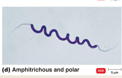
⚡ Arrangements of Bacterial flagella
Peritrichous → flagella all over the cell.
Arrangements of Bacterial Falgella
Polar → flagella only at the ends.
Monotrichous → 1 flagellum at one end.
Arrangements of Bacterial Falgella
Lophotrichous → a tuft (bunch) of flagella at one end.
Arrangements of Bacterial Falgella
Amphitrichous → one or more flagella at both ends.
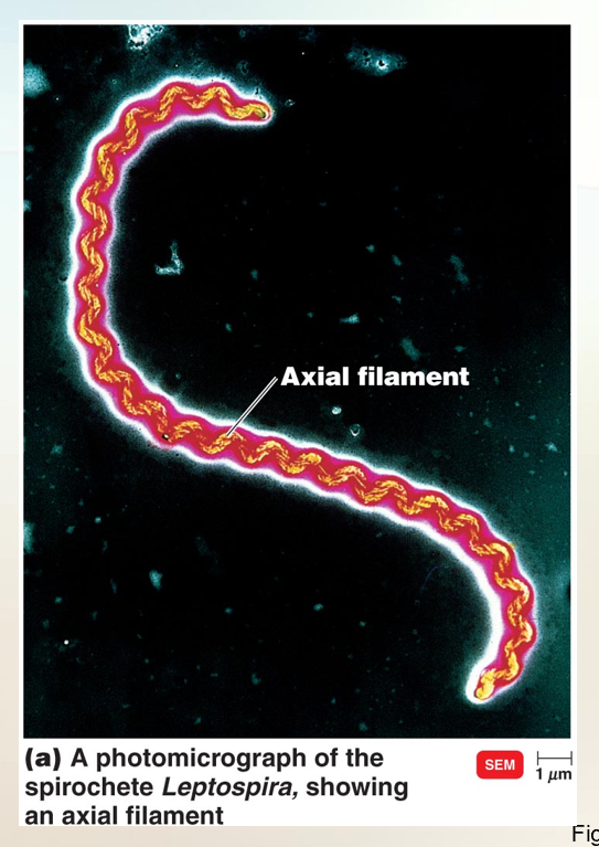
Axial Filaments (Endoflagella)
Also called endoflagella
Found in spirochetes
Anchored at one end of a cell
Propels the bacteria in a spiral motion
Found inside spirochetes, between cell wall and outer membrane.
Covered by sheath → protected.
Provides strong corkscrew movement.
Side note: Impossible to stain because of the protective covering.
🌀 Axial Filaments (Endoflagella)
Found in spirochetes (a special group of spiral bacteria).
Examples:
Treponema pallidum → causes syphilis.
Borrelia burgdorferi → causes Lyme disease.
🔧 Structure
Axial filaments are inside the cell wall, under the outer sheath.
They are bundles of tiny fibers (fibrils) anchored at each end.
They twist around the bacterium.
🚀 Movement
When the axial filaments rotate → the whole bacterium twists like a corkscrew (spiral drill).
This corkscrew motion helps spirochetes move through thick fluids (like mucus or tissue).
👉 Super simple:
Flagella = outside tails (like a propeller).
Axial filaments = hidden tails inside that make the bacteria twist like a corkscrew.
This helps diseases like syphilis & Lyme disease spread through the body.
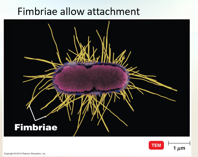
Fimbriae and Pili
Fimbriae allow attachement
Short, thin, hair-like projections.
Used for attachment, not movement.
Example: Neisseria gonorrhoeae → attaches to genital tissue with fimbriae.
Teacher’s image: “like the fuzzy skin of a kiwi.”
Even shorter and thinner than flagella.
Fimbriae and PiliBoth are short, hair-like structures on bacteria.
Made of a protein called pilin.
They are shorter, thinner, and straighter than flagella.
✨ Fimbriae
Many per cell (can be a few to several hundred).
Found at poles or spread all over the cell.
Function → help bacteria stick/attach to each other and to surfaces.
Important for forming biofilms (slimy layers).
Example:
Neisseria gonorrhoeae → uses fimbriae to attach to mucous membranes (causes gonorrhea).
E. coli O157 → uses fimbriae to attach to the intestine → severe watery diarrhea.
Without fimbriae → bacteria cannot attach, colonize, or cause disease.
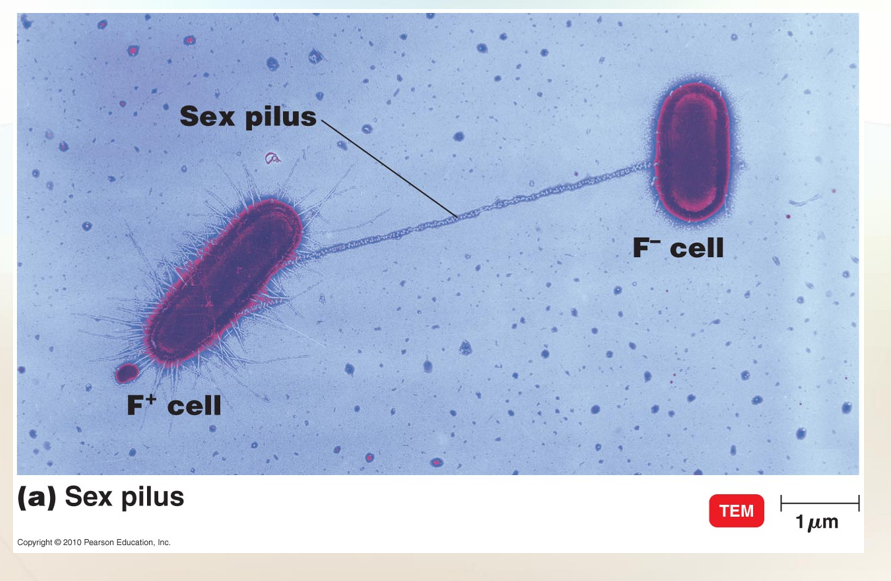
✨ Pili
Usually longer than fimbriae, but fewer in number.
Function → transfer DNA between bacteria (called conjugation).
Special pili used in DNA transfer = sex pili.
👉 Super simple:
Fimbriae = Velcro hairs (for sticking).
Pili = DNA bridge (for sharing genes).
Philli - Facilitate transfer of DNA from one cell to another
Longer than fimbriae but fewer in number.
Form a bridge between two bacteria → transfer DNA.
Known as sex pili.
Example: E. coli.
Pili (singular: Pilus)Longer than fimbriae.
Only 1–2 per cell (not hundreds like fimbriae).
Two main jobs: motility (movement) and DNA transfer.
🚀 1. Pili and Motility a) Twitching motility
Works like a grappling hook 🪝.
Pilus extends and grabs onto a surface/another cell.
It retracts (pulls back), dragging the bacterium forward.
Movement is jerky and short.
Seen in: Pseudomonas aeruginosa, Neisseria gonorrhoeae, some E. coli.
b) Gliding motility
Smooth sliding movement (not jerky).
Helps bacteria move in low-water places like soil or biofilms.
Example: myxobacteria (some use pili to glide).
🧬 2. Pili and DNA Transfer
Some pili are conjugation pili (sex pili).
Act like a bridge between two bacteria.
DNA moves from donor (F+ cell) → recipient cell.
New DNA can give the recipient:
Antibiotic resistance
Ability to digest new nutrients
Other survival advantages
👉 Super simple:
Fimbriae = Velcro hairs (stick).
Pili = Grappling hooks (move) + Bridges (share DNA).

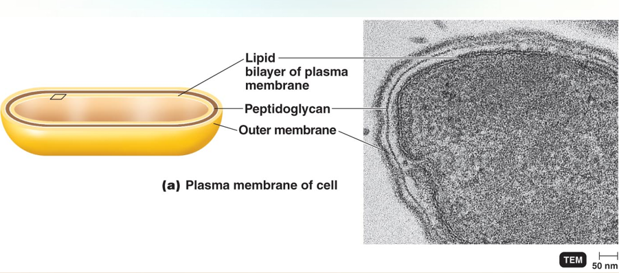
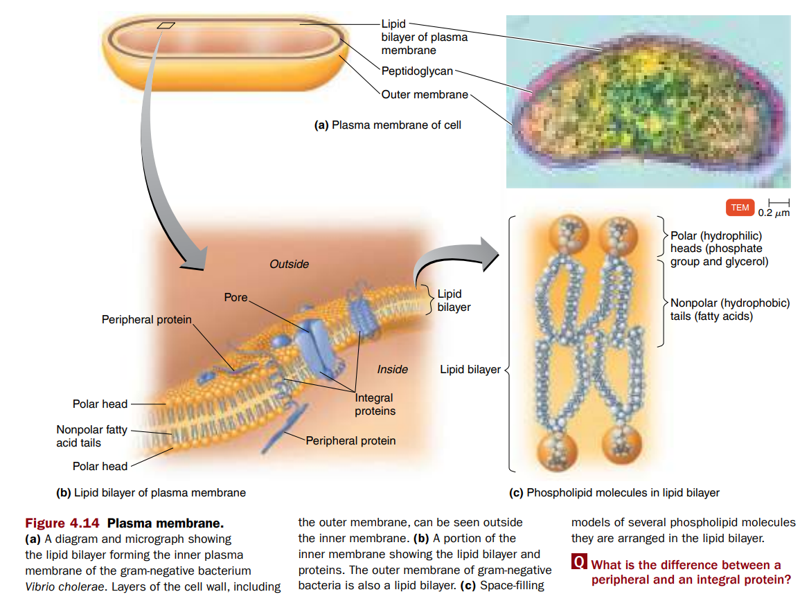
The Cell Wall
Compare and contrast the cell walls of gram-positive bacteria, gram-negative bacteria, acid-fast bacteria, archaea, and mycoplasmas.
Cell Wall
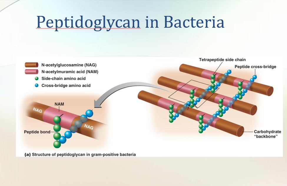
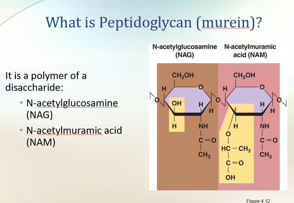
Made of peptidoglycan (PG).
Peptido → peptide (protein cross-links).
Glycan → sugar chains (NAG and NAM).
Bacterial Cell Wall
A tough, semirigid structure around the plasma membrane.
Found in almost all prokaryotes.
🔧 Functions of the Cell Wall
Prevents rupture → keeps the cell from bursting when water pressure inside is higher than outside.
Gives shape → rod, sphere, spiral.
Anchor for flagella → flagella are attached to the wall.
Helps bacteria cause disease → contributes to virulence.
Target for antibiotics → many drugs attack the cell wall.
Used for identification → chemical composition helps distinguish Gram-positive vs Gram-negative bacteria.
⚖ Comparison with Eukaryotic Cell Walls
Some eukaryotes (plants, algae, fungi) also have cell walls.
But eukaryotic walls are:
Chemically different
Simpler
Less rigid than bacterial cell walls.
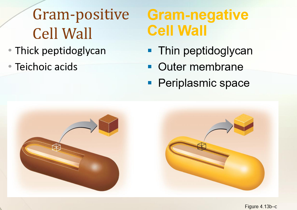
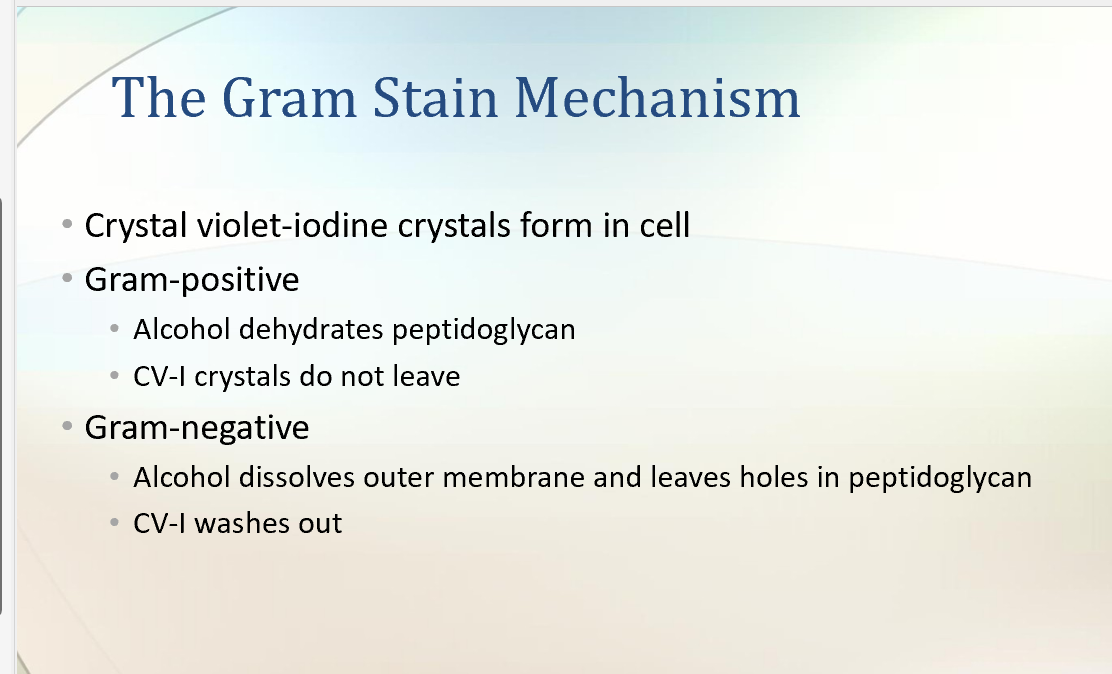
Gram-Positive Cell Wall
Thick layers of PG.
Contains teichoic acid (alcohol + phosphate).
Functions: structural support, cell wall maintenance.
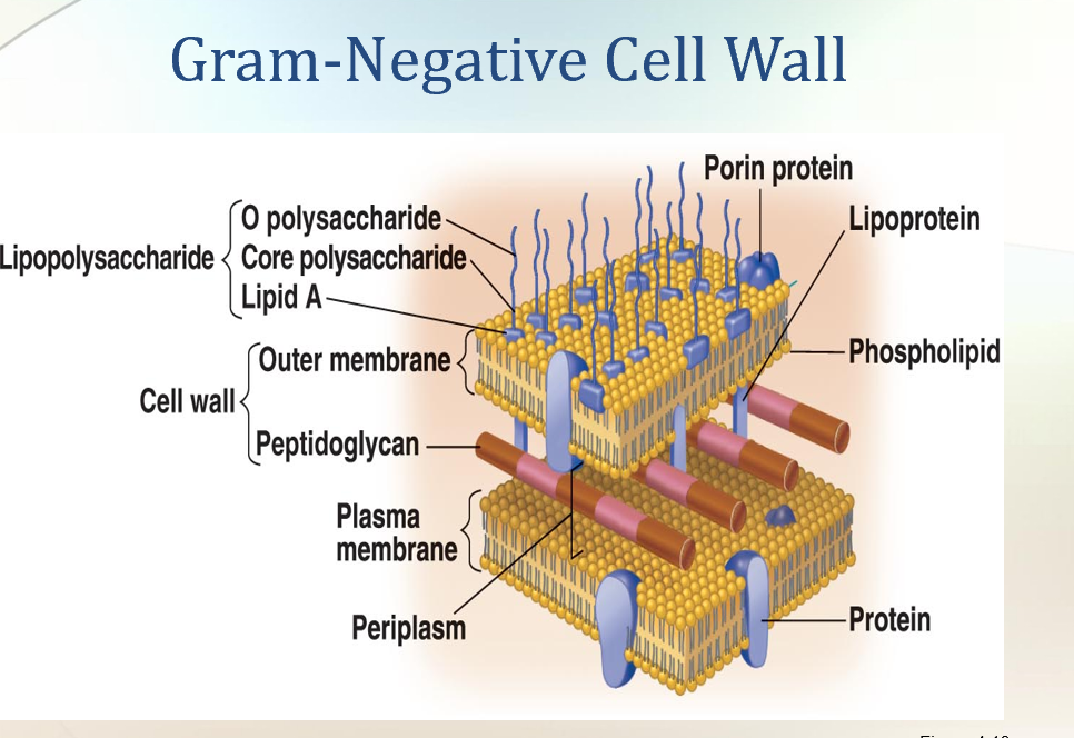
Gram-Negative Cell Wall
Thin PG layer.
Outer membrane present (lipids, proteins, LPS).
More resistant to antibiotics.
+GRam
Gram-Positive Cell Walls
Thick peptidoglycan layer (many layers → rigid wall).
Have teichoic acids:
Lipoteichoic acid → spans PG and attaches to plasma membrane.
Wall teichoic acid → linked only to PG.
Functions of teichoic acids:
Bind/regulate movement of cations (positive ions).
Help in cell growth.
Prevent cell wall breakdown/lysis.
Provide antigenic specificity (used to ID bacteria).
Example: Gram-positive streptococci → wall polysaccharides used for medical typing.
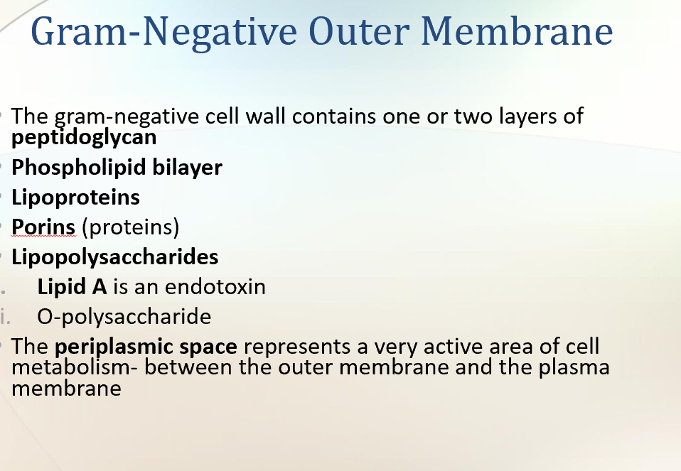
🧱 Gram-Negative Cell Walls
Thin peptidoglycan (only 1–2 layers).
Surrounded by an outer membrane made of:
Lipopolysaccharides (LPS)
Lipoproteins
Phospholipids
Periplasmic space → between plasma membrane & outer membrane.
Contains degradative enzymes & transport proteins.
No teichoic acids.
Because PG is thin → more easily broken mechanically.
🔒 Functions of the Outer Membrane (Gram-negative only)
Protects against:
Phagocytosis (immune attack).
Complement system (immune proteins).
Detergents, heavy metals, bile salts, dyes, antibiotics (like penicillin), and lysozyme.
Still allows nutrients in via porins (channel proteins).
Let in nucleotides, sugars, peptides, amino acids, vitamin B12, iron.
⚠ Lipopolysaccharide (LPS) structure (Gram-negative only)
Lipid A
Embedded in outer membrane.
Released when bacteria die → acts as an endotoxin (causes fever, shock).
Core polysaccharide
Provides structural stability.
O polysaccharide
Outer part; functions as an antigen → helps identify different Gram-negative species.
👉 Super simple comparison:
Feature | Gram-Positive | Gram-Negative |
|---|---|---|
Peptidoglycan | Thick (many layers) | Thin (1–2 layers) |
Teichoic acids | Present (lipoteichoic & wall) | Absent |
Outer membrane | No | Yes (LPS, lipoproteins, phospholipids) |
Periplasm | Small | Large, with enzymes & transport proteins |
Resistance | More sensitive to penicillin | More resistant (outer membrane barrier) |
Toxins | Exotoxins (proteins) | Endotoxin (Lipid A of LPS) |
Example diseases | Strep throat, anthrax | E. coli infections, Salmonella |
Atypical Cell Walls
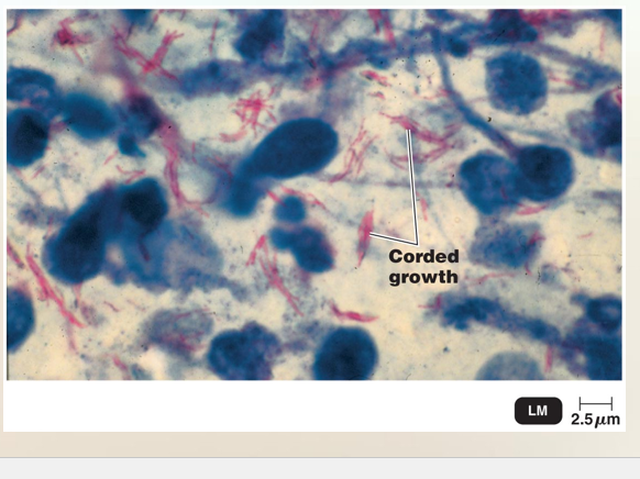
Acid-Fast
Like Gram-positive but with a waxy mycolic acid layer.
Example: Mycobacterium tuberculosis, M. leprae.
Function: protects against drying and chemicals.
Mycoplasma
No cell wall → only plasma membrane.
Example: Mycoplasma pneumoniae.
Exam tip: If no cell wall → only plasma membrane remains.
Archaea
No cell wall, or have pseudo-peptidoglycan.
Atypical Cell WallsSome prokaryotes don’t have normal cell walls.
1⃣ Mycoplasma
Smallest known bacteria that can live and grow without a host.
Have no cell wall → only plasma membrane.
Their membrane has sterols (special lipids) → help protect from rupture (lysis).
Because they have no walls, they can pass through many bacterial filters → originally mistaken for viruses.
2⃣ Archaea
May lack walls or have unusual walls.
Their walls are made of polysaccharides and proteins (NOT peptidoglycan).
Instead, they have pseudomurein:
Similar to peptidoglycan but different chemistry.
Contains N-acetyltalosaminuronic acid (instead of NAM).
Lacks D-amino acids.
Cannot be Gram-stained (no peptidoglycan).
Usually appear Gram-negative under a microscope.
👉 Super simple summary:
Normal bacteria = peptidoglycan walls.
Mycoplasma = no wall, sterols in membrane.
Archaea = no peptidoglycan, instead pseudomurein
Acid-Fast Cell Walls
Found in Mycobacterium (TB, leprosy) and Nocardia.
Contain lots of mycolic acid (a waxy lipid, ~60% of wall).
🛡 Structure
Thin peptidoglycan layer inside.
Covered by a thick, waxy mycolic acid layer outside.
Mycolic acid + PG held together by polysaccharides.
⚡ Properties
Waxy wall = hydrophobic → bacteria stick together in clumps.
Prevents uptake of many dyes (like Gram stain).
Makes bacteria resistant to drying, disinfectants, and some antibiotics.
🎨 Acid-Fast Stain
Uses carbolfuchsin dye (red).
Heat helps dye penetrate the waxy wall.
Even after washing with acid-alcohol, the dye stays (bacteria remain red).
Non–acid-fast bacteria lose the red and turn blue with counterstain.
👉 If you remove the mycolic acid layer, the bacteria would stain Gram-positive (because of the peptidoglycan).
🧩 Super simple summary
Acid-fast bacteria = waxy shield (mycolic acid).
Normal dyes don’t work → need acid-fast stain.
Appear red after staining (because carbolfuchsin resists washing).
Examples: TB (Mycobacterium tuberculosis), leprosy (M. leprae).
Atypical Cell Walls
1) Acid-fast cell walls
waxy lipid (mycolic acid) bound to peptidoglycan
Mycobacterium species
2) Mycoplasma
Lack cell walls
Sterols in plasma membrane
3) Archaea
Wall-less or
Walls of pseudomurein
Damage to the Cell Wall
Antibiotics (e.g., penicillin): break peptide bonds in PG.
More effective against Gram-positive.
Lysozyme (enzyme in tears, saliva, etc.): breaks glycan bonds.
Outcomes:
Gram-positive → protoplast → cell death.
Gram-negative → spheroplast → cell death.
What is Peptidoglycan (murein)?
Structures Internal to the Cell Wall
Plasma membrane → barrier, transport, selective permeability.
Cytoplasm → gel-like fluid with proteins, enzymes, etc.
Nucleoid → region containing bacterial DNA (no nucleus).
Ribosomes → protein synthesis (different from eukaryotes, target for antibiotics
Damage to the Cell Wall
Lysozyme digests disaccharide in peptidoglycan
a. Protoplast: formed is a wall-less gram-postive cell
b. Spheroplast: is formed by the action of lysozyme on a gram-negative cell
Penicillin inhibits peptide bridges in peptidoglycan
How cell walls are damaged
Bacterial cell walls are different from human cells, so many antibiotics can attack them without harming us.
Example: penicillin → blocks cell wall synthesis.
Example: lysozyme (an enzyme in tears, saliva, mucus) → breaks bonds in peptidoglycan.
🧪 Effects of Lysozyme
Gram-positive bacteria: almost completely destroyed by lysozyme.
If only the plasma membrane remains → the cell is called a protoplast (round, still alive if it doesn’t burst).
Gram-negative bacteria: not fully destroyed because the outer membrane is still there.
What’s left is called a spheroplast (plasma membrane + outer membrane + some wall).
To help lysozyme work, EDTA can be added to weaken the outer membrane.
🌀 Special Cases
Some bacteria (like Proteus) can naturally lose their walls → form irregular shapes called L forms.
L forms can grow, divide, or return to normal walled state.
💧 Osmotic Lysis
Protoplasts and spheroplasts can burst in pure water or dilute salt/sugar solutions.
Why? → Water rushes in (osmosis) since the cell has no rigid wall to protect it.
👉 Super simple summary:
Penicillin = stops wall building.
Lysozyme = cuts peptidoglycan (like scissors).
Protoplast = wall-less Gram+.
Spheroplast = partly wall-less Gram–.
L forms = wall-free bacteria (can still live).
Osmotic lysis = cell bursts if water rushes in. \
How Penicillin Works
Penicillin stops bacteria from making peptide cross-bridges in peptidoglycan.
Without cross-bridges → no strong cell wall → bacteria burst and die.
⚖ Gram+ vs Gram– Susceptibility
Gram-positive bacteria → very sensitive to penicillin.
Thick peptidoglycan → penicillin easily attacks.
Gram-negative bacteria → less sensitive.
Outer membrane blocks penicillin.
Also, fewer peptide cross-bridges.
🧪 Other β-lactam antibiotics
Some antibiotics (like ampicillin) can cross the Gram– outer membrane.
These are more effective against Gram-negative bacteria than plain penicillin.
👉 Super simple summary:
Penicillin = breaks wall building.
Gram+ = easy target.
Gram– = harder to kill (outer shield), but special β-lactams can work.
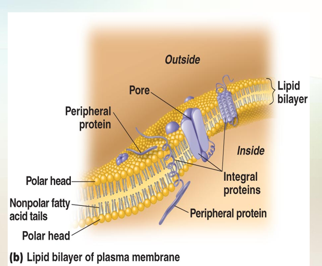
The Plasma Membrane
Has:
Phospholipid bilayer
Peripheral proteins
Integral Proteins
Exhibits: Selective permeability allows passage of some molecules
Movement of Material across Membranes
Materials move across the plasma membranes of both prokaryotic and eukaryotic cells by two kinds of processes: passive and active. In passive processes, substances across the membrane from an area of high concentration to an area of low concentration (move with the concentration gradient, or difference), without any expenditure of energy by the cell. In active processes, the cell must use energy to move substances from areas of low concentration to areas of high concentration (against the concentration gradient).
Passive processes include simple diffusion, facilitated diffusion, and osmosis.
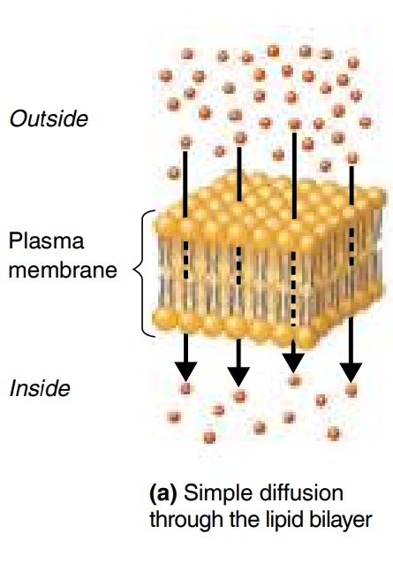
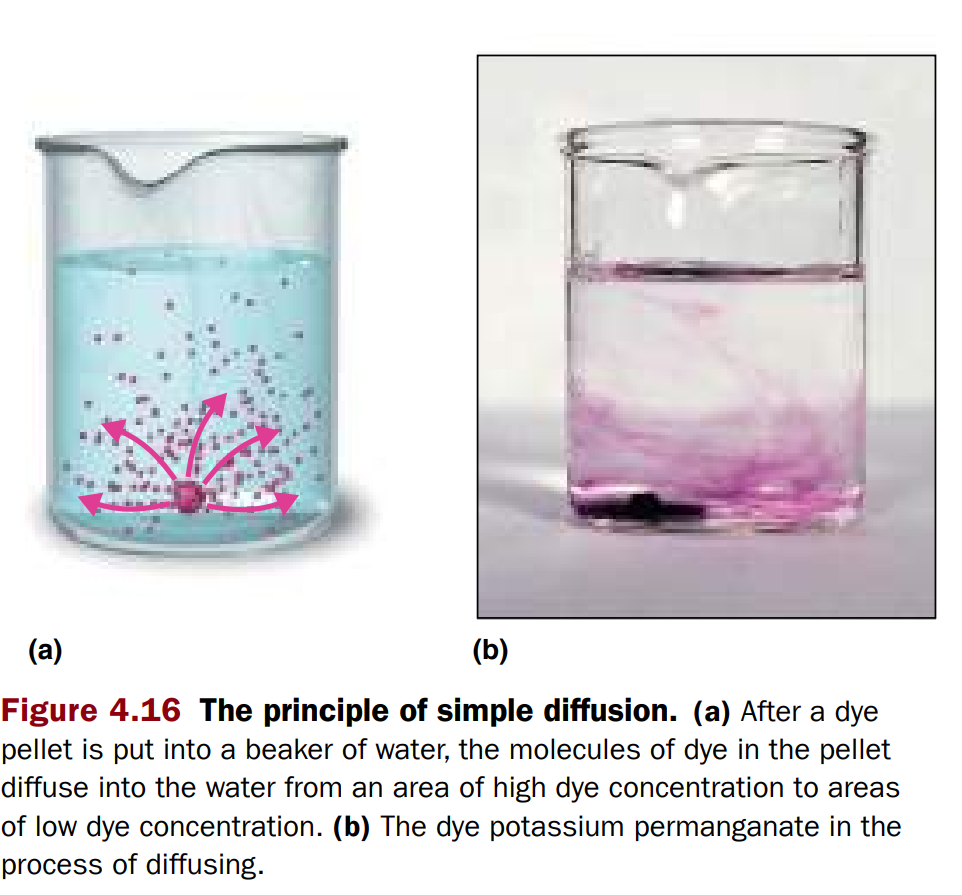
Simple diffusion is the net (overall) movement of molecules or ions from an area of high concentration to an area of low concentration (Figure 4.16 and Figure 4.17a). The movement continues until the molecules or ions are evenly distributed. The point of even distribution is called equilibrium. Cells rely on simple diffusion to transport certain small molecules, such as oxygen and carbon dioxide, across their cell membranes.
Facilitated diffusion: Solute combines with a transporter protein in the membrane
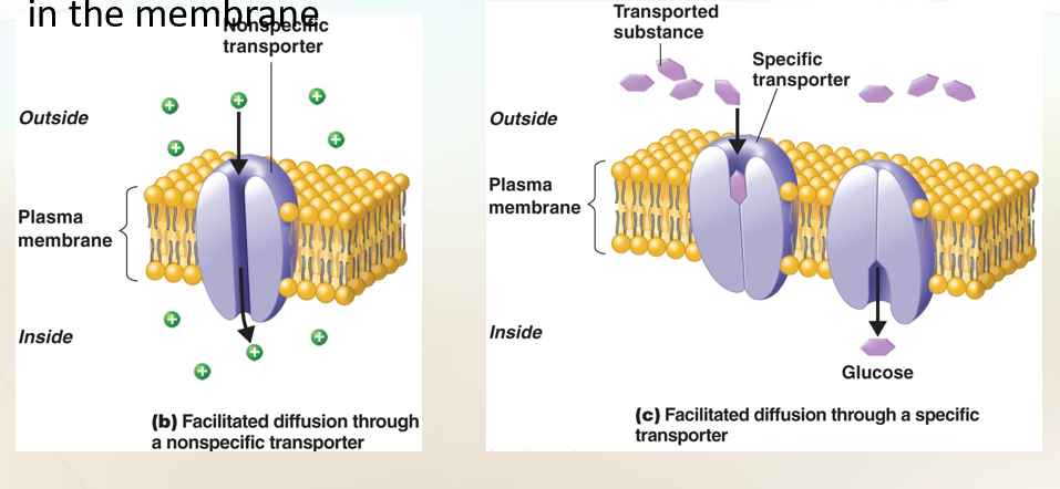
Facilitated diffusion
Facilitated Diffusion
A way substances cross the plasma membrane with the help of transport proteins (channels or carriers).
No energy needed → still goes high → low concentration.
Proteins used: transporter proteins / permeases.
🔹 How it works
Molecule (like sugar or ion) binds to a transporter protein.
Protein changes shape.
Molecule is released on the other side of the membrane.
🔹 Types of Transporters
Prokaryotes (bacteria):
Transporters are often nonspecific → allow many ions or small molecules through (like open channels).
Eukaryotes (our cells):
Transporters are specific → only certain molecules fit (like glucose, fructose, galactose, vitamins).
🔹 Problem: Big Molecules
Some molecules (like proteins, polysaccharides) are too big to enter directly.
Solution: bacteria release extracellular enzymes into the environment.
These enzymes break big molecules into smaller subunits.
Example: proteins → amino acids, polysaccharides → simple sugars.
The smaller subunits are then transported into the cell using transport proteins.
👉 Super simple summary:
Facilitated diffusion = doors/channels in the membrane.
No energy needed, just help for things that can’t cross easily.
Bacteria use enzymes outside the cell to cut big food into small pieces → then bring them in.
Movement of Materials across Membranes
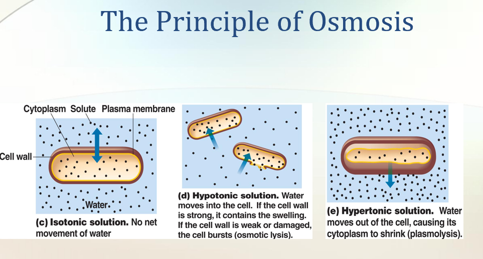
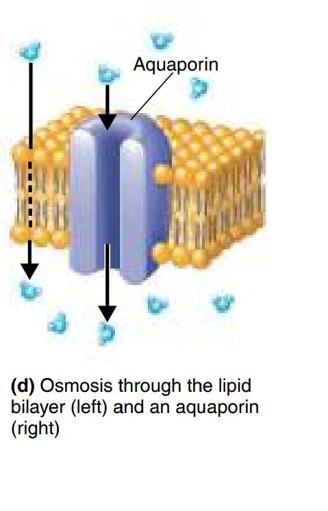
Osmosis: The movement of water across a selectively permeable membrane from an area of high water to an area of lower water concentration.
💧 Osmosis = Water Movement
Water moves across a selectively permeable membrane.
Moves from:
High water concentration (low solutes) → to
Low water concentration (high solutes).
Water can pass:
Directly through lipid bilayer.
Through special protein channels = aquaporins.
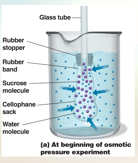
🧪 Experiment Example (Cellophane Sack)
Sack filled with sugar solution, placed in pure water.
Water enters the sack (since water is higher outside, lower inside).
Sugar cannot leave (too big).
Sack swells until pressure pushes water out again.
Balance point = equilibrium.
Pressure created = osmotic pressure.
Osmosis
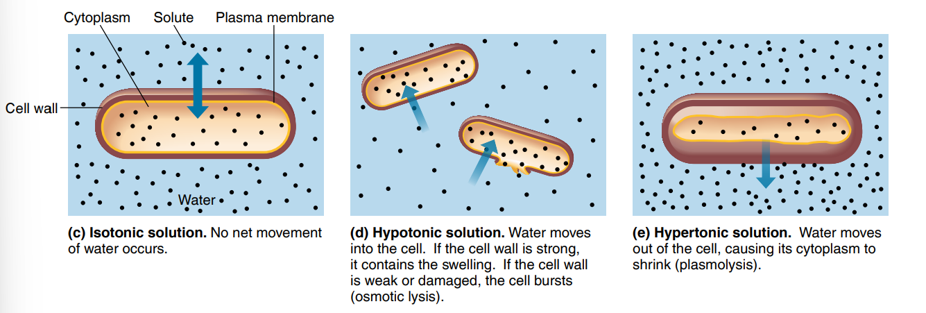
Types of Osmotic Solutions for Cells
Isotonic solution
Solute concentration = same inside & outside.
No net water movement.
Cell stays the same.
Hypotonic solution
Solute lower outside (more water outside).
Water moves into the cell.
Cell swells; if wall weak/damaged → osmotic lysis (bursting).
Hypertonic solution
Solute higher outside (less water outside).
Water moves out of the cell.
Cell shrinks = plasmolysis.
👉 Super simple summary:
Osmosis = water moves to where there’s more solute.
Isotonic = happy cell.
Hypotonic = cell swells (may burst).
Hypertonic = cell shrinks.
Active trasnport
Active Transport
Unlike diffusion (which is passive), active transport uses energy (ATP).
Moves substances against the concentration gradient (low → high).
Needs transporter proteins in the plasma membrane.
Each substance usually has its own transporter (specific).
Substances moved: ions (Na⁺, K⁺, H⁺, Ca²⁺, Cl⁻), amino acids, simple sugars.
🔹 Group Translocation (special to prokaryotes like bacteria)
A form of active transport.
While moving across the membrane, the substance is chemically changed so it cannot leave once inside.
Example:
Glucose enters → gets a phosphate group added → becomes glucose-phosphate.
Trapped inside & can be used in metabolism.
Energy comes from high-energy phosphate compounds (like PEP).
🔹 In Eukaryotes (not bacteria)
Two other active processes exist:
Phagocytosis = "cell eating" (engulfs solids).
Pinocytosis = "cell drinking" (engulfs liquids).
👉 Super simple summary:
Active transport = energy + transporter proteins.
Group translocation = bacteria trick → chemically change substance (like adding phosphate to glucose) so it stays inside.
Eukaryotes can also eat/drink with phagocytosis & pinocytosis.
The plasma (Cytoplasmic) Membrane
Plasma (Cytoplasmic) Membrane
Also called the inner membrane.
Thin layer just inside the cell wall, surrounding the cytoplasm.
🧱 Composition
Made mostly of phospholipids + proteins.
In eukaryotes, membranes also contain:
Carbohydrates
Sterols (like cholesterol) → make them more rigid.
In prokaryotes:
Usually no sterols → less rigid than eukaryotic membranes.
Exception: Mycoplasma (no cell wall) → has sterols in its membrane for strength.
👉 Super simple summary:
Plasma membrane = thin barrier inside wall, holds cytoplasm in.
Prokaryotes = phospholipids + proteins (not very rigid).
Eukaryotes = phospholipids + proteins + carbs + sterols (stronger).
Mycoplasma = special → sterols in membrane (for extra protection).
🧱 Plasma Membrane Structure
Lipid Bilayer
Made of phospholipids in 2 layers.
Each phospholipid has:
Polar head → phosphate + glycerol (water-loving, hydrophilic).
Nonpolar tails → fatty acids (water-fearing, hydrophobic).
Heads face outside/inside (water).
Tails face each other (inside layer, away from water).
👉 Looks like a sandwich: heads = bread, tails = filling.
🔹 Proteins in the Membrane
Peripheral proteins → sit on the surface (inner or outer).
Functions: enzymes, scaffolds, movement helpers, mediators.
Integral proteins → go deep into the membrane.
Harder to remove (need detergents).
If they cross fully → called transmembrane proteins.
Some form channels/pores → let substances in/out.
🔹 Carbohydrates in the Membrane
Glycoproteins = proteins + carbohydrates.
Glycolipids = lipids + carbohydrates.
Functions:
Protect & lubricate the cell.
Help in cell-to-cell recognition (ID tags).
Viruses & toxins use them as “handles” to enter cells.
Example: influenza virus, cholera toxin, botulism toxin bind to glycoproteins.
👉 Super simple summary:
Plasma membrane = phospholipid sandwich + proteins + carbs.
Lipid bilayer = barrier.
Proteins = helpers (enzymes, channels, anchors).
Carbs (glyco-) = protection + recognition (but also used by viruses/toxins).
Fluid Mosaic Model (Plasma Membrane)
The membrane isn’t stiff → it’s fluid.
Phospholipids and proteins can move around, like boats floating in olive oil.
Because fatty acid tails stick together, the bilayer is self-sealing:
If it tears, it can fix itself.
The membrane is about as viscous (thick) as olive oil → flexible but stable.
This movement is important → proteins can shift and do their jobs without breaking the membrane.
🧩 Why “Fluid Mosaic”?
Fluid = lipids and proteins move around.
Mosaic = made of many different parts (phospholipids, proteins, carbohydrates).
👉 Super simple summary:
Cell membrane = like olive oil with floating proteins.
Flexible, self-healing, dynamic.
Called the Fluid Mosaic Model.
⚡ Functions of the Plasma Membrane
. Selective Permeability (semipermeability)
Acts like a gatekeeper → controls what enters and leaves.
Small molecules (water, O₂, CO₂, simple sugars) → pass easily.
Large molecules (proteins, big polysaccharides) → cannot pass.
Nonpolar molecules (lipid-soluble) → pass easily (because membrane = lipids).
Ions (charged) → pass slowly, often need transport proteins.
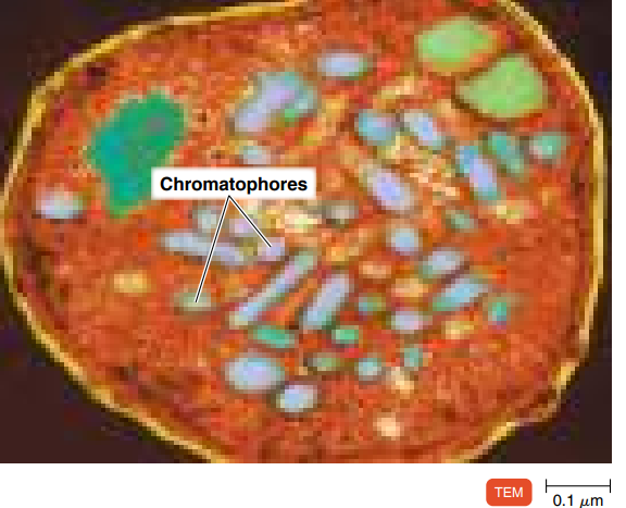
2. Nutrient Breakdown & Energy Production
Contains enzymes for chemical reactions → break down nutrients → make ATP.
In photosynthetic bacteria → infoldings of membrane contain pigments & enzymes.
These folds = chromatophores.
3. Mesosomes (old idea)
Seen in early electron microscopy → thought to be real structures.
Now known to be artifacts from sample prep, not true parts of the cell.
❌ Damage to the Plasma Membrane
Plasma membrane is vital → if destroyed → cell death.
Agents that damage it:
Alcohols
Quaternary ammonium compounds (disinfectants)
Polymyxins (antibiotics) → disrupt phospholipids → leakage of contents → death.
👉 Super simple summary:
Plasma membrane = gate + energy site.
Functions: controls entry/exit, makes energy, supports photosynthesis in some bacteria.
Damaged by alcohol, detergents, or antibiotics (causes cell death).
Cytoplasm
The substance inside the plasma membrane
Cytoplasm
Inside the plasma membrane.
Mostly water (about 80%).
Contains:
Proteins (enzymes)
Carbohydrates, lipids, ions, small compounds
Thick, jelly-like, elastic.
Main parts inside:
Nucleoid (DNA)
Ribosomes
Inclusions (storage deposits).
🧵 Cytoskeleton (in prokaryotes)
Network of tiny rods & fibers inside the cytoplasm.
Used to think bacteria had no cytoskeleton – but they do!
Similar to eukaryotic cytoskeleton.
Components:
MreB, ParM → like microfilaments.
Cresetin → like intermediate filaments.
FtsZ → like microtubules.
Functions:
Helps with cell division.
Maintains cell shape.
Assists in growth, DNA movement, protein targeting, organelle alignment.
👉 Super simple summary:
Cytoplasm = watery soup inside cell (with enzymes, DNA, ribosomes, storage).
Cytoskeleton = hidden scaffolding in bacteria → keeps shape, helps divide, and organizes internal parts.
The Nucleiod
Contains the bacterial chromosme
Nucleoid
Region inside prokaryotic cells that holds DNA.
Contains bacterial chromosome:
One long, circular, double-stranded DNA thread.
Carries all instructions for cell’s structure & function.
Differences from eukaryotes:
No nuclear membrane.
No histones (special proteins in eukaryotic DNA).
Shape: can be round, oval, or dumbbell-like.
In fast-growing bacteria, DNA can take up 20% of cell volume.
DNA attached to plasma membrane.
Helps with replication & separating DNA into new cells during division.
🧪 Plasmids
Small, circular DNA pieces (extra DNA, not part of main chromosome).
Can replicate independently.
Usually 5–100 genes.
Not essential for basic survival but give extra abilities:
Antibiotic resistance
Tolerance to toxic metals
Toxin production
Enzyme synthesis
Plasmids can move from one bacterium to another → spread traits (like antibiotic resistance).
Used in biotechnology (plasmid DNA in gene manipulation).
👉 Super simple summary:
Nucleoid = main instruction book (chromosome).
Plasmids = extra mini-booklets with bonus tricks (like resistance).
Ribosomes
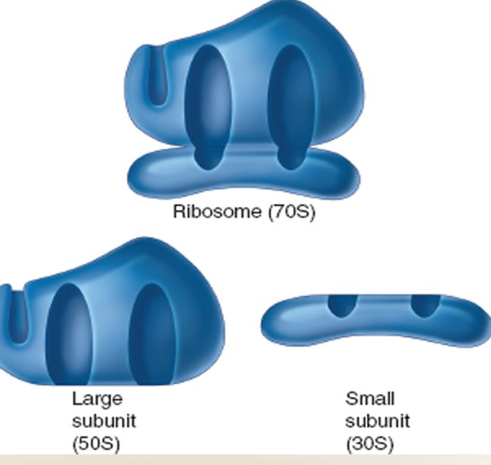
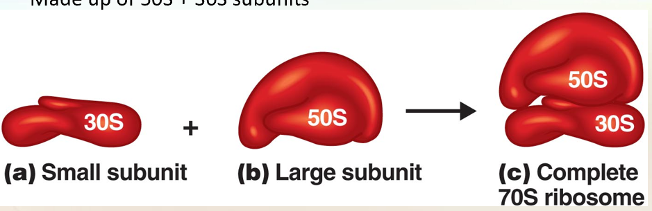
The Prokaryotic Ribosome
Is ussed for protein synthesis
Size is 70s:
Made up of 50s + 30s subunits
What Ribosomes Do
Found in all cells (prokaryotic + eukaryotic).
Job: protein synthesis (make proteins).
Actively growing cells = lots of ribosomes.
In prokaryotes, ribosomes make the cytoplasm look grainy.
🔹 Structure
Ribosomes = 2 subunits (protein + rRNA):
Small subunit (30S) → 1 rRNA + proteins.
Large subunit (50S) → 2 rRNAs + proteins.
Together: 70S ribosome (not 80, because “S” is about shape/speed, not math).
🔹 Prokaryotic vs Eukaryotic Ribosomes
Prokaryotic: 70S ribosomes.
Eukaryotic: 80S ribosomes.
Difference in size/density helps medicine target bacteria.
💊 Why It Matters for Antibiotics
Some antibiotics kill bacteria by stopping protein synthesis.
Examples:
Streptomycin, gentamicin → bind to 30S subunit.
Erythromycin, chloramphenicol → bind to 50S subunit.
Human ribosomes are different (80S), so antibiotics can kill bacteria without harming us.
👉 Super simple summary:
Ribosomes = protein factories.
Bacteria = 70S (30S + 50S).
Humans = 80S.
Antibiotics use this difference to attack bacteria, not us.
Inclusions = Storage Units in Bacteria
Reserve deposits in the cytoplasm.
Store nutrients when abundant → use later when scarce.
Help avoid too much osmotic pressure (water flooding cell).
Some are common to many bacteria, others unique (used for identification).
🔹 Types of Inclusions
Metachromatic Granules (Volutin)
Store inorganic phosphate → used to make ATP.
Found in bacteria, algae, fungi, protozoa.
Seen in Corynebacterium diphtheriae (diagnostic clue).
Stain red with blue dyes (like methylene blue).
Polysaccharide Granules
Store glycogen & starch.
Test: add iodine →
Glycogen = reddish brown.
Starch = blue.
Lipid Inclusions
Store lipids (fats).
Found in Mycobacterium, Bacillus, Azotobacter, Spirillum.
Special polymer: poly-β-hydroxybutyric acid.
Seen with fat-soluble stains (e.g., Sudan dyes).
Sulfur Granules
Store sulfur for energy.
Found in sulfur bacteria (e.g., Acidithiobacillus).
👉 Super simple summary:
Inclusions = bacterial storage bins.
Can store phosphate (ATP fuel), starch/glycogen (carbs), lipids (fats), or sulfur (energy).
Some are diagnostic (like metachromatic granules in diphtheria).
Carboxysomes
Contain enzyme RuBisCO (ribulose 1,5-bisphosphate carboxylase).
Function: fix carbon dioxide for photosynthesis.
Found in nitrifying bacteria, cyanobacteria, Acidithiobacillus.
🔹 Gas Vacuoles
Found in many aquatic bacteria (cyanobacteria, photosynthetic bacteria, halobacteria).
Made of rows of gas vesicles (hollow protein cylinders).
Function: act like a floatation device → control buoyancy.
Keeps bacteria at right water depth for light, oxygen, nutrients.
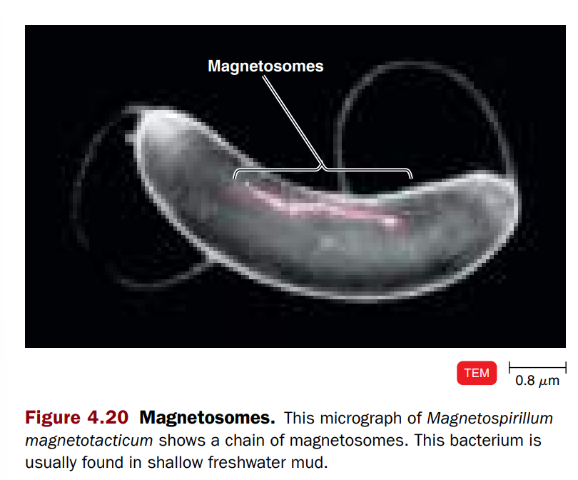
🔹 Magnetosomes
Inclusions of iron oxide (Fe₃O₄) inside the plasma membrane.
Found in some Gram-negative bacteria (e.g., Magnetospirillum).
Function: act like tiny magnets → help bacteria orient/move along Earth’s magnetic field to reach proper depth.
May also break down hydrogen peroxide → protects cell from damage.
👉 Super simple summary:
Carboxysomes = carbon-fixing enzyme bags (for photosynthesis).
Gas vacuoles = floating balloons (for buoyancy in water).
Magnetosomes = compass needles (for orientation + protection).
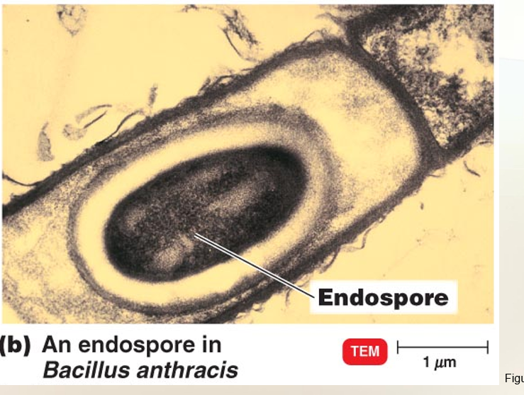
Endospores
Resting cells
Resistant to desiccation, heat, chemicals
Formed by Bacillus, Clostridium speices
Exception: ?
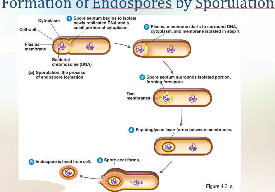
Sporulation: Endospore formation
Germination: Return to vegtative state
Endospores
Special resting cells formed when nutrients are low.
Found in Gram-positive bacteria (e.g., Clostridium, Bacillus).
Function: survival, not reproduction.
Extremely resistant: survive heat, drying, chemicals, radiation.
Can stay dormant for thousands of years and still germinate.
🔹 Formation (Sporulation / Sporogenesis)
DNA copied + cytoplasm isolated by plasma membrane → spore septum.
Double membrane forms around DNA + cytoplasm → forespore.
Thick peptidoglycan layer forms.
Spore coat (protein) forms → chemical resistance.
Original cell breaks down → endospore released.
🔹 Structure
Inside = DNA, little RNA, ribosomes, enzymes.
Surrounded by spore coat + peptidoglycan layers.
Contains dipicolinic acid (DPA) + calcium ions → protect DNA from damage.
Very little water inside = makes them resistant.
🔹 Germination
When conditions improve → spore returns to vegetative (active) cell.
Triggered by heat or chemicals.
Germination = resuming metabolism, not reproduction.
🔹 Location in Cell (before release)
Terminal = at one end.
Subterminal = near one end.
Central = in the middle.
Location can help identify bacteria.
🔹 Why Important?
Clinically: cause diseases → anthrax, tetanus, botulism, gas gangrene.
Food industry: survive boiling water → need special sterilization methods.
👉 Super simple summary:
Endospores = bacterial survival capsules.
Form when food is scarce, survive extreme conditions, restart growth when safe.
Protected by spore coat + DPA + dehydration.
Not for reproduction, just survival.
✓ 4-10 Where is the DNA located in a prokaryotic cell? ✓ 4-11 What is the general function of inclusions? ✓ 4-12 Under what conditions do endospores form?
👉 In the nucleoid region of the cytoplasm (not inside a nucleus, since prokaryotes don’t have one).
👉 Storage – inclusions are reserve deposits where cells keep nutrients (like glycogen, lipids, sulfur, etc.) to use when the environment lacks them.
👉 Endospores form when essential nutrients are depleted (like carbon or nitrogen), or under harsh/unfavorable conditions (extreme heat, lack of water, toxic chemicals).
4-9 How are simple diffusion and facilitated diffusion similar? How are they different?
Similarities:
Both move substances from high → low concentration (down the gradient).
Both are passive → no energy (ATP) is used.
How are they different?
👉 Differences:
Simple diffusion: Molecules pass directly through the membrane (like small gases: O₂, CO₂).
Facilitated diffusion: Molecules need transport proteins (channels or carriers) to cross (like glucose, ions).
✨ Super short version to memorize:
Both: passive, high → low
Simple: no help needed
Facilitated: needs protein helper
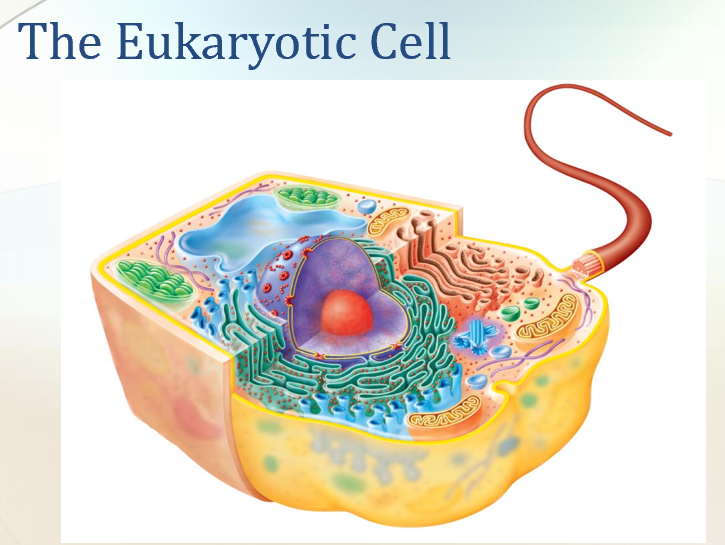
The Eukaryotic Cell vs Prokartoic cell
Prokaryotes (Bacteria, Archaea)
Size: Small (0.2–2.0 µm)
Nucleus: ❌ No true nucleus (DNA floats in nucleoid)
Organelles: ❌ Few (no membrane-bound organelles)
Flagella: Simple (made of 2 protein parts)
Glycocalyx: Capsule or slime layer
Cell Wall: Usually present; made of peptidoglycan
Membrane: No sterols (except some special ones)
Cytoplasm: Cytoskeleton but no cytoplasmic streaming
Ribosomes: Small (70S)
DNA: One circular chromosome, no histones
Cell Division: Binary fission (simple split)
Reproduction: No sexual recombination; DNA transfer only
🧫 Eukaryotes (Plants, Animals, Fungi, Protists)
Size: Big (10–100 µm)
Nucleus: ✅ True nucleus with nuclear membrane + nucleoli
Organelles: ✅ Many (mitochondria, ER, Golgi, chloroplasts, etc.)
Flagella: Complex (made of microtubules)
Glycocalyx: Present only in some (esp. without cell wall)
Cell Wall: If present → simple (cellulose in plants, chitin in fungi)
Membrane: Has sterols & carbs (for receptors)
Cytoplasm: Cytoskeleton + cytoplasmic streaming
Ribosomes: Large (80S), but organelles may have 70S
DNA: Multiple linear chromosomes with histones
Cell Division: Mitosis
Reproduction: Sexual (meiosis)
👉 Shortcut Memory Trick:
Prokaryotes = Simple, small, no nucleus, no organelles.
Eukaryotes = Complex, big, true nucleus, many organelles.
The Evolution of Eukaryotes (pp. 102–103)
1. According to the endosymbiotic theory, eukaryotic cells evolved from symbiotic prokaryotes living inside other prokaryotic cells.
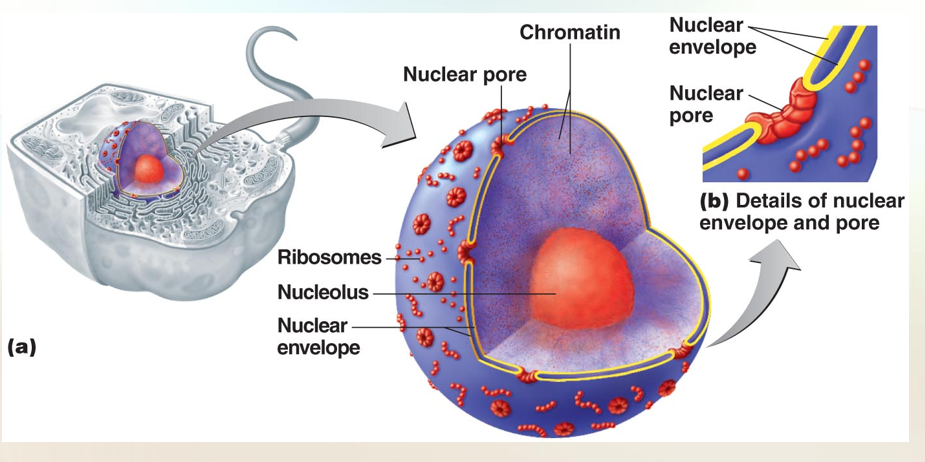
The Eukaryotic Nucleus
The nucleus is frequently the largest structure in the cell, and contains almost all of the cell’s hereditary information (DNA).
The nucleus is surrouned by a double membrane called the nuclear envelope. Tiny channels in the membrane called nuclear pores allow the nucleus to communicate with the cytoplasm Nuclear pores control the movement of substances between the nucleus and cytoplasm. Within the nuclear envelope are one or more spherical bodies called nucleoli (singular: nucleolus).
Nucleoli are actually condensed regions of chromosomes where ribosomal RNA is being synthesized. Ribosomal RNA is an essential component of ribosomes.
The nucleus also contains most of the cell’s DNA, which is combined with several proteins, including some basic proteins called histones and nonhistones. The combination of about 165 base pairs of DNA and 9 molecules of histones is referred to as a nucleosome.
When the cell is not reproducing, the DNA and its associated proteins appear as a threadlike mass called chromatin. During nuclear division, the chromatin coils into shorter and thicker rodlike bodies called chromosomes. Prokaryotic chromosomes do not undergo this process, do not have histones, and are not enclosed in a nuclear envelope.
Eukaryotic cells require two elaborate mechanisms: mitosis and meiosis to segregate chromosomes prior to cell division. Neither process occurs in prokaryotic cells.
Flagella & Cilia
1. Flagella are few and long in relation to cell size; cilia are numerous and short.
2. Flagella and cilia are used for motility, and cilia also move substances along the surface of the cells.
3. Both flagella and cilia consist of an arrangement of nine pairs and two single microtubules.
The Plasma (Cytoplasmic) Membrane (p. 97)
1. Like the prokaryotic plasma membrane, the eukaryotic plasma membrane is a phospholipid bilayer containing proteins.
2. Eukaryotic plasma membranes contain carbohydrates attached to the proteins and sterols not found in prokaryotic cells (except Mycoplasma bacteria).
3. Eukaryotic cells can move materials across the plasma membrane by the passive processes and by active transport used by prokaryotes and endocytosis (phagocytosis, pinocytosis, and receptor-mediated endocytosis).
Cytoplasm (p. 98)
1. The cytoplasm of eukaryotic cells includes everything inside the plasma membrane and external to the nucleus.
2. The chemical characteristics of the cytoplasm of eukaryotic cells resemble those of the cytoplasm of prokaryotic cells.
3. Eukaryotic cytoplasm has a cytoskeleton and exhibits cytoplasmic streaming.
The Cell Wall and Glycocalyx (pp. 96–97)
1. The cell walls of many algae and some fungi contain cellulose.
2. The main material of fungal cell walls is chitin.
3. Yeast cell walls consist of glucan and mannan.
4. Animal cells are surrounded by a glycocalyx, which strengthens the cell and provides a means of attachment to other cells.
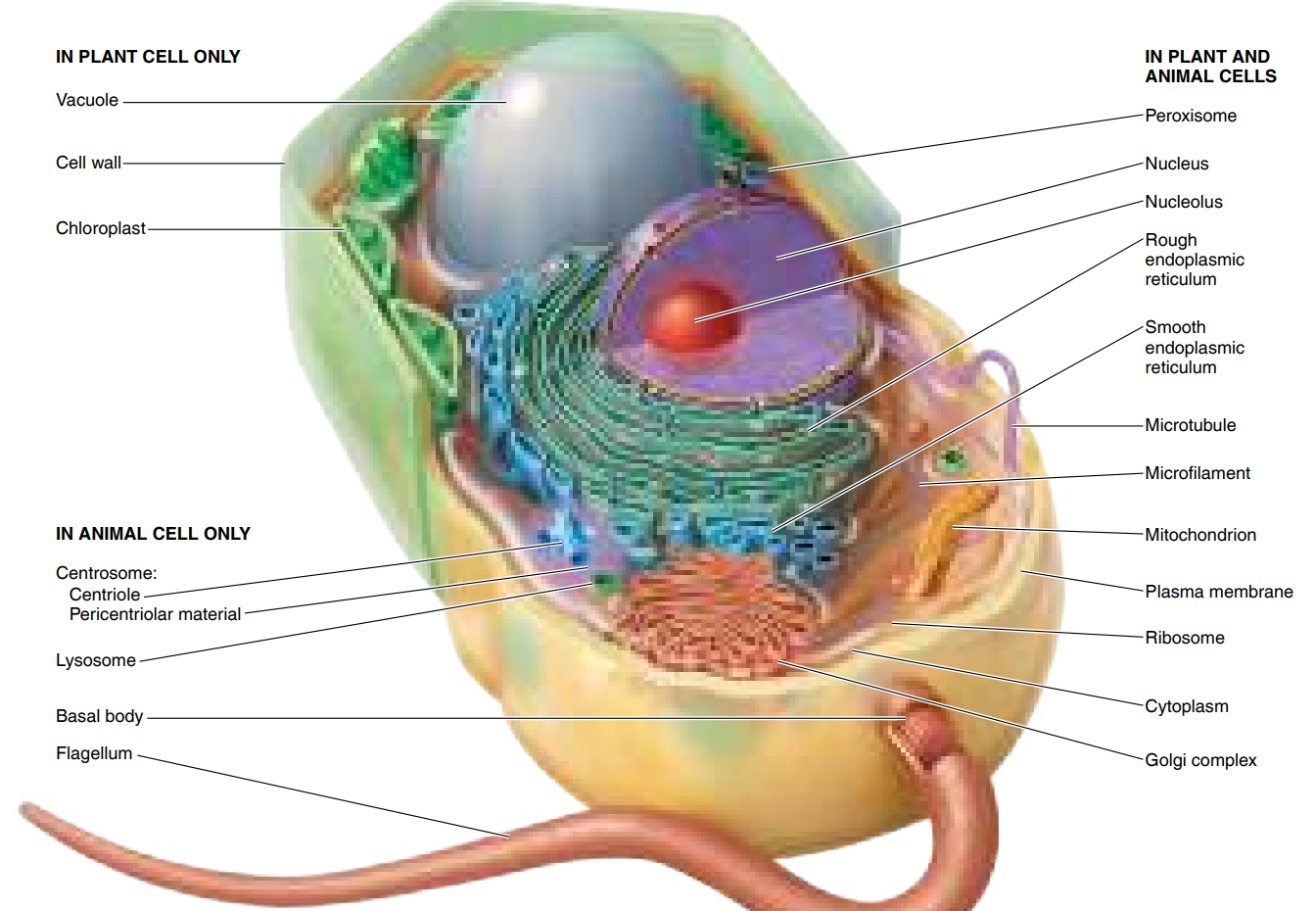
Organelles (pp. 98–102)
1. Organelles are specialized membrane-enclosed structures in the cytoplasm of eukaryotic cells.
2. The nucleus, which contains DNA in the form of chromosomes, is the most characteristic eukaryotic organelle.
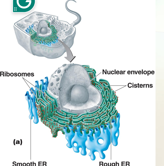
3. The nuclear envelope is connected to a system of membranes in the cytoplasm called the endoplasmic reticulum (ER).
4. The ER provides a surface for chemical reactions and serves as a transport network. Protein synthesis and transport occur on the rough ER; lipid synthesis occurs on the smooth ER.
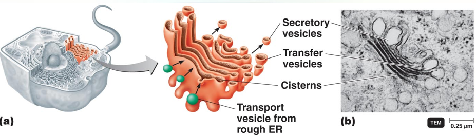
5. The Golgi complex consists of flattened sacs called cisternae. It functions in membrane formation and protein secretion.
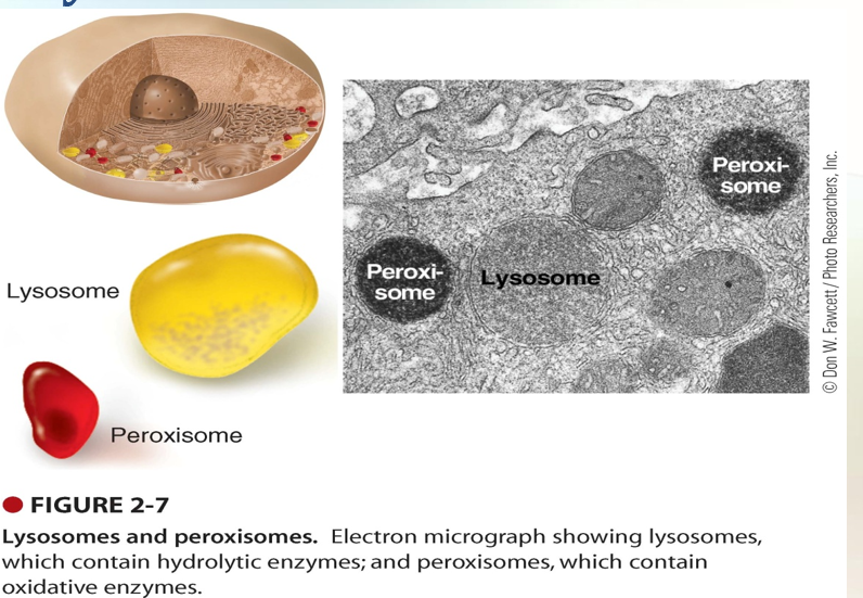
6. Lysosomes are formed from Golgi complexes. They store digestive enzymes.
7. Vacuoles are membrane-enclosed cavities derived from the Golgi complex or endocytosis. They are usually found in plant cells that store various substances and provide rigidity to leaves and stems.
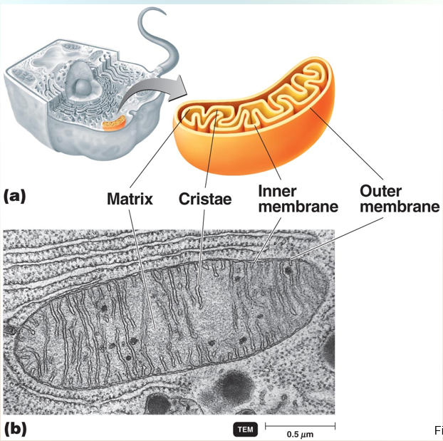
8. Mitochondria are the primary sites of ATP production. They contain 70S ribosomes and DNA, and they multiply by binary fission.
9. Chloroplasts contain chlorophyll and enzymes for photosynthesis. Like mitochondria, they contain 70S ribosomes and DNA and multiply by binary fission.
10. A variety of organic compounds are oxidized in peroxisomes. Catalase in peroxisomes destroys H2O2.
11. The centrosome consists of the pericentriolar material and centrioles. Centrioles are 9 triplet microtubules involved in formation of the mitotic spindle and microtubules.
Endoplasmic Reticulum
Golgi Complex
Lysosomes & Peroxisomes
Mitochondria
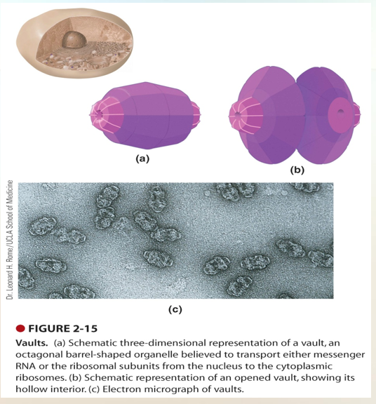
Vaults
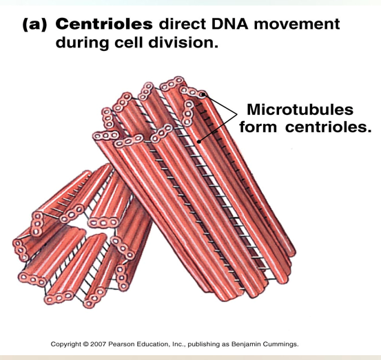
Centrioles
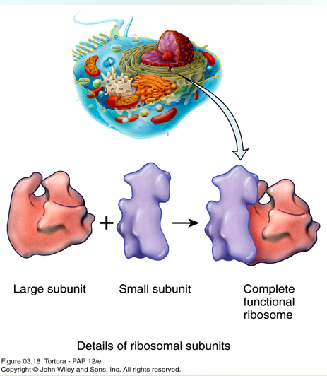
Ribosome
80S ribosomes are found in the cytoplasm or attached to the rough endoplasmic reticulum.