C2.1 Chemical Signaling | IB Biology HL
1/43
Earn XP
Description and Tags
Name | Mastery | Learn | Test | Matching | Spaced | Call with Kai |
|---|
No study sessions yet.
44 Terms
Outline the structure and function of receptor molecules
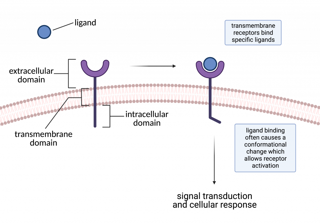
C2.1.1: Receptors as proteins with binding sites for specific signaling chemicals.
The protein that the ligand binds to (induced fit).
The receptor’s binding site is complementary to the shape of the ligand
The binding of the ligand causes the signal transduction pathway (series of steps that the cell responds to the binding).
Define ligand.
C2.1.1: Receptors as proteins with binding sites for specific signaling chemicals.
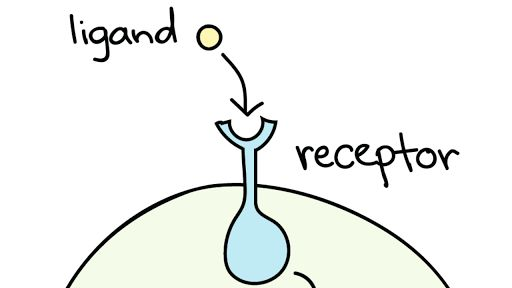
The signaling chemical - the molecule that binds reversibly to specific proteins.
It is typically smaller than the protein. Once it unbinds, its structure is unchanged, so it can bind to other receptors.
Outline the relationship between receptors and a specific ligand.
C2.1.1: Receptors as proteins with binding sites for specific signaling chemicals.
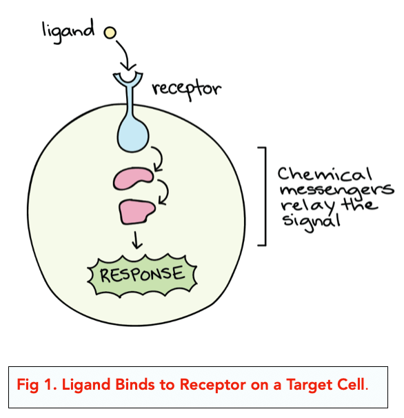
The receptor is the protein that the specific ligand binds to.
The shape of the ligand is complementary to the shape of the receptor binding site (there is specificity).
The fewer ligands a protein can bind to, the greater the specificity.
The binding causes the signal transduction pathway.
Define target cell.
C2.1.1: Receptors as proteins with binding sites for specific signaling chemicals.
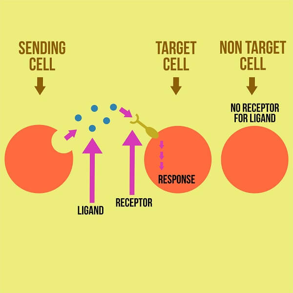
The cells which have the receptors for a specific ligand.
If a cell doesn’t have the receptors for a specific ligand, it is not affected by that ligand’s signal.
Define signal transduction pathway.
C2.1.1: Receptors as proteins with binding sites for specific signaling chemicals.
The series of steps that allows the cell to respond to ligands bonding.
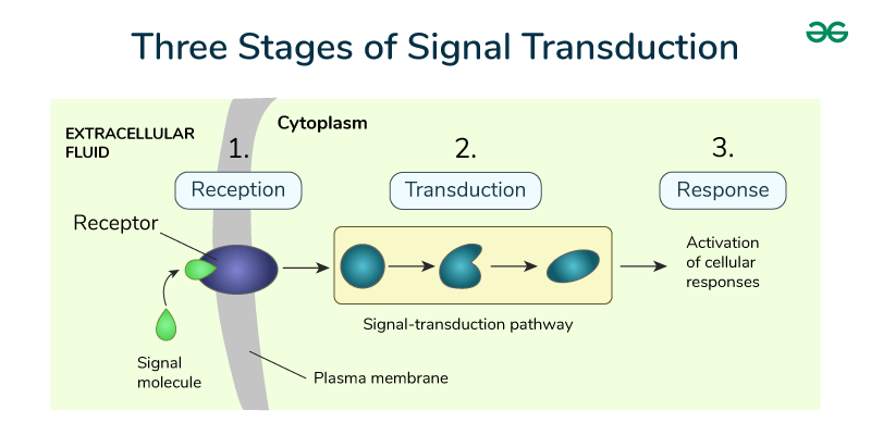
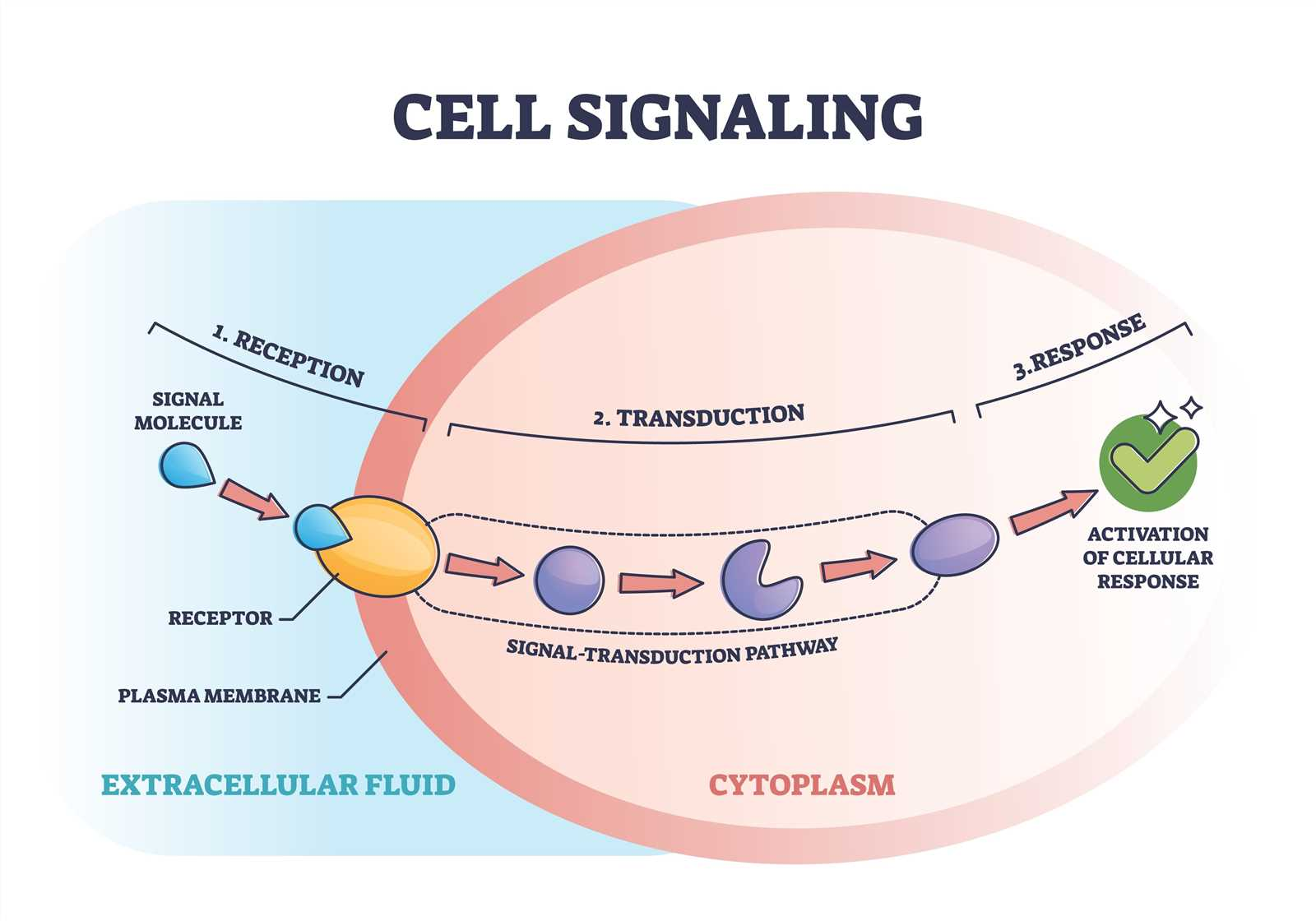
Outline the stages of cell signaling.
C2.1.1: Receptors as proteins with binding sites for specific signaling chemicals.
First, the ligand binds with the receptor.
Next, the binding sets the signal transduction pathway into motion.
Finally, the cellular response to the ligand bonding occurs.
Define quorum sensing.
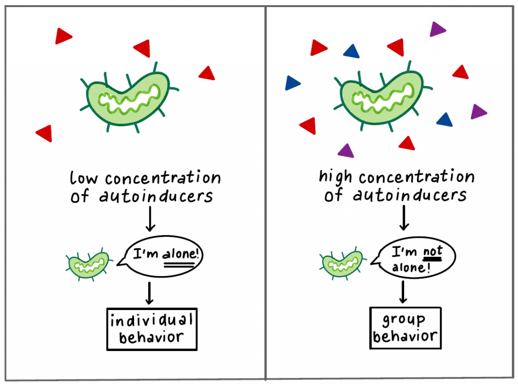
C2.1.2: Cell signaling by bacteria in quorum sensing.
A mechanism by which bacteria’s group behavior is based on population density.
It is a method for individual bacteria to coordinate actions with colonies.
Describe the process of quorum sensing in a population of bacteria, including the role of signaling molecules, receptors, and a threshold for gene expression.
C2.1.2: Cell signaling by bacteria in quorum sensing.
As the number of bacteria increases (bacteria reproduce), the concentration of autoinducers (a type of ligand produced by bacteria) increases.
Once the autoinducer concentration reaches a certain threshold, the entire bacterial population’s gene expression changes.
The autoinducer binds to a receptor which changes the gene expression of the bacteria.
Outline the process of bioluminescence in Vibrio fischeri as an example of quorum sensing.
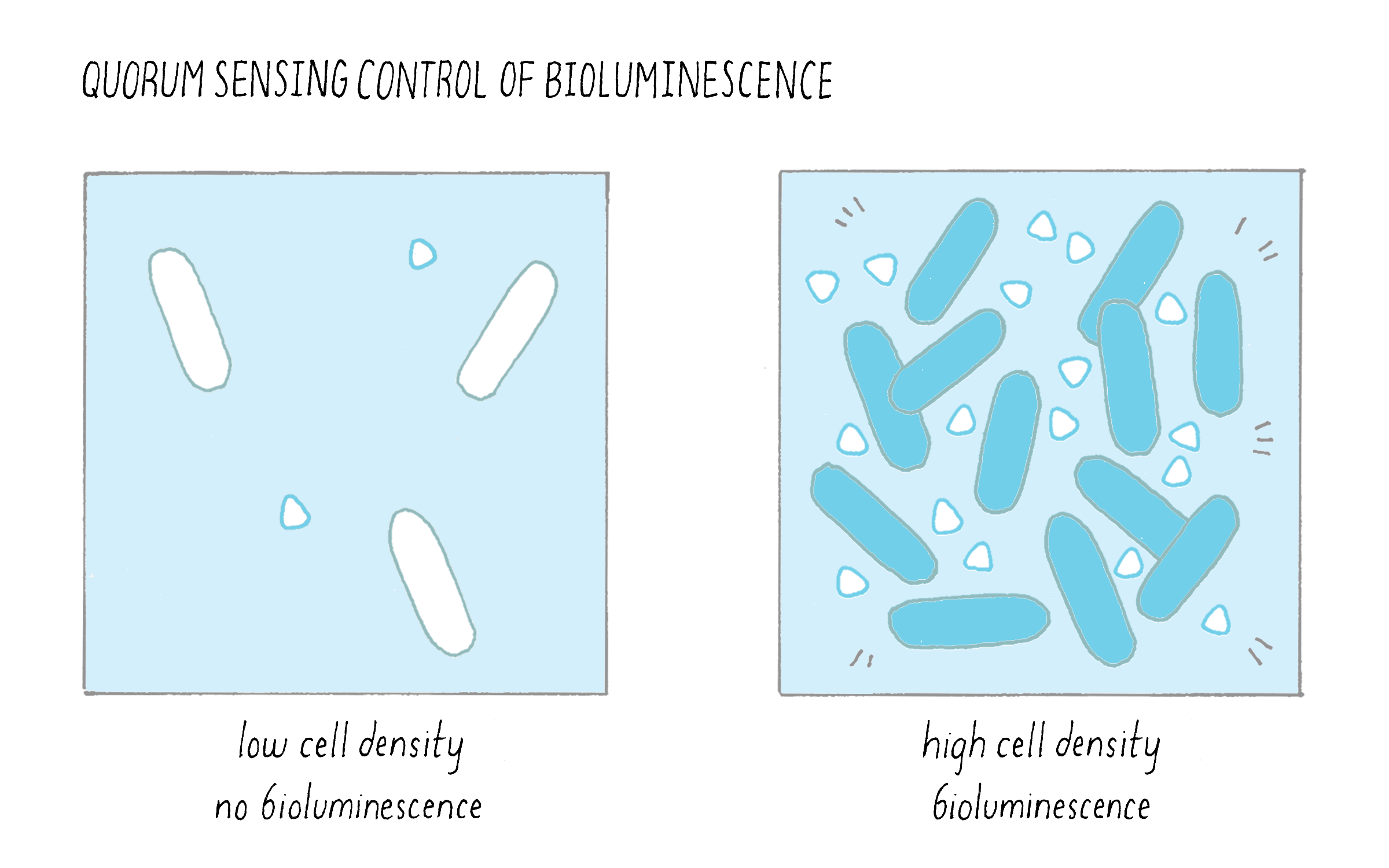
C2.1.2: Cell signaling by bacteria in quorum sensing.
Quorum sensing was first discovered in marine bacterium Vibrio fischeri. They have a symbiotic relationship with marine animals, especially the Hawaiian bobtail squid (Euprymna scolopes).
Free-floating V. fischeri are captured by the squid, where they are housed in the light organ.
Individual V. fischeri reproduce. In their reproductive cycle, they also produce autoinducer molecules.
Autoinducer molecules pass through the cell membrane and cell wall to reach the exterior environment (inside the light organ).
As bacteria continue to reproduce, the V. fischeri population increases, and the concentration of the autoinducer also increases.
Once the concentration of the autoinducer reaches a threshold level, they move back into bacterial cells and bind to the LuxR protein (the receptor).
The LuxR protein binds to a DNA binding site known as the Lux Box.
The Lux Box is activated through this binding, so it produces luciferase (luminescent protein) which causes bioluminescence and illuminates the squid.
State why there is a wide range of signaling chemicals.
C2.1.3: Hormones, neurotransmitters, cytokines and calcium ions as examples of functional categories of signaling chemicals in animals.
Signaling systems have repeatedly evolved. As a result, a wide range of chemical are suitable as signaling chemicals.
Define a hormone. List examples.
C2.1.3: Hormones, neurotransmitters, cytokines and calcium ions as examples of functional categories of signaling chemicals in animals.
Chemical signal secreted by endocrine glands into the bloodstream to reach target tissues.
They bind to receptors on plasma membrane or in the cytoplasm.
Examples:
Epinephrine (adrenaline)
Insulin
Oestradiol (estrogen)
Testosterone
Define a neurotransmitter. List examples.
C2.1.3: Hormones, neurotransmitters, cytokines and calcium ions as examples of functional categories of signaling chemicals in animals.
Chemicals that transmit signals across a synapse (junction between 2 neurons).
Enables communication between neurons & between neurons and effectors.
Example:
Acetylcholine
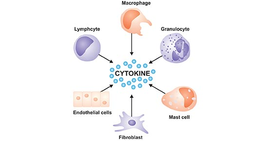
Define a cytokine. List important roles.
C2.1.3: Hormones, neurotransmitters, cytokines and calcium ions as examples of functional categories of signaling chemicals in animals.
Glycoproteins made by immune cells, functioning as a messenger by diffusing between cells.
They are released by immune cells (T cells, macrophages) & travel to receptors on other cells
Important roles:
Control activity & growth of immune cells
Activate cells to attack viruses & bacteria
Trigger inflammation (help fight infection)
Trigger activation, division, inhibition of immune cells
Define a calcium ion. List important roles.
C2.1.3: Hormones, neurotransmitters, cytokines and calcium ions as examples of functional categories of signaling chemicals in animals.
Secondary messengers in cellular processes
They are triggered by something else & stored in gated calcium ions in response to stimuli
Important roles:
Muscle contractions
Neurotransmitter release
Triggers within a cell (muscle fiber, neurons)
Compare the structure and function of different categories of animal chemical signaling molecules, including hormones, neurotransmitters, cytokines, and chemical ions.
C2.1.3: Hormones, neurotransmitters, cytokines and calcium ions as examples of functional categories of signaling chemicals in animals.
Hormones are proteins secreted by endocrine glands. Neurotransmitters are amines, gasses, or amino acids. Cytokines are glycoproteins, and calcium ions are simply ions.
Hormones signal cells that are far apart by altering gene expression. Neurotransmitters excite or inhibit nerve impulses by diffusing across very short distances (a sypantic gap). Cytokines alter gene expression through diffusion. Calcium ions are used for muscle contraction or exocytosis.
State the properties shared by all signaling chemicals.
C2.1.4: Chemical diversity of hormones and neurotransmitters.
Signaling chemical rely on the activation of specific receptors and cause cells to effect change.
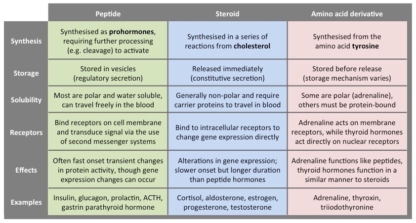
Outline the chemical categories of hormones.
C2.1.4: Chemical diversity of hormones and neurotransmitters.
Amine hormones are small molecules that are modifications of amino acids. This includes epinephrine.
Peptide & protein hormones are polypeptide chains (including peptides, proteins, and glycoproteins). Insulin is a protein hormone.
Steroid hormones are lipids derived from carbohydrates that function as chemical messengers, including oestradiol, progesterone, testosterone.
Outline the chemical categories of neurotransmitters.
C2.1.4: Chemical diversity of hormones and neurotransmitters.
Amino acids can act as neurotransmitters.
Peptides are chains of amino acids functioning as neurotransmitter.
Amiens are modified amino acids.
Nitrous oxides are gases that act as a neurotransmitter.
Contrast the location of effect relative to the location of release between hormones and neurotransmitters.
C2.1.5: Localized and distant effects of signaling molecules.
Hormones are transported through bloodstream, while neurotransmitters diffuse across a synaptic gap.
Hormones are slow, widespread, and long-lasting. This allows them to target distant cells and tissues.
On the other hand, neurotransmitters work rapidly and localized, so they target nearby neurons or effectors.
Distinguish between structure transmembrane receptors and intracellular receptors, including distribution of hydrophilic and hydrophobic amino acids.
C2.1.6: Differences between transmembrane receptors in a plasma membrane and intracellular receptors in the cytoplasm or nucleus.
Transmembrane receptor proteins are embedded in the plasma membrane. The inner and outer surfaces of the protein are hydrophilic, while the middle section is hydrophobic, allowing them to be embedded into the wall.
On the other hand, intracellular receptors proteins are in the cytoplasm or nucleus. They are completely hydrophobic, which allows them to diffuse through the bilayer
Compare the ligands ability to enter the target cell for transmembrane and intracellular receptors.
C2.1.6: Differences between transmembrane receptors in a plasma membrane and intracellular receptors in the cytoplasm or nucleus.
Ligands that bind to transmembrane receptors proteins are hydrophilic, so they cannot diffuse through the lipid bilayer membrane. They must go through transmembrane receptors to penetrate the cell.
Ligands that bind to intracellular receptors are hydrophobic, so they can simply diffuse through the lipid bilayer (interact with hydrophobic tails) to penetrate the cell. The intracellular receptors is also hydrophobic.
Outline the effect of binding a signalling chemical’s binding.
C2.1.7: Initiation of signal transduction pathways by receptors.
When a signalling chemical binds to a receptor, it results in a cascade, a series of reactions inside the cell.
The ligand binds to intracellular protein receptors, changing the gene expression.
Outline the mechanism by which the receptor for the neurotransmitter acetylcholine changes membrane potential in a postsynaptic cell membrane.
C2.1.8: Transmembrane receptors for neurotransmitters and changes to membrane potential.
The binding of a receptor causes ion channels in the receptor to open. As a result, positively charged ions (Na+) flow into the cell. These receptor channels across the cell membrane. As ions enter the cell, voltage travels across the plasma membrane, and the action potential (change in electricity)travels down the cell.
Describe the structure and function of the G-protein coupled receptors (GPCRs) and of the G-proteins.
C2.1.9: Transmembrane receptors that activate G proteins.
G-protein coupled receptors (GPCRs) are a type of transmembrane receptors that act indirectly on enzymes or ion channels with a G protein. Change cannot be activated without the intermediary GPCR. Extracellular loops form the binding site that signalling molecules attach to.
G proteins are coupled to GPCR and activates change in the target cell.
Outline activation of G-protein coupled receptors (GPCRs).
C2.1.9: Transmembrane receptors that activate G proteins.
When the ligand activates the G protein-coupled receptors, the GPCR functions as an intermediary protein, activating the G protein.
Outline how binding of a signaling ligand to a G-protein coupled receptor (GPCR) can cause change in the target cell.
C2.1.9: Transmembrane receptors that activate G proteins.
G proteins bind to GTP. When GPCR is activated, G proteins are activated, and GDP (guanosine diphosphate) becomes GTP, which can do work and open channels.
Without GPCR as the intermediary protein, the signal could not be carried into the cell.
List example signaling ligands that target G-protein coupled receptors (GPCRs).
C2.1.9: Transmembrane receptors that activate G proteins.
Peptides
Proteins
Ions
Lipids
Taste Molecules
Odor molecules
Pheromones
Hormones
Glucagon
epinephrine
Gonadotropin releasing hormone
Oxytocin
Neurotransmitters
Acetylcholine
State that epinephrine is secreted by adrenal glands in preparation for vigorous activity.
C2.1.10: Mechanism of action of epinephrine (adrenaline) receptors.
Epinephrine is secreted by adrenal glands.
The term “adrenaline” comes from the Latin words ad (at) + ren (kidney).
The term “epinephrine” comes from the Greek epi (above) + nephros (kidney).
Describe the activation of the cAMP second messenger system by the epinephrine receptor.
C2.1.10: Mechanism of action of epinephrine (adrenaline) receptors.
GPCRs use a secondary messenger to produce a cellular response.
Cyclic AMP (cAMP) functions as a secondary messenger in the pathway.
When the ligands epinephrine (adrenaline) binds to the appropriate receptor, G proteins are activated. This activated G protein activates a signaling cascade with cAMP as the secondary messenger, with phosphorylation in each step. The final cellular response is the breakdown of glycogen (the storage form of glucose), allowing for individual glucose molecules available for energy.
Outline the effects of epinephrine on a body.
C2.1.10: Mechanism of action of epinephrine (adrenaline) receptors.
When the ligands epinephrine (adrenaline) binds to the appropriate receptor, G proteins are activated. This activated G protein activates a signaling cascade with cAMP as the secondary messenger, with phosphorylation in each step.
The final cellular response is the breakdown of glycogen (the storage form of glucose), allowing for individual glucose molecules available for energy.
Define kinase.
C2.1.11: Transmembrane receptors with tyrosine kinase activity.
An enzyme that catalyzes the transfer of a phosphate group from aTP to another substance.
Define phosphorylation.
C2.1.11: Transmembrane receptors with tyrosine kinase activity.
Adding a phosphate molecule.
Describe the action of receptors with tyrosine kinase activity.
C2.1.11: Transmembrane receptors with tyrosine kinase activity.
Tyrosine kinase is a transmembrane receptor in pairs with 3 domains (extracellular, transmembrane, and intracellular).
When an extracellular domain binds with an insulin hormone ligand, the intracellular domain functions as a kinase to phosphorylate something, leading to a sequence of reactions.
State where and when insulin is secreted.
C2.1.11: Transmembrane receptors with tyrosine kinase activity.
Insulin is secreted by the pancreas cells when blood glucose levels are high.
Describe the cause & effect of the activation of the insulin receptor.
C2.1.11: Transmembrane receptors with tyrosine kinase activity.
When the protein hormone insulin binds to a receptor with tyrosine kinase activity, tyrosine is phosphorylated inside the cell. As a result, a series of events occurs. The final effect is the movement of vesicles (Glut-4) containing certain glucose transporters to the plasma membrane, which releases glucose into the bloodstream.
Describe the mechanism of steroid hormone action.
C2.1.12: Intracellular receptors that affect gene expression.
Steroid hormones are a form of chemical signaling.
Steroid hormones are hydrophobic. They diffuse across the plasma membrane to bind to intracellular receptor.
Once they bind to the receptor, they form a hormone-receptor complex.
This complex attaches to the DNA on a specific gene.
The complex acts as a transcription factor, turning on the transcription of DNA into mRNA.
The mRNA is translated into a protein at the ribosome, which then affects action in the cell.
List examples of steroid hormones.
C2.1.12: Intracellular receptors that affect gene expression.
Oestradiol
Progesterone
Testosterone
Outline one example of a steroid hormone promoting transcription of a specific gene.
C2.1.12: Intracellular receptors that affect gene expression.
Testosterone, a hydrophobic steroid hormone, passes through the cell membrane.
Inside the cell, it binds to a specific receptor protein in the cytoplasm or nucleus, forming a receptor-signal complex.
The complex enters the nucleus and binds to specific DNA sequences to promote gene transcription (copy in mRNA to make amino acids)
The hormone controls the synthesis of enzymes or other proteins to regulate cell activity.
State that oestradiol & progesterone are steroid hormones.
C2.1.13: Effects of the hormones oestradiol and progesterone on target cells.
Both oestradiol & progesterone are steroid hormones that produce female traits (in addition to other functionalities)
Describe the role of oestradiol in the regulation of the release of FSH and LH from the anterior pituitary, including the role of the hypothalamus, the oestradiol receptor, transcription factor and gonadotropin releasing hormone.
C2.1.13: Effects of the hormones oestradiol and progesterone on target cells.
Oestradiol is a type of estrogen produced by the ovaries.
Oestradiol binds to the hypothalamus (forms hormone-receptor complex, activates transcription), which triggers the release of gonadotropoin-releasing hormone (GnRH). GnRH travels through the bloodstream to the pituitary to trigger the release of follicle-stimulating hormone (FSH).
This leads to a growth in follicles (sack that holds & protects eggs).
The FSH encourages the production of more eggs.
Describe the role of progesterone in the formation and maintenance of the endometrium, including the progesterone receptor, transcription factor and growth factor protein.
C2.1.13: Effects of the hormones oestradiol and progesterone on target cells.
Progesterone binds to receptors in the endometrial cell (binds to receptors, forms hormone-receptor complex, acts as transcription factor) to trigger the production of Insulin-like Growth Factor.
The Insulin-like Growth Factor leads to cellular proliferation and growth of the endometrium during the menstrual cycle and pregnancy (thickens the endometrium).
Compare the processes and consequences of positive and negative feedback.
C2.1.14: Regulation of cell signaling pathways by positive and negative feedback.
Positive feedback amplifies a change (driving a response further). This leads to a rapid & large response within a cell.
Negative feedback stops changes that go too far to maintain homeostasis within the cell by counteracting those changes. This leads to the maintenance of stable intracellular conditions.
Outline one example each of hormonal regulation via positive feedback.
C2.1.14: Regulation of cell signaling pathways by positive and negative feedback.
Oxytocin, a positive feedback hormone, increases as the uterus contracts. This triggers more oxytocin production.
As a result, stronger contractions & more oxytocin continues.
This cycle continues until the baby is born.
Outline one example each of hormonal regulation via negative feedback.
C2.1.14: Regulation of cell signaling pathways by positive and negative feedback.
The body’s response to temperature changes is an example of negative feedback. As the body temperature drops, blood vessels constrict so that the heat is conserved, sweat glands don’t secrete fluid, and shivering generates heat.
If the temperatures is too high, blood vessels dilate, resulting in heat loss, and sweat glands secrete fluid.