ANAT 100 - Block Theory 2
1/233
There's no tags or description
Looks like no tags are added yet.
Name | Mastery | Learn | Test | Matching | Spaced | Call with Kai |
|---|
No analytics yet
Send a link to your students to track their progress
234 Terms
Functions of the nervous system
1. Sensory Input
2. Integration
3. Motor Output
1. Sensory Input
- sensory info from the external environment (ex. Temp) and the internal enviroment (ex. having to pee) is detected from sensory nerves and travel through the body to communicate with other nerve cells
2. Integration
- puts all in the incoming sensory signals together to create a complete picture of the enviroment
- occurs in the spinal cord or brain
3. Motor Output
- the nervous system then produces an appropriate response by sending output signals from the brain and spinal cord to the skeletal, cardiac, or smooth muscle of the body, causing them to contract or relax
Organization of the Nervous System
Central Nervous System (CNS) and Peripheral Nervous System (PNS)
Central Nervous System (CNS)
- brain and spinal cord
- control centre of the body
- where are the signals come in, are integrated, and where info can be stored
Peripheral Nervous System (PNS)
- made up of nerves outside the brain and spinal cord
- cranial nerves, spinal nerves, autonomic nervous system, and special sense organs
- the system that brings those signals to the CNS
Afferent Neruons
- part of the sensory division and carry signals to the CNS
Smell/Sight/Touch/Pain
Efferent Neurons
- part of the motor division and carry signals away from the CNS
- voluntary or involuntary
- 2 divisions - somatic and autonomic
- think effort
Cells in The nervous system
- neurons (nerve cells) and supporting cells
- allow for communication between the CNS and PNS
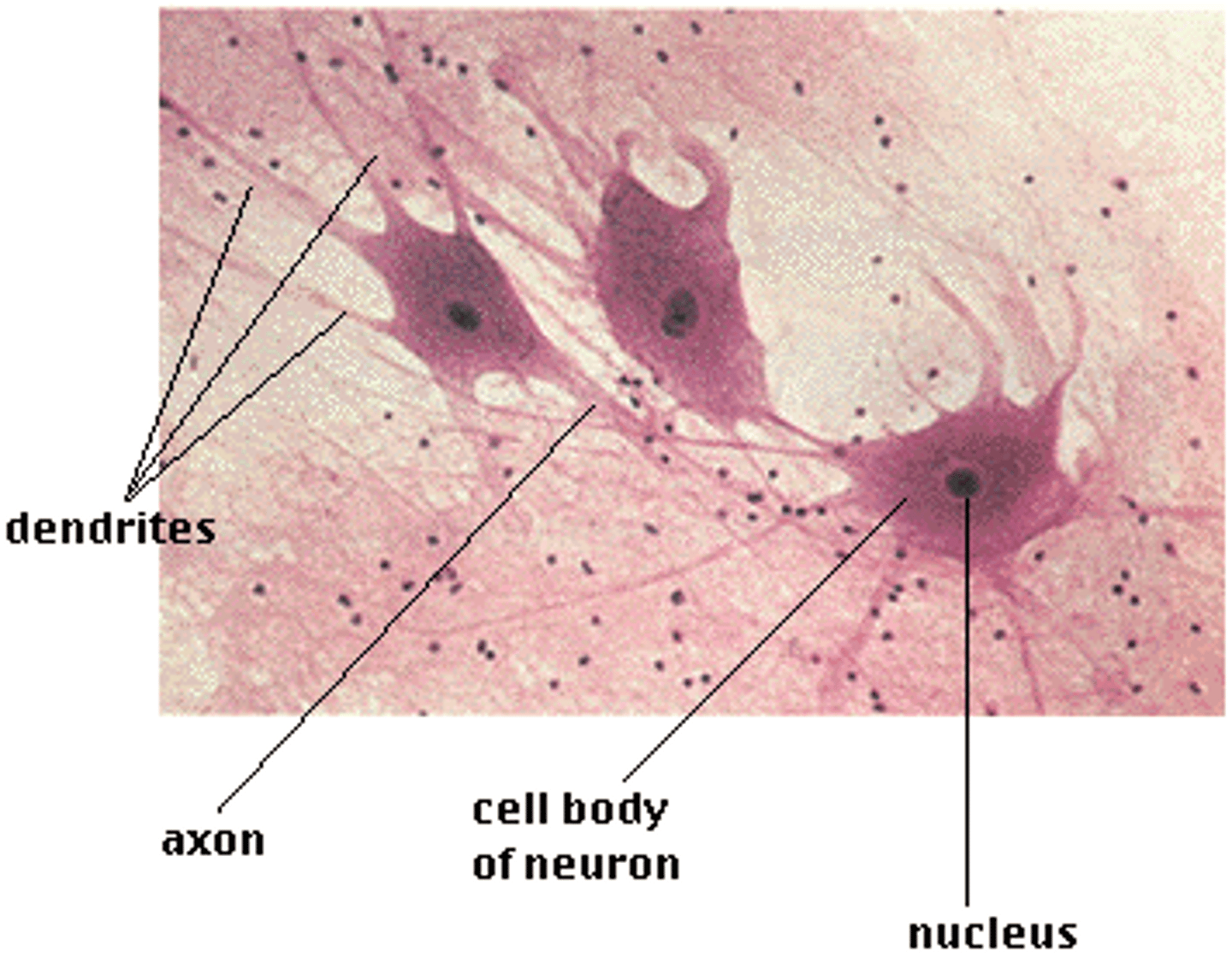
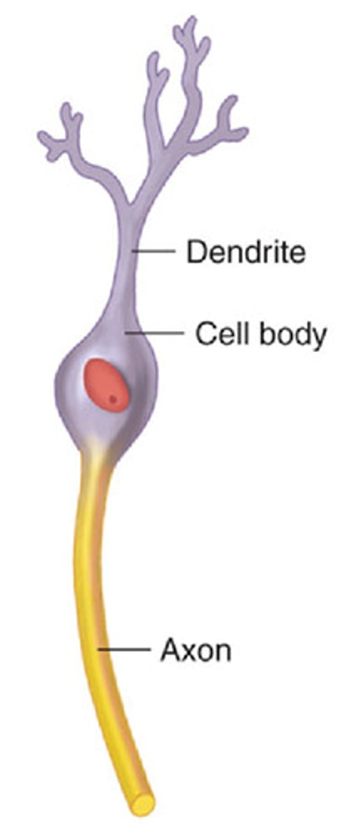
Neuron structure
- cell body
- cytoplasmic processes (dendrites, axon)
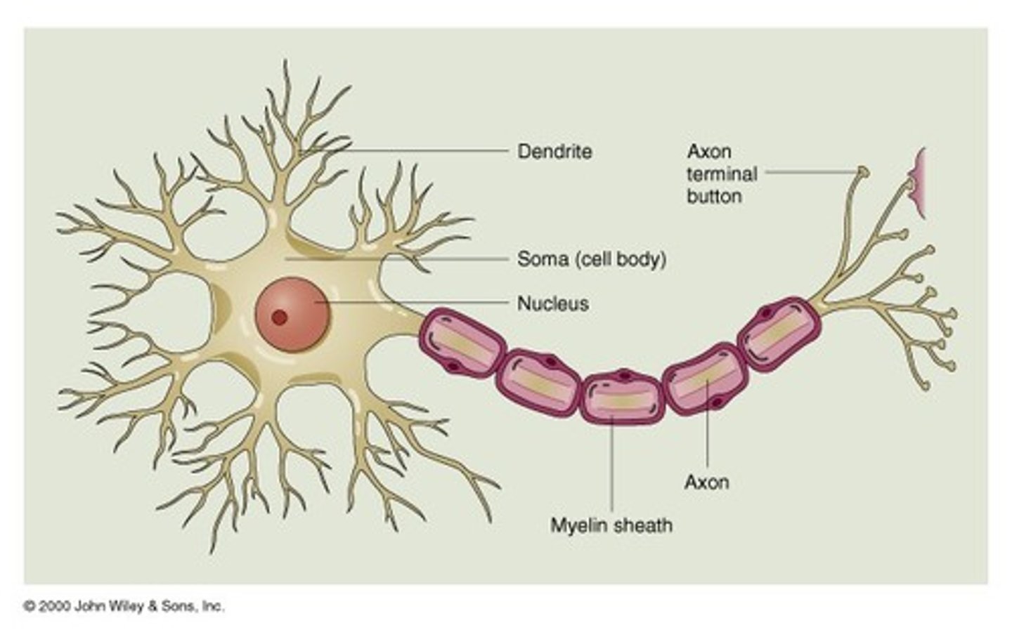
Neuron cell body
AKA soma
nucleus and other organelles
transfers electrical signals from dendrites to the axon
Neuron Dendrites
receive neuronal signal from other neurons
transfers signals to cell body
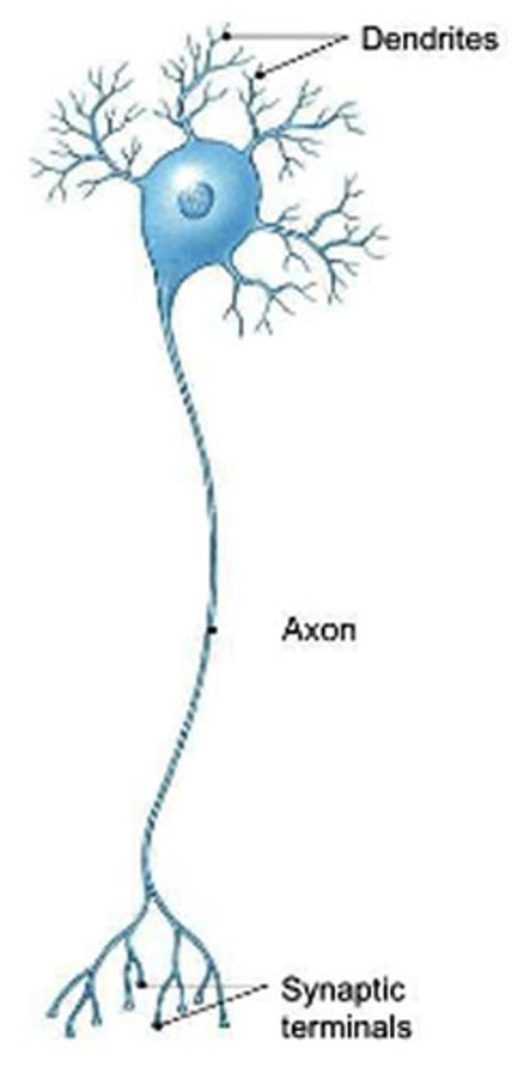
Neuron axon
only 1
carry signal from cell body to axon terminals
signal then transferred to other neurons/organs
direction of nerve impulse
- nerve impulses are rapid electrical signals that allow communication
- UNIDIRECTIONAL (they only go in 1 direction)
- signals come in the dendrites to the cell body, down through the axon, and then reach the dendrites of the next cell
DENDRITES -> SOMA -> AXON
classification of Neurons
- pseudo unipolar (unipolar) neurons
- bipolar neurons
- multipolar neurons
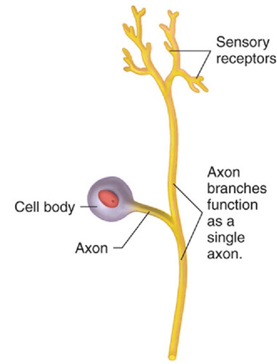
pseudounipolar neurons
- 1 cellular process that extends from the cell body (splits into a dendrite and an axon)
- the cellular process is the little part of the axon coming off the cell body
- ex. general sensory nerves
bipolar neurons
- 2 cellular processes
- 1 dendrite and 1 axon that extends directly from the cell body
only in special senses (sight/hearing)
multipolar neurons
- 3+ cellular processes
- multiple dendrites and 1 axon
- most common type
- ex. motor neurons
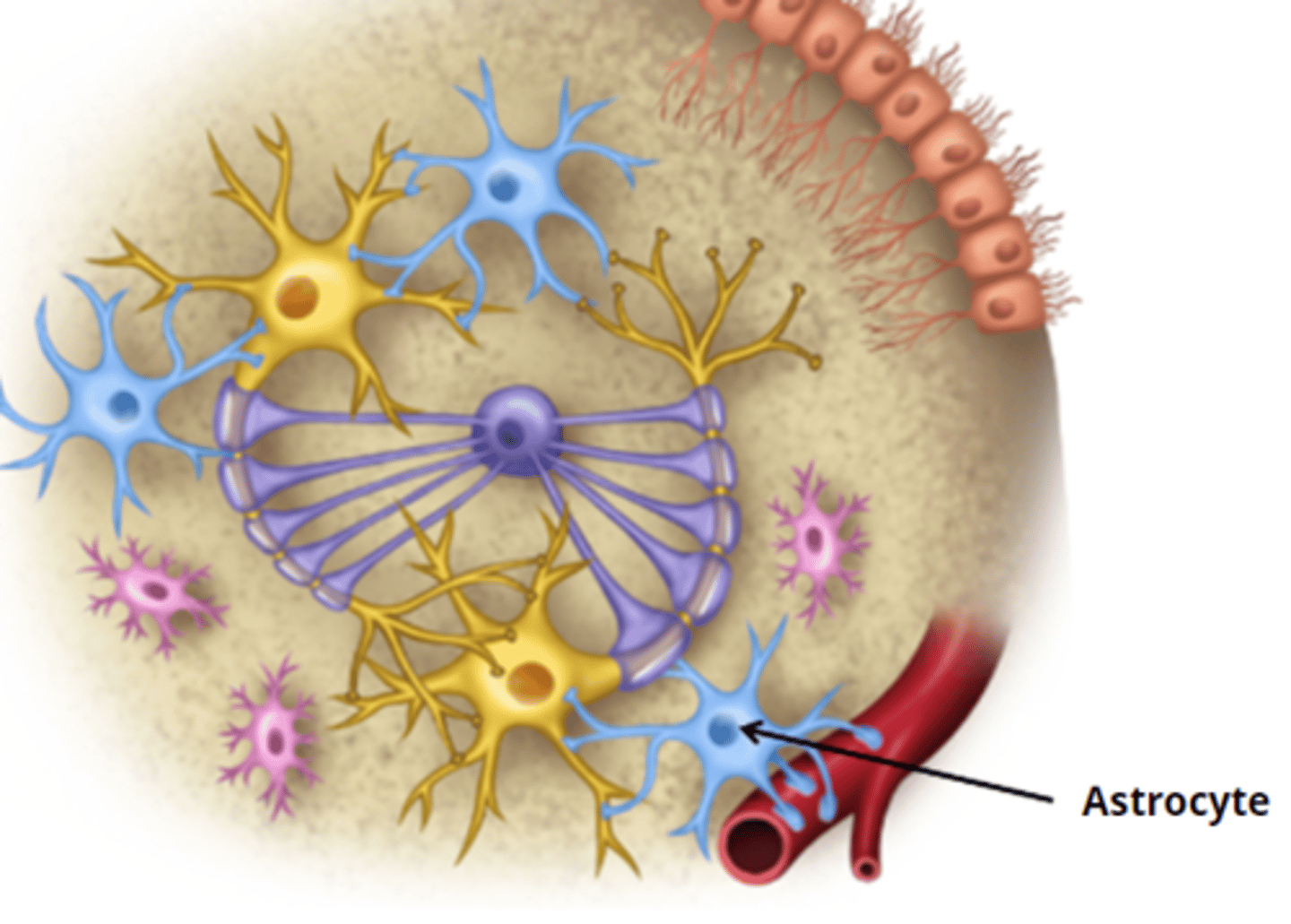
Supporting cells of the nervous system
- neuroglia (glia)
function:
- protect and support neurons
- improve communication between nerve cells
- no nerve impulses
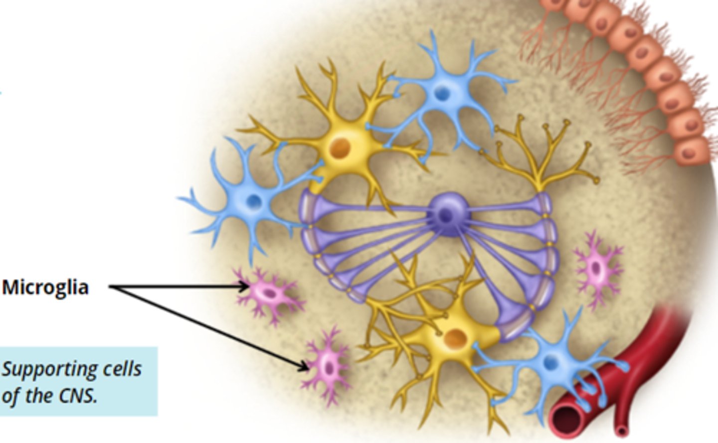
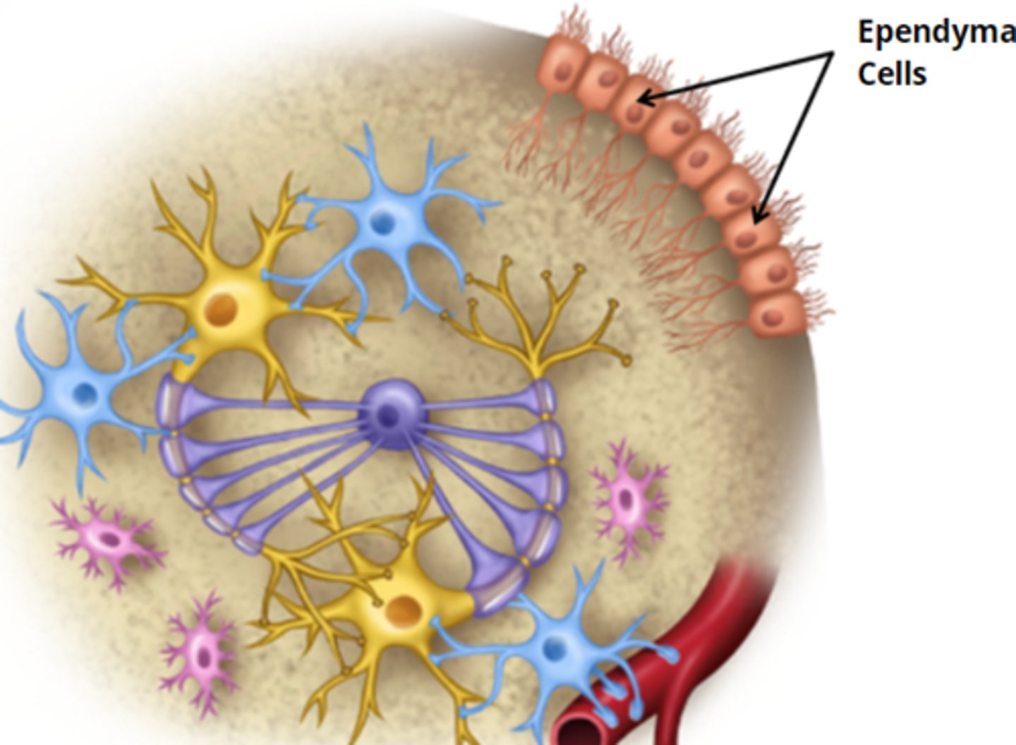
4 types of CNS glia
- astrocytes
- microglia
- ependymal
- oligodendrocytes
astrocytes
- large star-shaped, many processes
- they communicate with neurons and blood vessels to maintain the integrity of the neural tissue
- most abundant glial cell in the CNS
microglia
- small glial cells, protective functions in the CNS
- they are able to engulf/destroy (phagocytose) pathogens and eliminate waste products from other cells
- least common type of glia cell
ependymal
- cuboidal shape that lines the cavity of the brain and spinal cord
- assist in production, circulation, and monitoring of cerebral spinal fluid
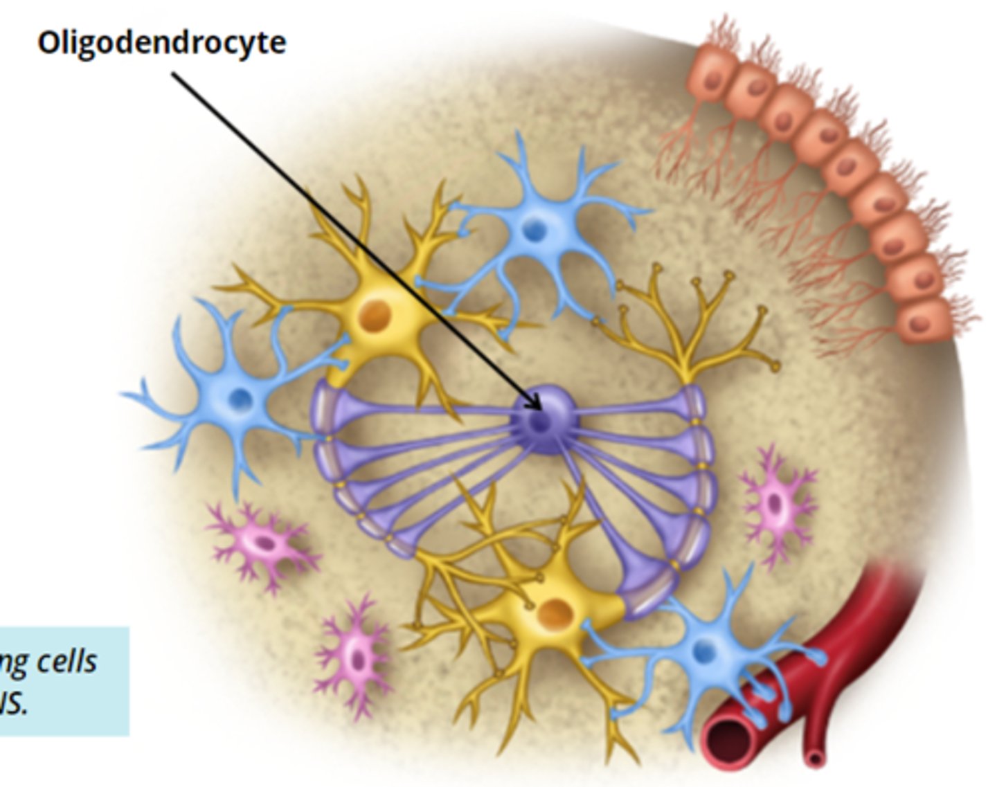
Oligodendrocytes
- important to myelinate parts of an axon (WRAPS MYELIN AROUND THE AXON)
- a single cell can myelinate up to 60 axons
- many processes extend to myelinate axons
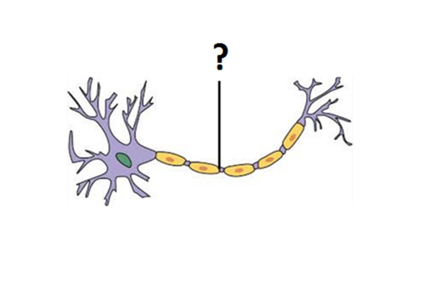
Myelination
- myelin is made up of mostly fats and looks white
- increases the speed of electrical signals that travel down the axon
Nodes of Ranvier
- the unmylinated gaps of the axon
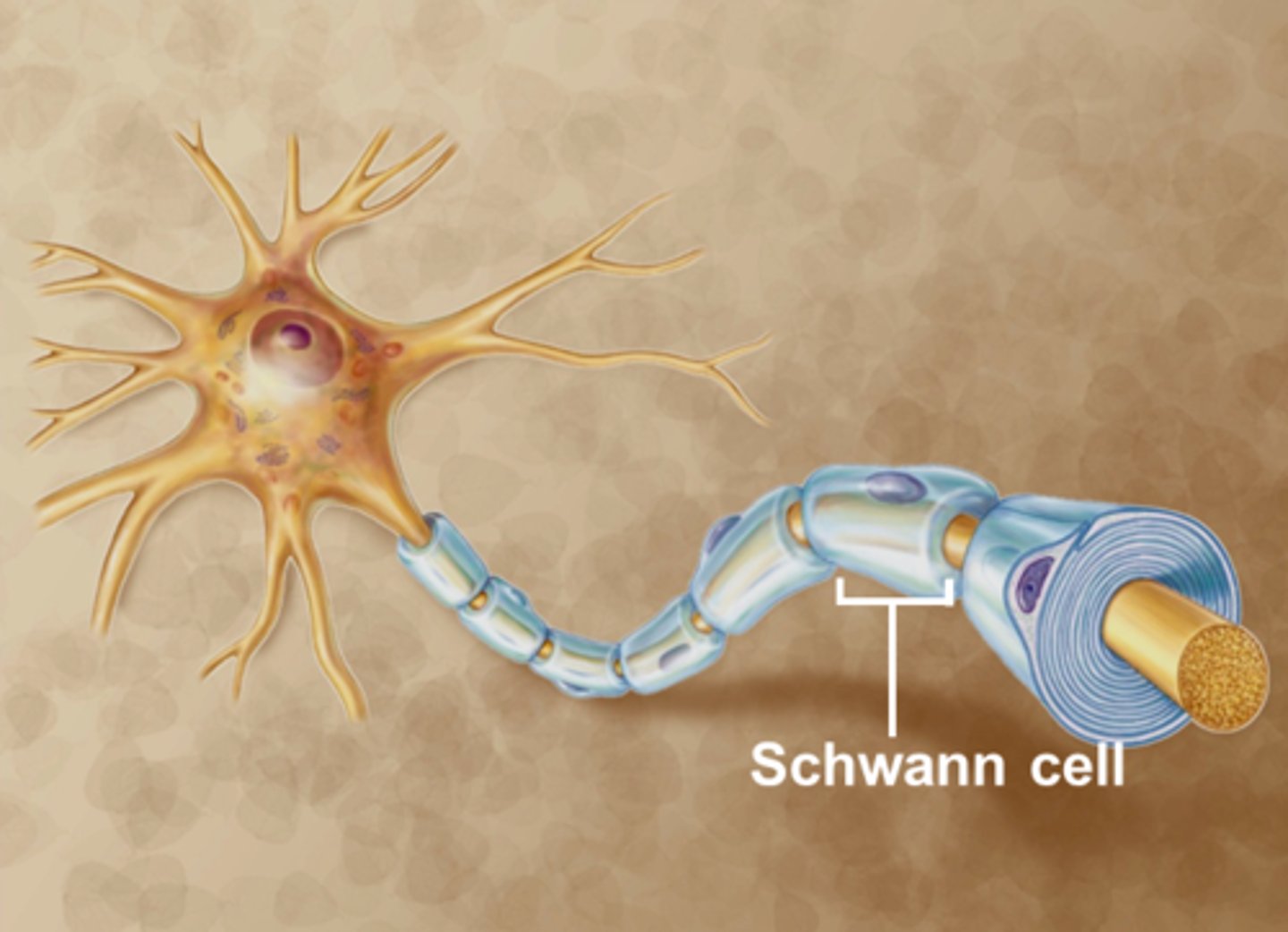
Supporting cells of the PNS
- (the glial cell of the PNS) Schwann cell
- the equivalent to the oligodendrocytes because they form the myelin sheath around the nerve fibres (axons) in the PNS
- a single Schwann cell can only myelinate a portion of 1 axon (they don't have processes like oligodendrocytes)
Parts of the brain
- cerebrum - telencephalon and diencephalon
- brain stem - midbrain, pons, medulla oblongata
- cerebellum
Parts of the brain: cerebrum
- the bulk of the brain
- made up of the telencephalon and the diencephalon
Telencephalon
- responsible for interpreting sensory signals, sending out motor signals, and decision making
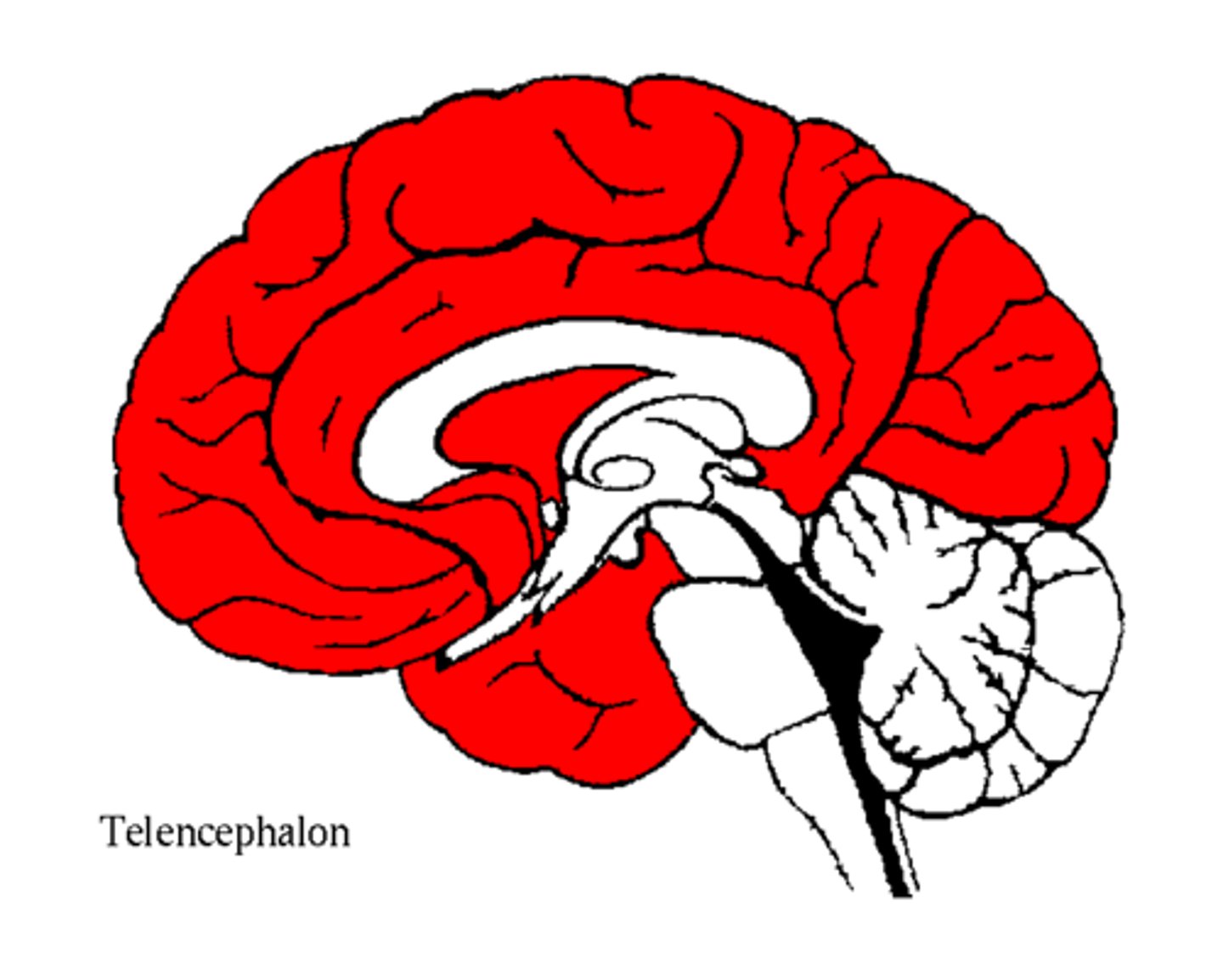
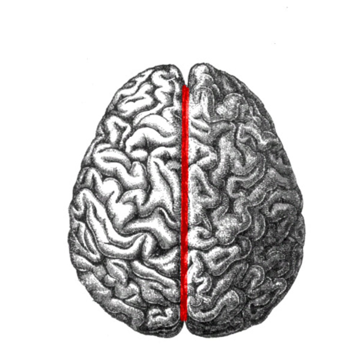
Telencephalon: Cerebral Hemisphere
- the cerebrum is made up of 2 paired hemispheres
(left and right)
- the 2 hemispheres are separated by a longitudinal fissure
parts of telencephalon
cerebral hemispheres
cerebral cortex
sulci and Gyri
lobes of the cerebrum
gray matter of the brain
white matter of the brain
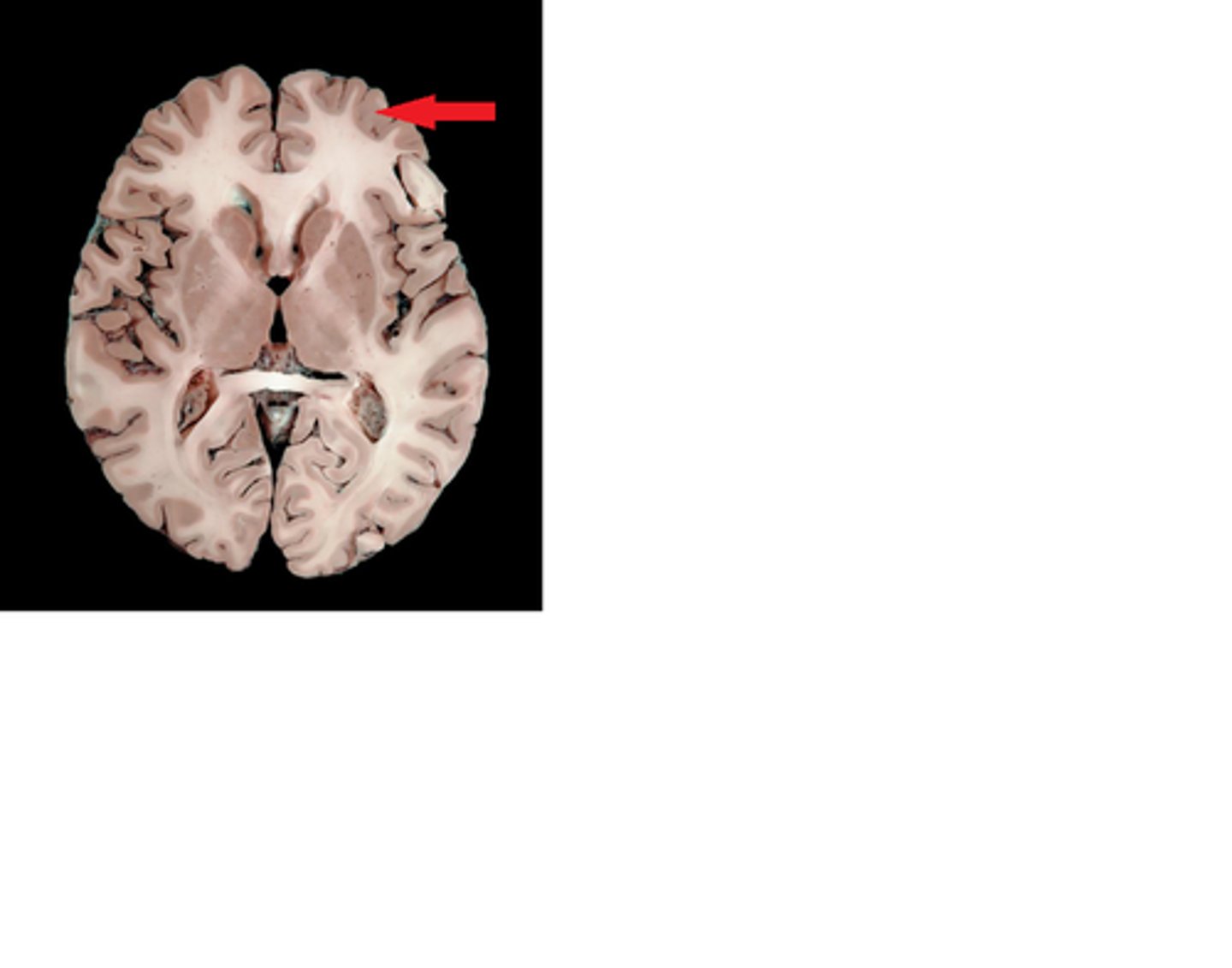
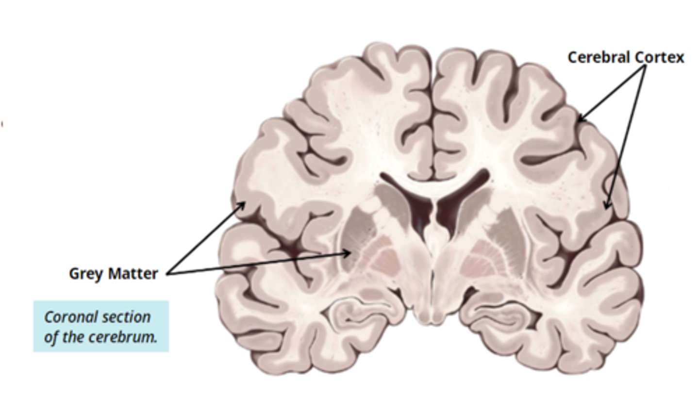
Telencephalon: Cerebral Cortex
- the outer layer of the cerebrum is the cerebral cortex
- grey matter
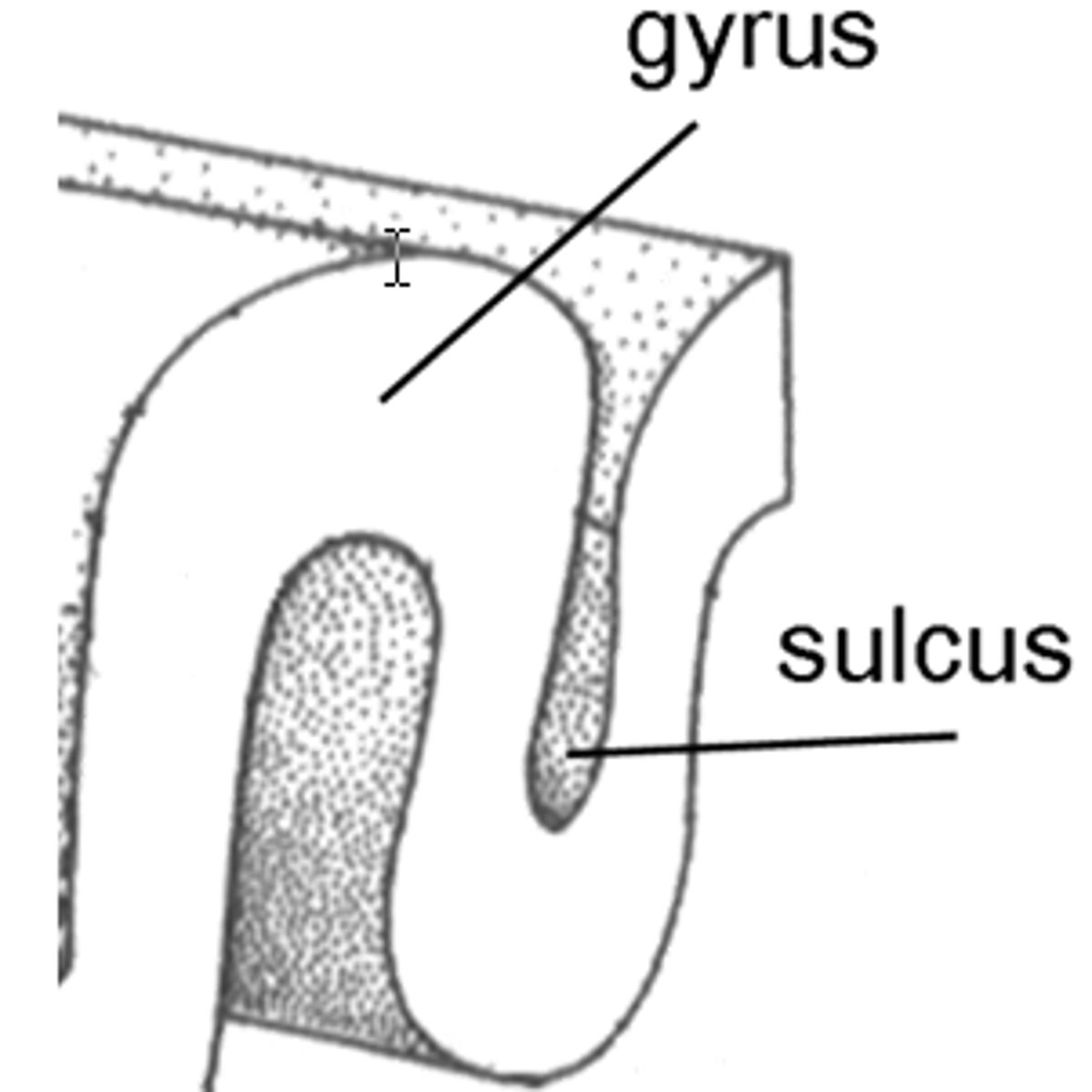
Telencephalon: Sucli and Gyri
- the cerebrum has small fissures (grooves) which create folds in the cerebrum
- this increases the surface area in the neural tissue, increasing the functional capacity of the tissue
fissures/grooves: Sulci
bumps/lumps: gyri
Different Sulci of the cerebrum
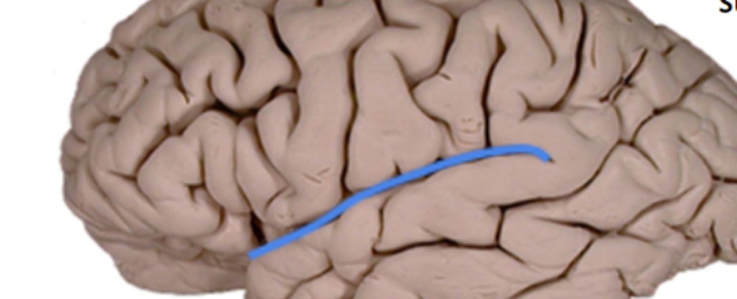
Lateral sulcus
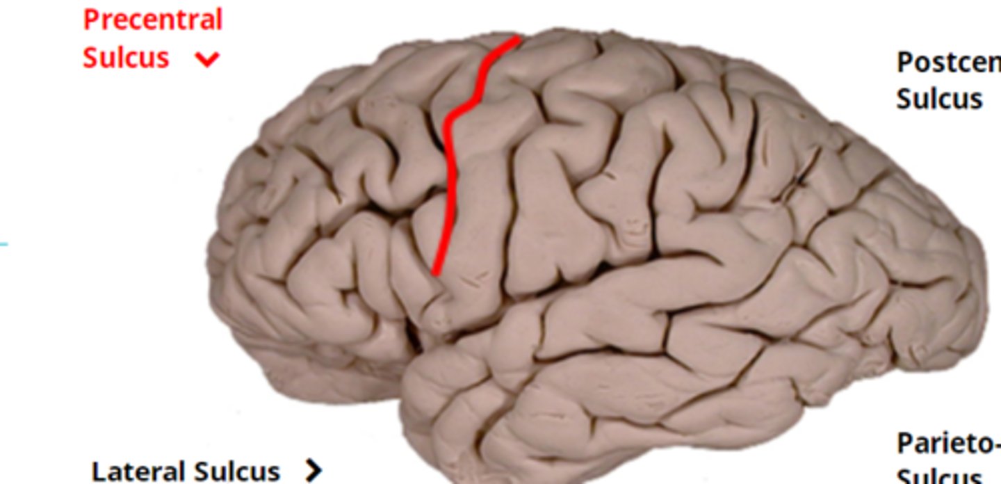
precentral sulcus
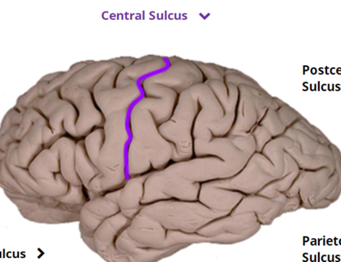
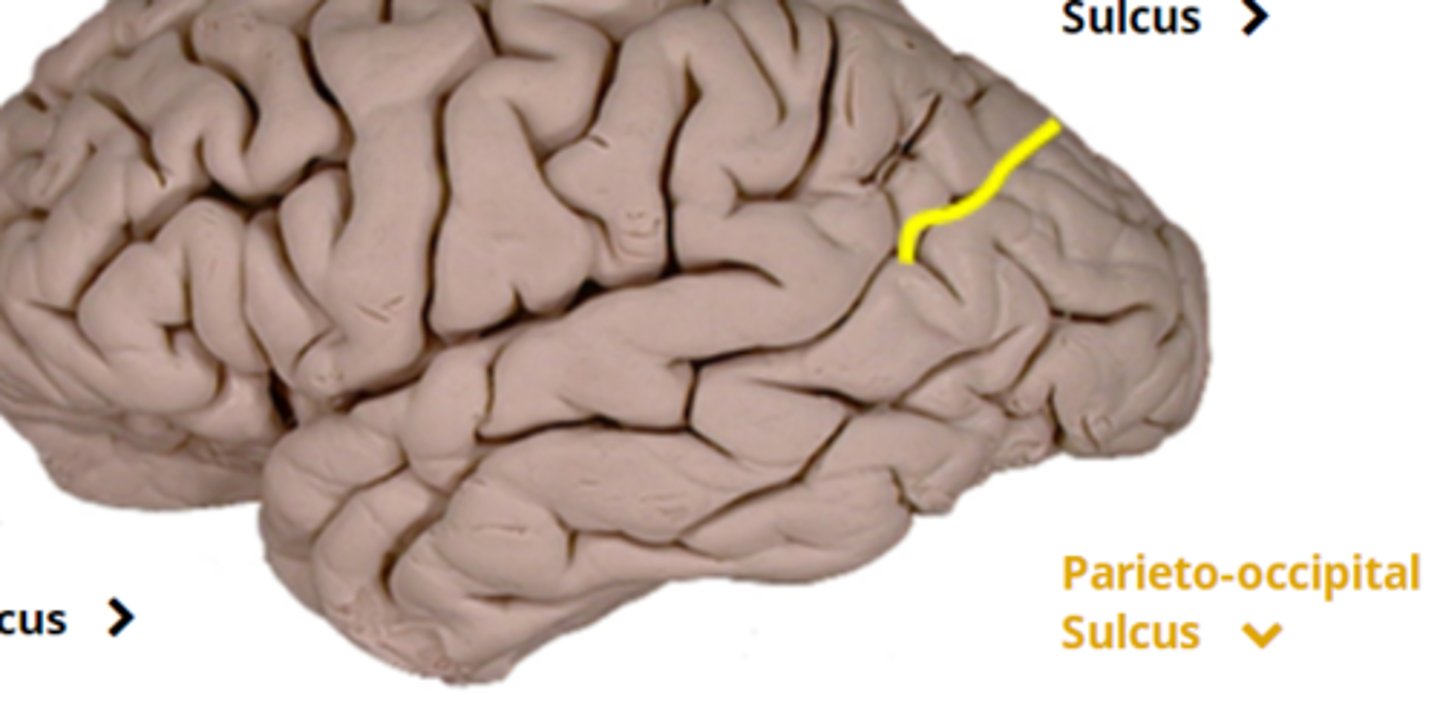
central sulcus
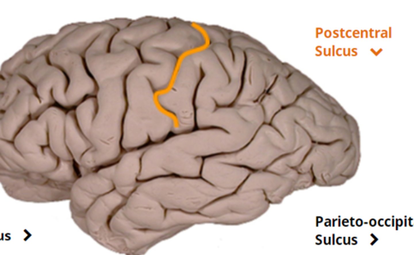
postcentral sulcus
parieto-occipital sulcus
lateral sulcus
Divides the temporal from frontal and parietal lobes.
- can't see it from the midsaggital section
precentral sulcus
- creates the anterior border of the motor cortex
central sulcus
- divides the frontal and parietal lobes
- also divides the motor and the sensory cortex
postcentral sulcus
creates posterior border of the sensory cortex
parieto-occipital sulcus
divides parietal and occipital lobes
- you can see It more from the medial view (inner view)
gyri of cerebrum
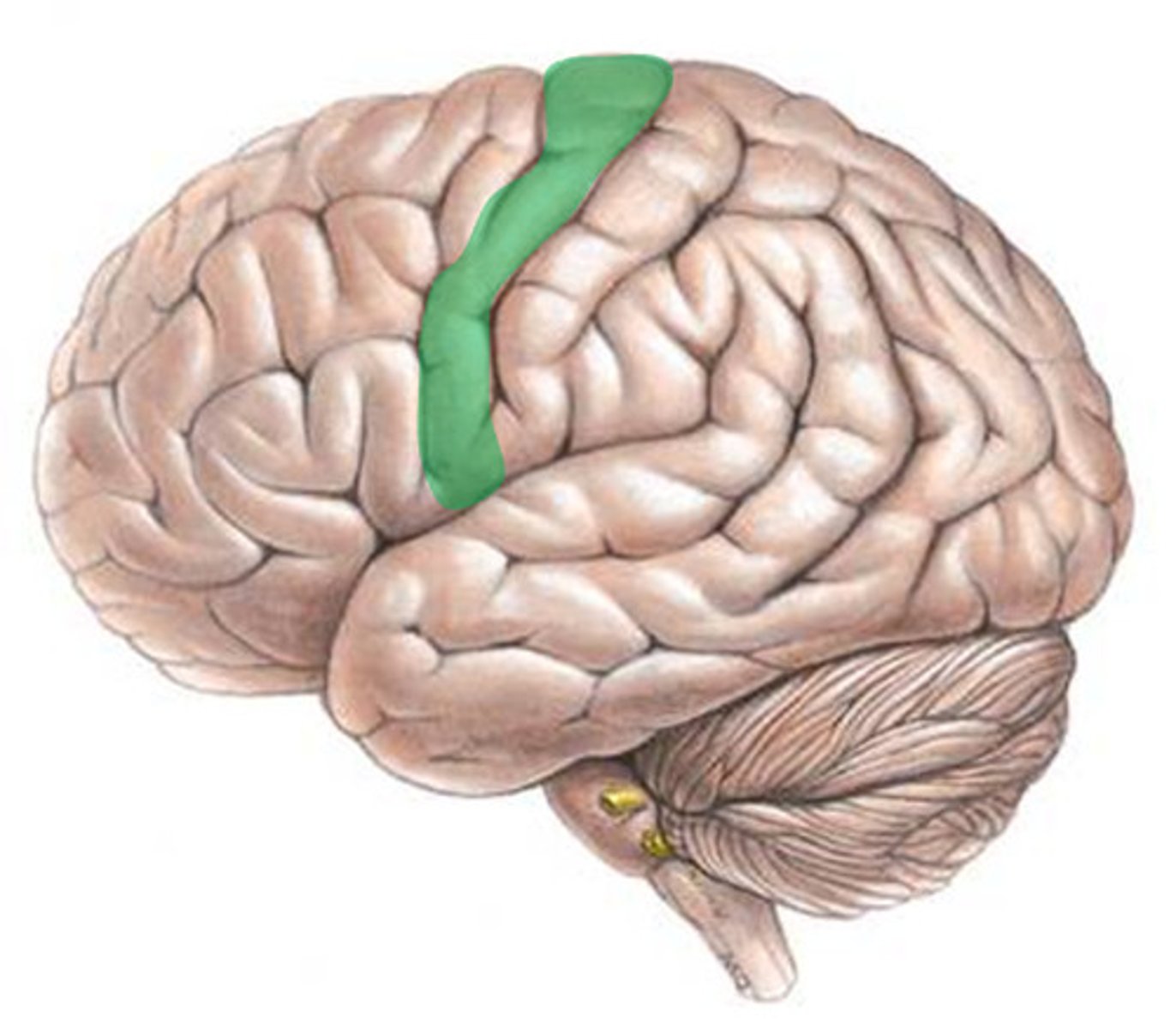
Precentral gyrus (motor cortex)
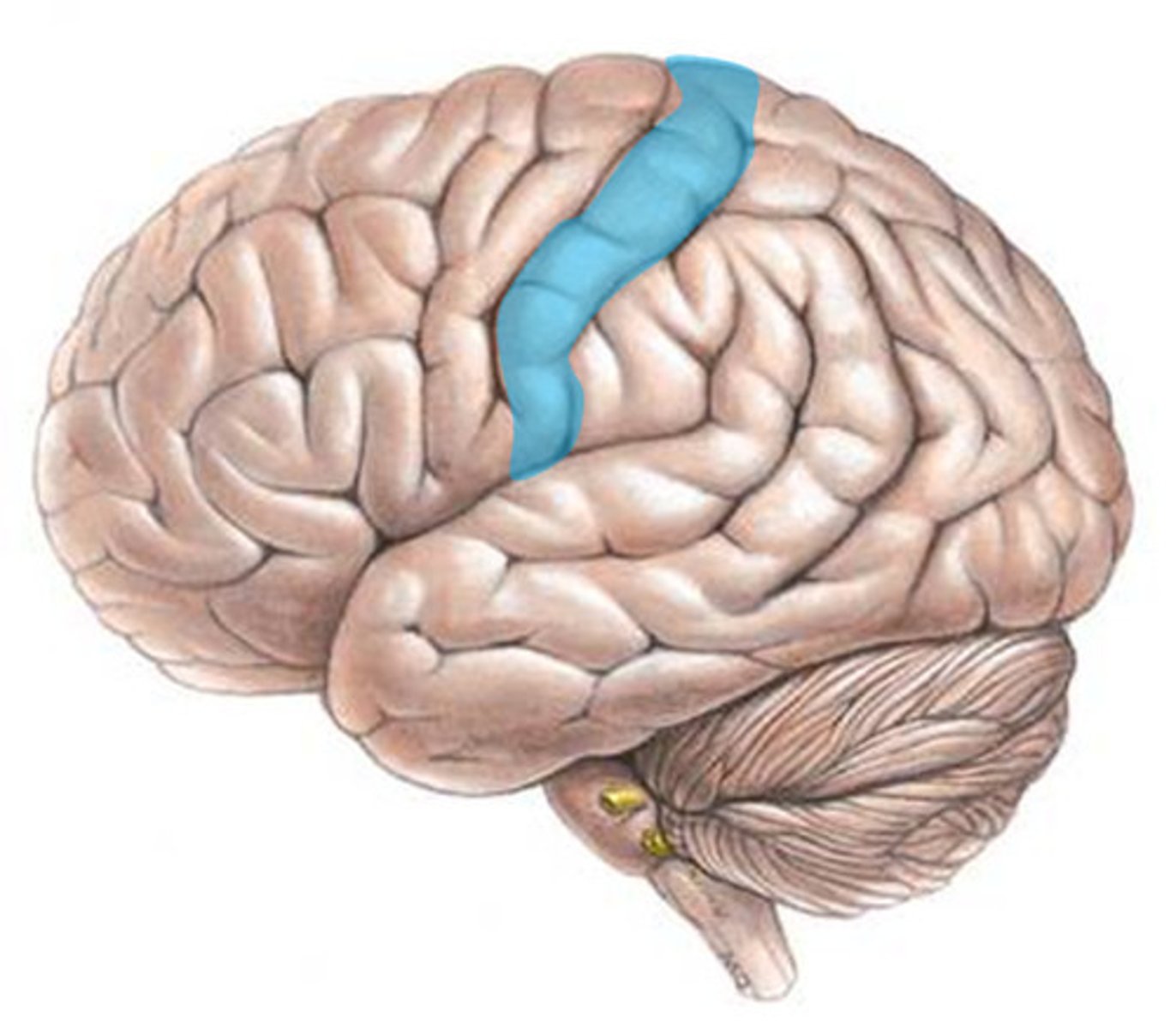
postcentral gyrus (somatosensory cortex)
Precentral gyrus (motor cortex)
- responsible for sending motor output signals
located anteriorly to the central sulcus
postcentral gyrus (somatosensory cortex)
- responsible for receiving sensory input related to touch and feel
- located posteriorly to the central sulcus
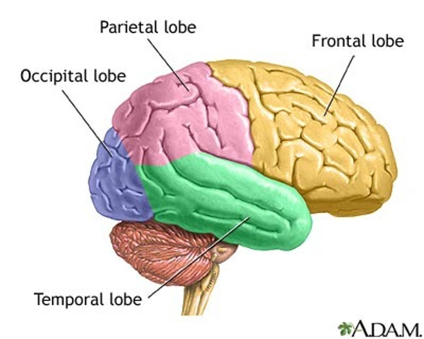
Telencephalon: Lobes of the cerebrum
- 4 lobes
- frontal, temporal, occipital, and parietal
Telencephalon: Grey matter of the brain
- made up of nerves cell bodies, short interneurons, and glial cells
- can be found in the cerebral cortex (outer) and the inner areas of the brain
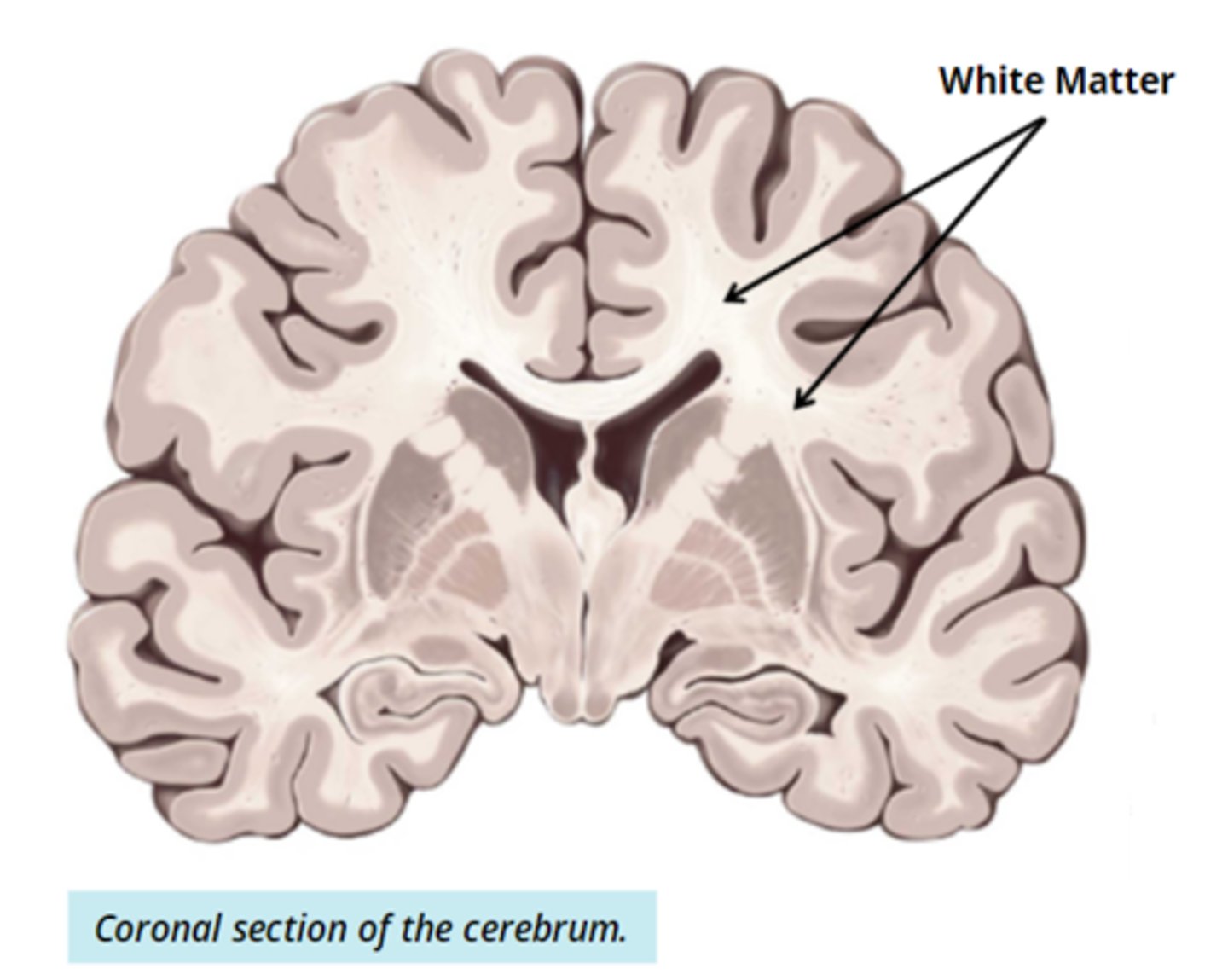
Telencephalon: White Matter of the brain
- made up of nerve fibres and axons
- found in the inner regions of the cerebrum
- ex. corpus callosum, connects the 2 hemispheres of the brain
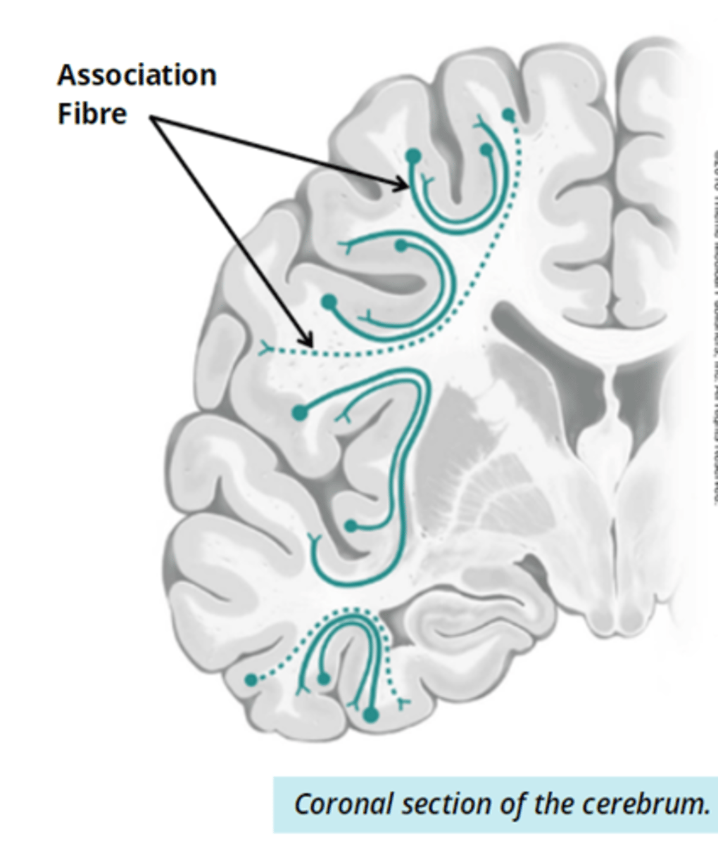
White Matter Tracts
3 categories of white matter tracts based on the structure and the type of communication they do
- associated fibres
- commissural fibres
- projection fibres
White Matter Tracts: Associated fibres
- facilitate communication within 1 hemisphere
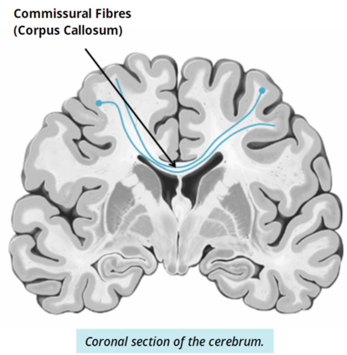
White Matter Tracts: Commissural fibres
- communication between the 2 hemispheres
-ex. corpus callosum
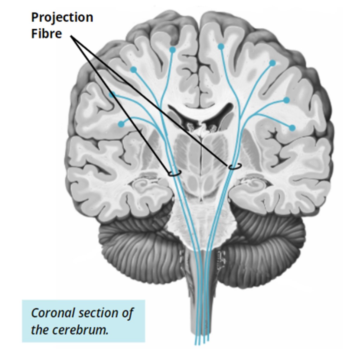
White Matter Tracts: Projection fibres
- facilitates communication between different levels of the CNS (like between the brain and the spinal cord)
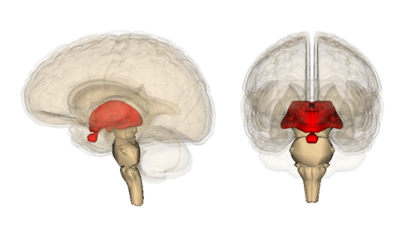
Diencephalon
- 2nd layer of the cerebrum (deep to the telencephalon)
- the core of the cerebrum, surrounded the cerebral hemispheres
- responsible for sensory and motor relay into and out of the brain, as well as control over the autonomic nervous system
parts of diencephalon
thalamus and hypothalamus
- encloses the 3rd ventricle
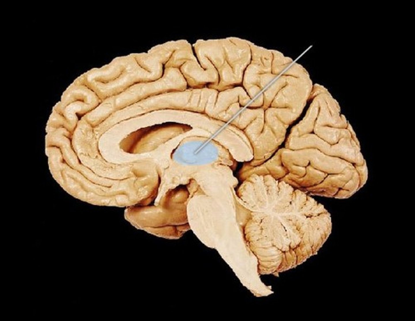
diencephalon: thalamus
- 2 bodies, 1 per hemisphere
- this part of the brain is responsible for relaying sensory information (except sense of smell) to other areas of the brain
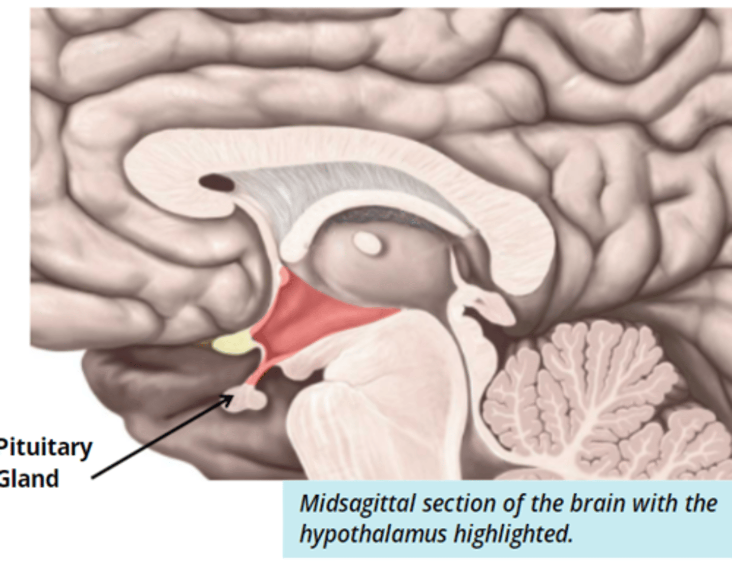
Diencephalon: Hypothalamus
- small structure below the thalamus that is connected to the pituitary gland
- controls the release of hormones through the pituitary gland
Parts of the brain: Brain Stem
- area of the brain that connects the brain to the spinal cord
- located just below the diencephalon and is made up of the midbrain, pons, and the medulla oblongata
- important because it contains many autonomic reflex centres and white matter tracts necessary for human functioning and survival
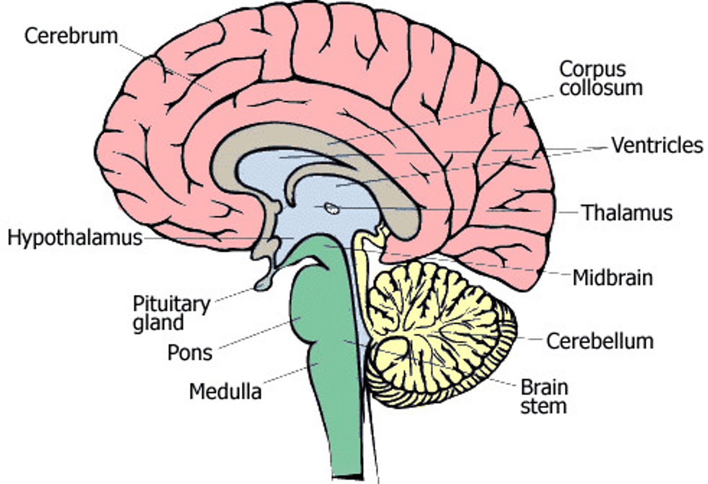
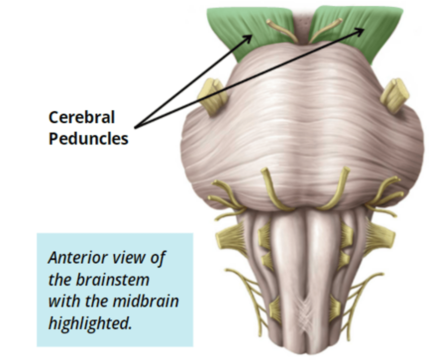
Brain stem: Midbrain
- most superior part of the brain stem
- anteriorly, the midbrain is made up of 2 large white matter tracts that make up the cerebral peduncles
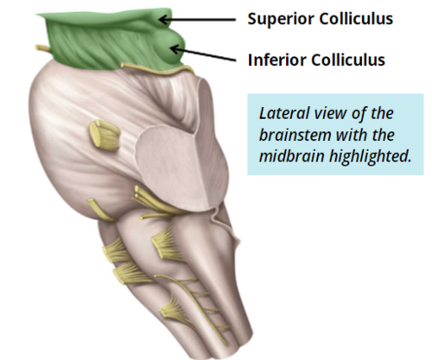
- posterior-laterally, the midbrain has 2 pairs of raised bumps known as superior and inferior colliculi
cerebral peduncles
anterior midbrain
superior and inferior colluculi
- posterior-laterally midbrain
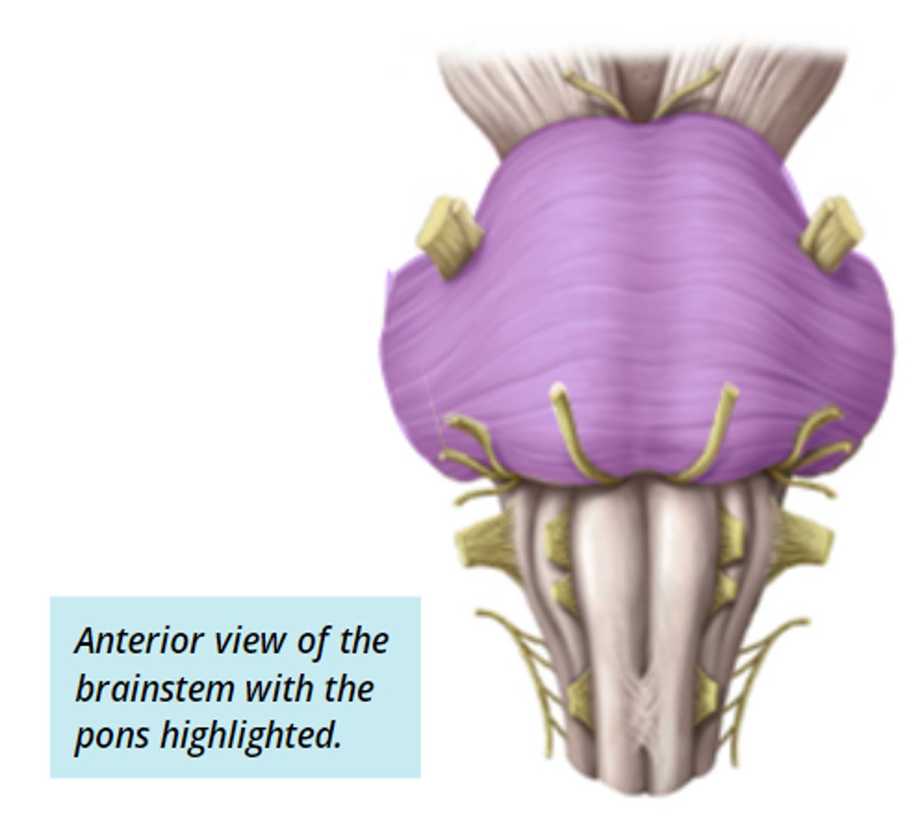
Brain stem: Pons
- structure in the middle of the brainstem
- located anteriorly to the 4th ventricle and the cerebellum
- pons have fibres connecting it to the cerebellum, allowing for communication between the 2 structures
- the red extends fully back
- the little bump is the 4th ventricle
Brain Stem: Medulla Oblongata
- AKA medulla
- most inferior structure of the brain stem
- continuous with the spinal cord
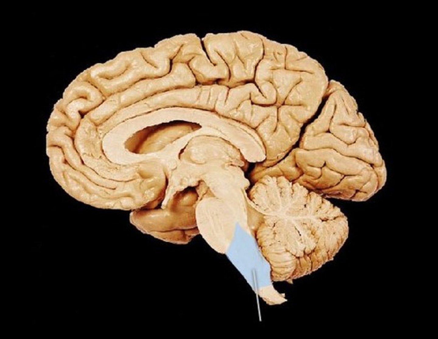
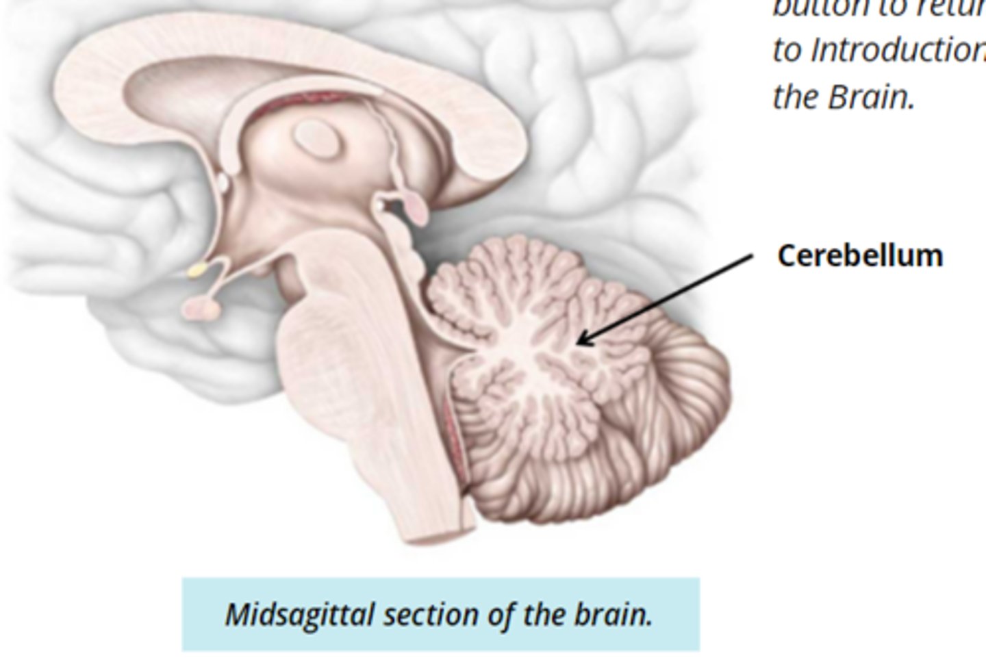
parts of the brain: cerebellum
- the structure that is appended to the back of the brainstem and sits underneath the occipital lobe of the cerebrum
function:
- coordinate motor activity
Support and Protection of the Brain
2 protective measures:
- meningies
- ventricles
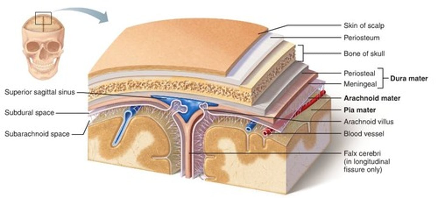
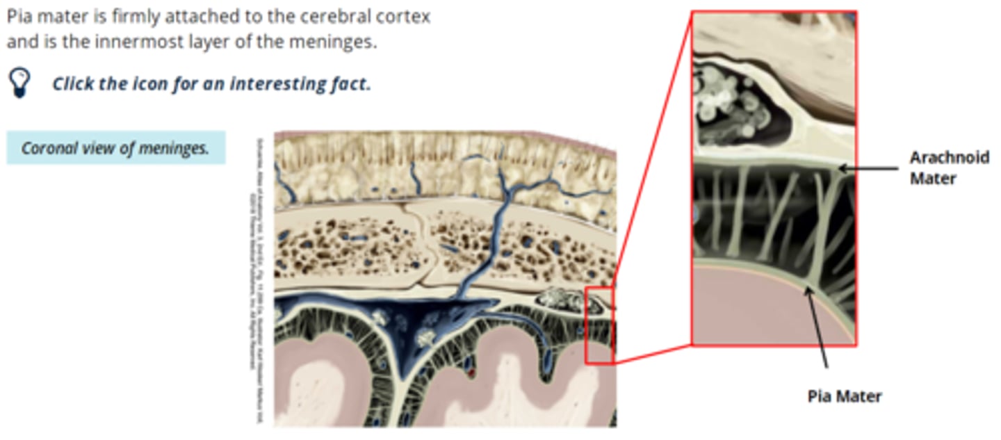
Support and Protection of the Brain: Meninges
3 connective tissue membranes portecting the structure of the CNS
- dura mater
- arachnoid mater
- pia mater
Dura Mater
- most superficial and tough layer of the meninges
- composed of a double layer of connective tissue that firmly attaches to the skull
projections of the Dura mater
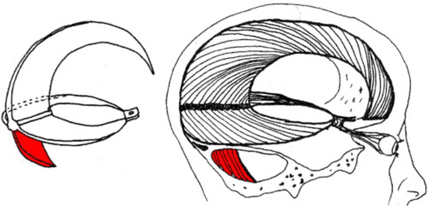
the dura mater also invaginate to:
- the longitudinal tissue, creating the FALX CEREBRI between the cerebral hemispheres
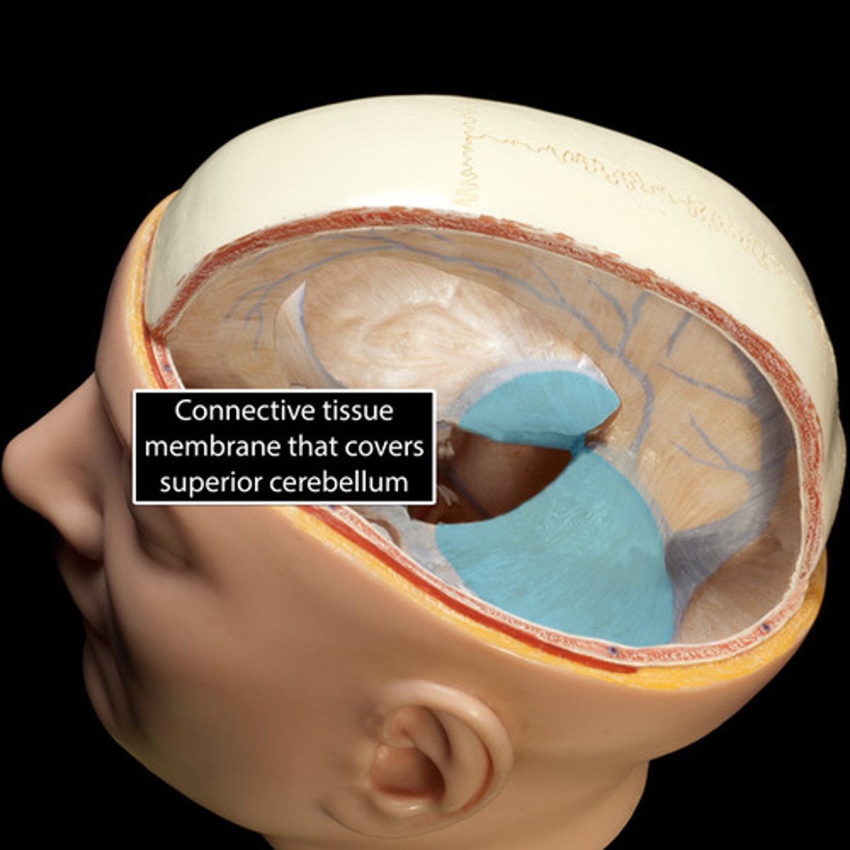
- the lateral fissure between the cerebrum and the cerebellum, known as the TENTORIUM CEREBELLI
- in the space at the middle of the cerebellum, known as the FALX CEREBELLI
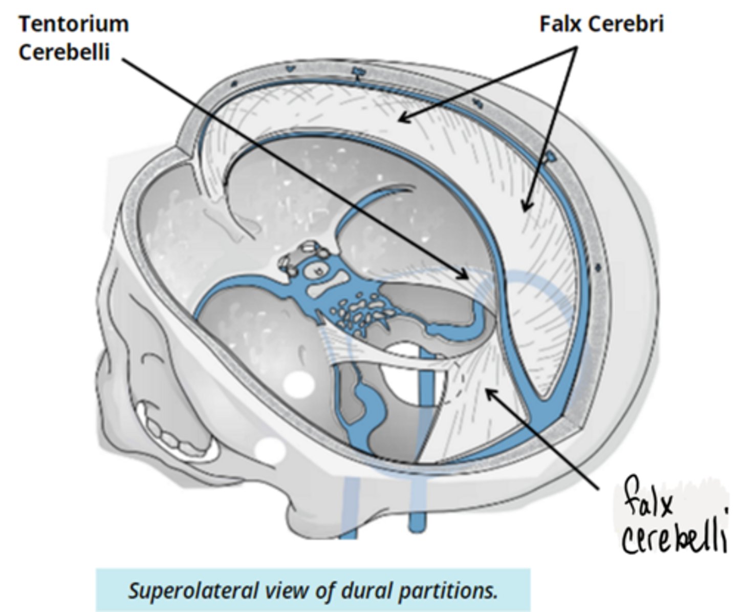
falx cerebri
separates the two cerebral hemispheres
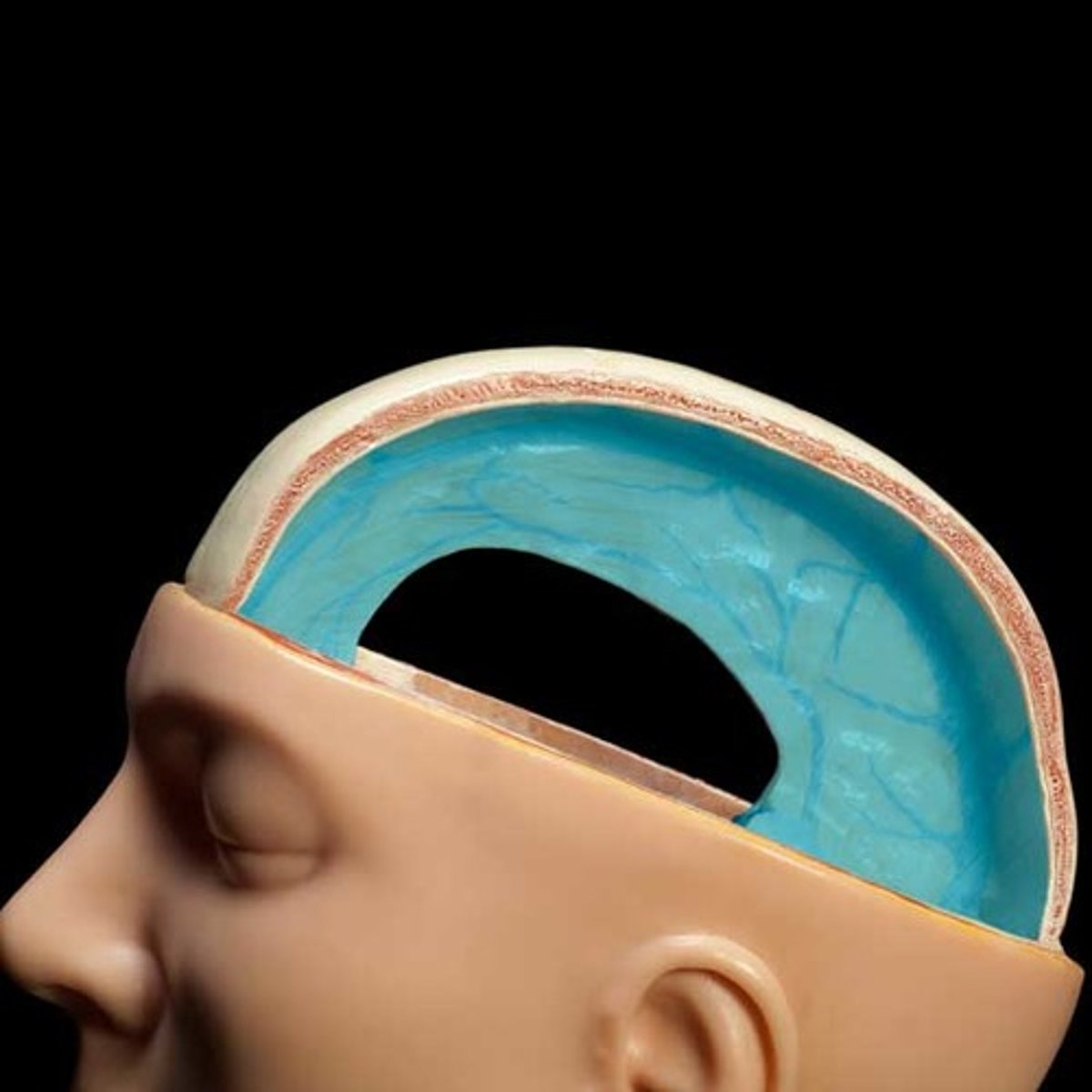
tentorium cerebelli
separates cerebrum from cerebellum
falx cerebelli
separates the two hemispheres of the cerebellum
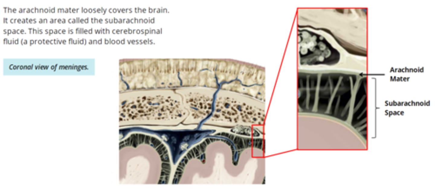
Arachnoid Mater
- losely covers the brain
- creates an area called the subarachnoid space
- this space is filled with cerebrospinal fluid (a protective fluid) and blood vessels
Pia Mater
- attached firmly to the cerebral cortex and is the innermost layer of the meninges
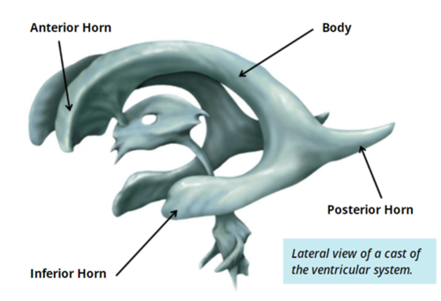
Support and Protection of the Brain: Ventricles
- also provides support and protection to the Brain
- made up of 4 hollow spaces that are continuous with each other and the spinal cord
- these spaces contain cerebral spinal fluid
lateral ventricles (2)
third ventricle
fourth ventricle
Ventricles: Lateral Ventricles
- paired "C" shapes spaces deep within the cerebral hemispheres
- a portion of the lateral ventricles can be found in each lobe
- 4 features - anterior horn, body, posterior horn, inferior horn
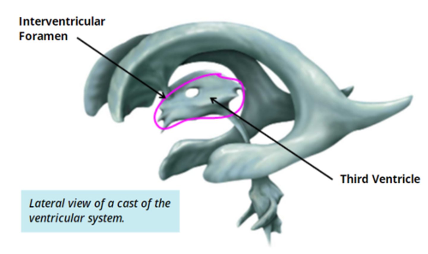
Ventricles: Third Ventricle
- 1
- located in the middle of the diencephalon between the 2 halves of the thalamus
- connected to the lateral ventricles by the
interventricular foramina
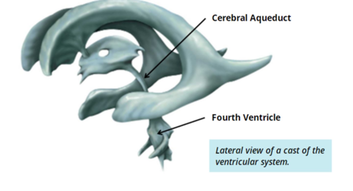
Ventricles: Fourth ventricle
- sits between the pons/medulla and the cerebellum
- connected to the third ventricle between the cerebral aqueduct
- it is continuous with the central canal of the spinal cord and the subarachnoid mater
cerebral spinal fluid (CSF)
- contained within the ventricles and flows from the lateral ventricle to the blood stream
function of CFS
- buoyancy effect to the brain and spinal cord because being surrounded by CFS makes these structures weigh less
- protection to the CNS (acts as a shock absorber)
- serves as a medium for transport of nutrients, waste, dissolved gases and other substances
production of CFS
- produced inside the ventricles by the choroid plexus
path flow of CFS
- produced in lateral ventricles
- flows down interventricular foramen
- third ventricle
- flows down the cerebral aqueduct
- fourth ventricle
- it leaves the ventricles, entering the subarachnoid space and the central canal of the spinal cord
- eventually will get absorbed into the blood stream
spinal cord
- located in the vertebral canal and supported by the vertebral column
- facilities communication between the CNS and PNS though SPINAL NERVES
spinal nerves
- spinal nerves exit the spinal cord to supply sensory and motor signals to the areas closest to where they exited
- ec. spinal nerves exiting the upper cervical region will serve the head and neck
- spinal nerves from the thoracic region will serve the thorax
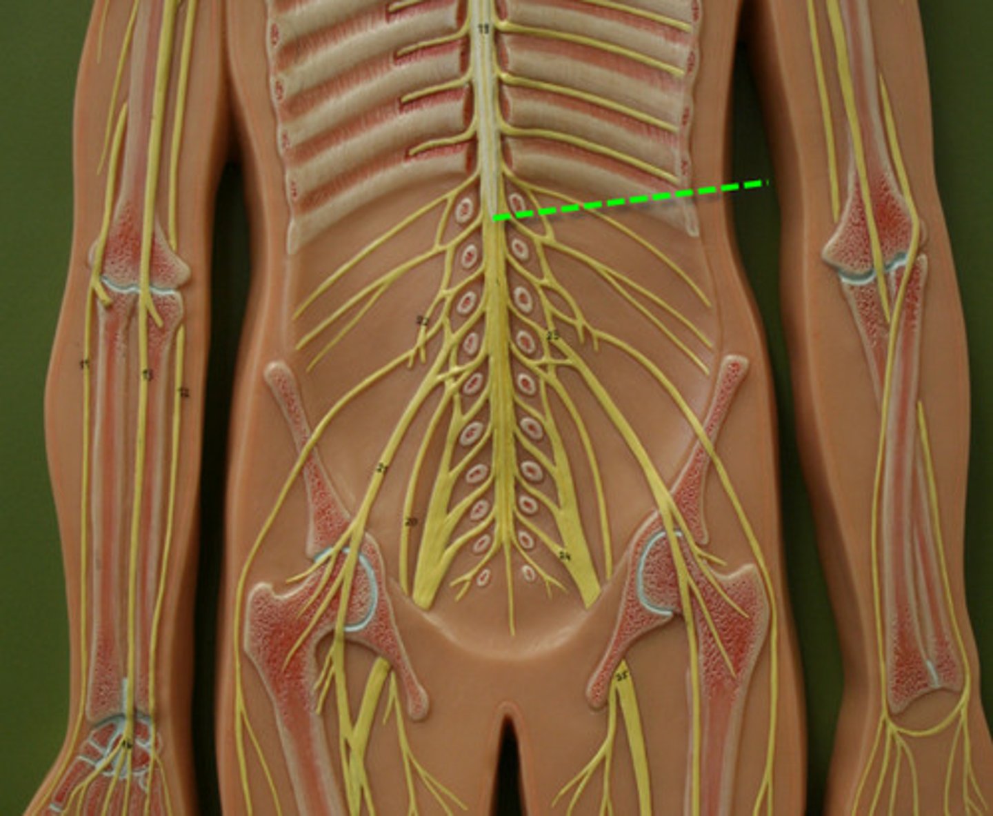
external topography of the spinal cord
- the spinal cord begins at the foramen magnum of the occipital bone
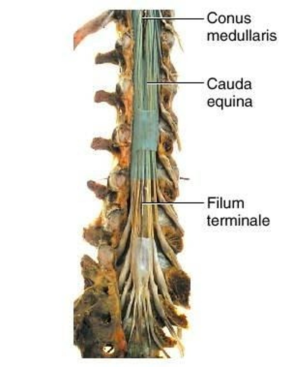
- extends inferiorly to the level of L2 (called CONUS MEDULLARIS)
- interior to L2, the axons emerging from the spinal cord are known as CAUDA EQUINA
- inside the cauda equina, a small thin stand of Pia mater (FILUM TERMINALE) anchor the spinal cord to the sacrum and coccyx
conus medullaris
cauda equina and filum terminale
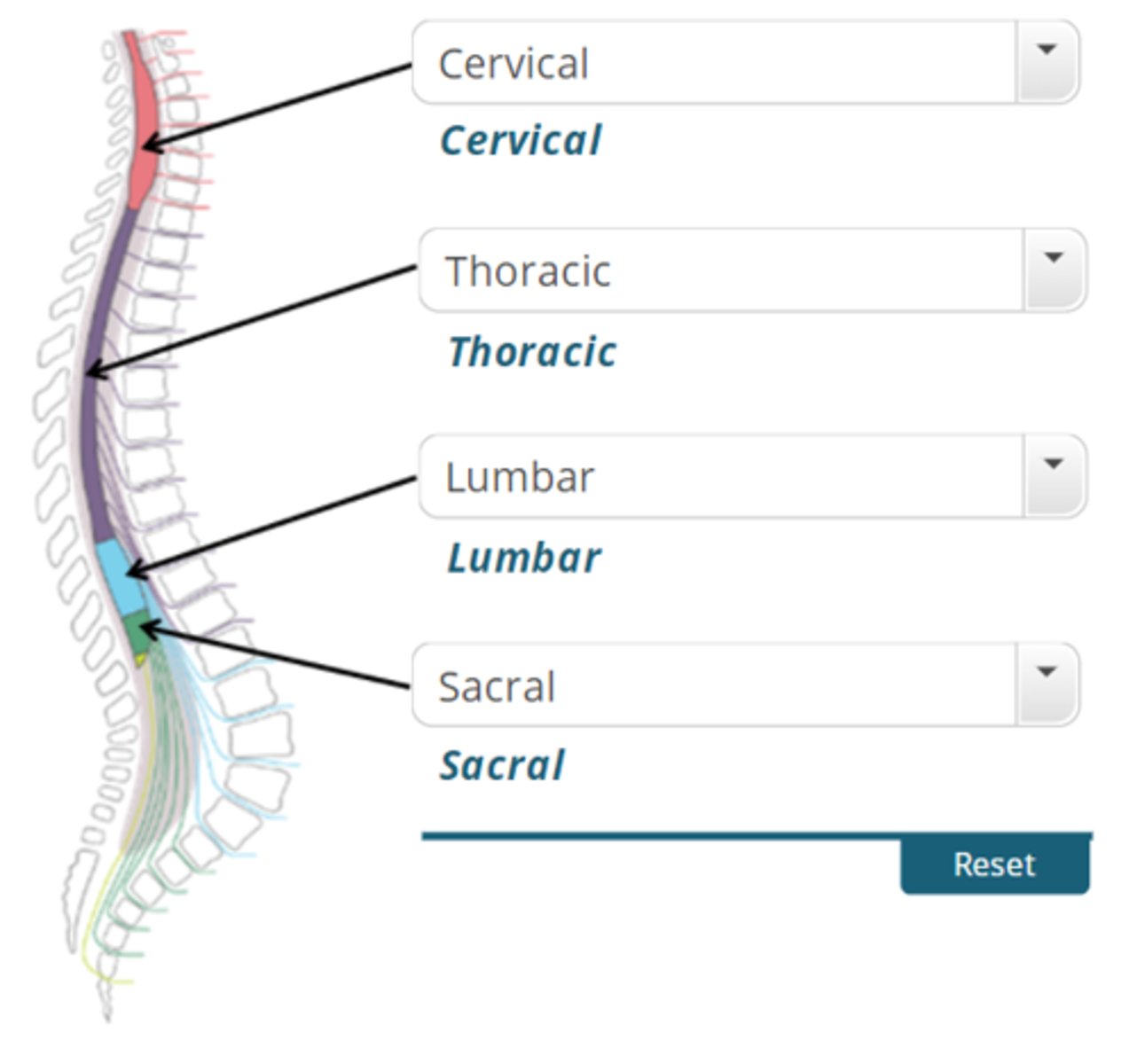
Regions of the Spinal cord
cervical, thoracic, lumbar, sacral
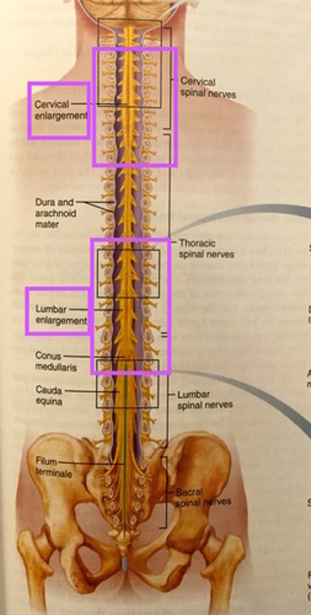
Spinal Enlargements
- the spinal cord is enlarged at the cervical and lumbar levels
- these enlargements are there because of additional nerves exiting the spinal cords to supply the limbs
- cervical enlargements supply the upper limbs
- lumbar enlargements supply the lower limbs
Spinal cord injury
- if the spinal cord is damaged, the messages from the brain and the rest of the body cannot go through those damaged areas
- the functions of the body above the point of injury will function normally, while the functions of the body below the point of injury will suffer impairment
- the higher the injury, the more severe
C1-C3 injury
- limited movement of the head and neck, can't move body
C3-C4 injury
- additionally some shoulder movement
C5 injury
- additionally can bend elbows and rotate hands
C6 and C7 injury
- additionally can extend elbows, and rotate palms
C8-T1 injury
- additionally can move fingers
T2-T6 injury
- normal upper Body
- impairment in the legs
T7- T12 injury
- more control than T2-T6
L injury
- upper body movement, can normally have some movement in the hips and knees
Support and Protection of the spinal cord: Meninges
- dura
- arachnoid
- pia
spinal meninges: Dura Mater
- (the brain dura is a double layer of connective tissue)
- the dura mater of the spinal cord only has 1 layer of tissue
- between the external surface of the dura mater and the bone is fat-filled external space
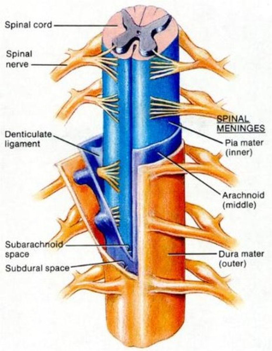
spinal meninges: arachnoid mater
- deep to the dura
- between the arachnoid and the pia mater is the subarachnoid space of the brain, and contains CFS

spinal meninges: Pia mater
- deep to the arachnoid mater
- directly attached to the spinal cord

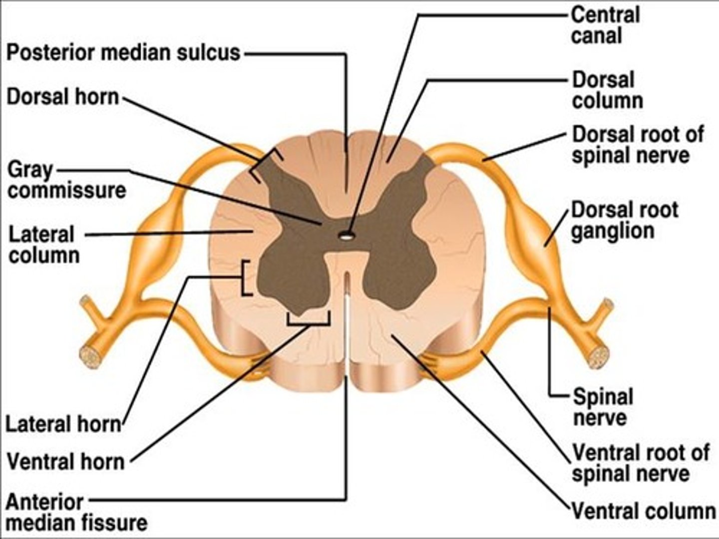
Internal Topography of the spinal cord
- unlike the brain, grey matter is on the inside and white matter is on the outside (this is due to different cell types)
grey matter: mainly composed of dendrites, neuronal cell bodies, glial cells, and unmyelinated axons
white matter: mainly composed of myelinated axons