Skin masses in equines SDL
1/50
There's no tags or description
Looks like no tags are added yet.
Name | Mastery | Learn | Test | Matching | Spaced | Call with Kai |
|---|
No analytics yet
Send a link to your students to track their progress
51 Terms
Give the 3 most common equine skin neoplasias
-melanoma
-Sarcoid
-Squamous cell carcinoma
what to consider in the history and clinical exam
Patient signalment (age/breed/sex)?
Where is the mass?
How long has it been there?
Colour?
Size?
Shape?
Ulcerated?
Painful to palpation ?
Speed of growth?
Normal hair / hair has fallen out?
what might be on you ddx list when looking at equine skin masses
Neoplasia
Abscess
Cyst
Eosinophilic granuloma
Papilloma (warts)
how is diagnosis achieved
Diagnosis achieved by history, clinical appearance and sometimes biopsy.
Describe a melanoma
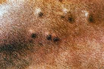
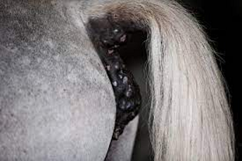
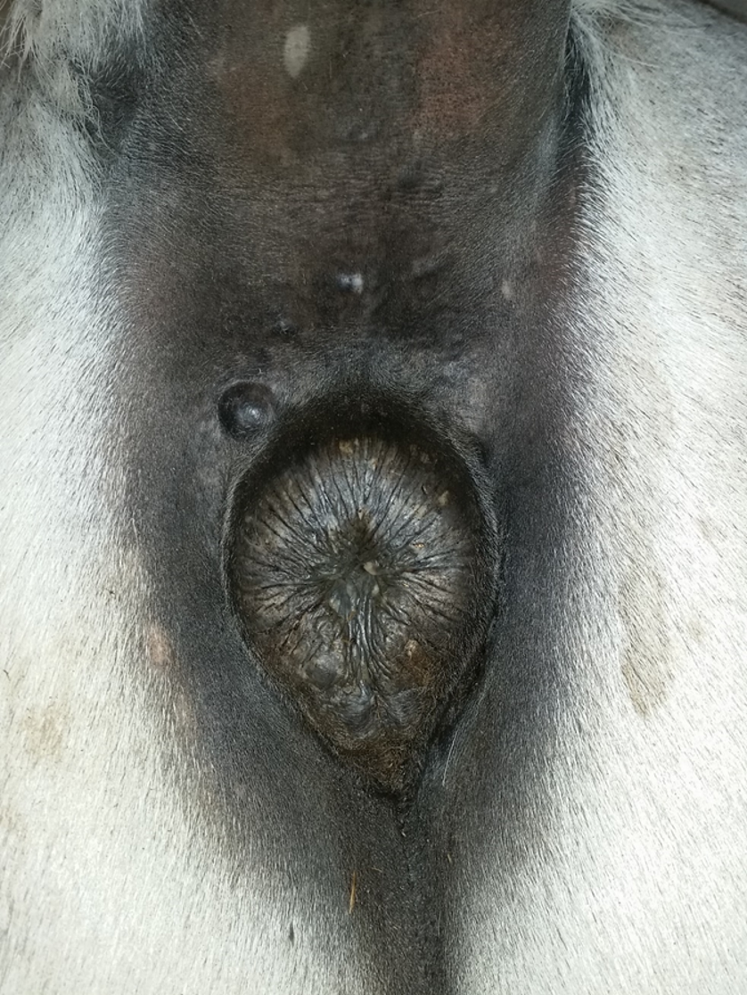
Black, spherical or plaque like masses that are most common around the perineum and parotid region on grey horses
Describe a sarcoid
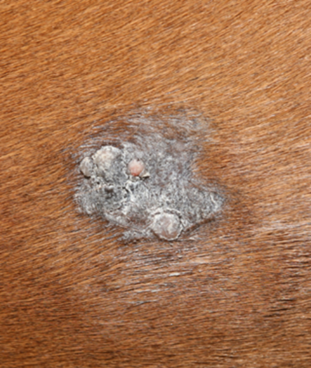
These masses are usually hairless, sometimes ulcerative and most commonly found in the groin, axilla and around the eyes
Describe squamous cell carcinoma
These masses can be described as raised, irregular, pink, locally invasive
-They are most common around the genitals and eyes
Describe sarcoids
-Most common skin tumour in horses
-Locally invasive
-Fibroblastic
-wart -like
-6 different types of sarcoids
-multiple lesions are more common
how to diagnose a sarcoid
Diagnosis is usually made by visual appearance alone, but can be confirmed by biopsy
Describe the histological appearance of sarcoids?
Increased density of dermal fibroblasts which form interlacing bundles and whorls within the dermis
What contributes to the development of equine sarcoids?
Bovine papillomavirus
How are sarcoids transmitted?
-flies act as vector for transmission of the virus allowing sarcoids to be passed from one horse to another
-Flies usually reside in the groin, around the axilla and eyes
What are the types of sarcoid?
-occult
-Verrucose
-nodular
-Fibroblastic
-Malignant
-Mixed
What is a mixed lesion?
Areas of two or more types of sarcoids with no distinguishable margin between them
Occult
Verrucose
Nodular
Fibroblastic
Malignant
Mixed (verrucose with fibroblastic parts)
Describe treatment for sarcoids
-Sarcoids do not always require treatment if they are small, slow growing, not ulcerated / bleeding and do not interfere with tack.
-Even so, early treatment is usually a good idea to prevent lesions becoming large and to reduce the risk of spread to other locations on the same horse and to other horses.
What are the treatment options of sarcoids?
1) Laser surgical removal - A laser is preferred to a scalpel as it reduces the risk of recurrence, bleeding and post operative pain.
2) Cryotherapy
3) Caustic cream application
4) Elastrator band application (causes hypoxic necrosis in nodular sarcoids).
5) Radiotherapy (particularly for periocular sarcoids)
Describe the treatment of melonomas
It is reasonable to leave many melanomas untreated, but sometimes early removal is beneficial. In particular those around the anus can be removed easily with a surgical laser, to prevent future defacation difficulties
describe melanomas
usually benign, black, nodular, slow growing, firm nodules
that occur most commonly on the perineum, sheath and parotid region of grey coloured horses.
how to diagnose melanomas
Diagnosis is usually made on visual appearance.
What are the most common locations of squamous cell carcinoma?
-Penis
-Third eyelid
what are squamous cell carcinomas
are locally invasive, pink, irregular, sometimes ulcerated lesions which can grow rapidly
How is squamous cell carcinoma treated?
All SCC should be excised as they can rapidly progress and spread,
this is usually curative - complete removal should be confirmed histologically
Describe eosinophilic granuloma
-Eosinophilic granuloma are small, non-itchy, firm, round, non-painful, raised nodules, with normal hair covering.
Where are eosinophilic granulomas located?
-They can be located anywhere on the body but are found most commonly on the withers and back.
What are the causes of eosinophilic granulomas?
-They are not neoplasia but their actual cause is unknown.
-There is some evidence that they are a type of hypersensitivity reaction to insect bites or may follow trauma e.g. ill fitting tack.
How are eosinophilic granulomas diagnosed?
Diagnosis is by clinical appearance, fine needle aspirate or biospy.
How are eosinophilic granulomas treated?
Treatment is often not required, but surgical excision or injection with corticosteroid may be performed if they are interfering with tack.
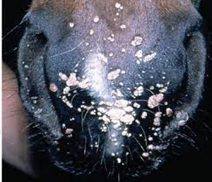
What are juvenile papillomas?
Multiple, small, irregular, verrucose, grey, proliferative lesions
Where are juvenile papillomas (warts) located?
Most commonly found on the muzzle, face and sheath of young horses
What are juvenile papillomas caused by?
Caused by equine papilloma virus
How are juvenile papillomas treated?
-diagnosis is by clinical appearance (and possibly biopsy).
- They are usually self-limiting so that treament is not required, but cryotherapy may be considered if they are severe.
Juvenile papilloma (warts)
Eosinophilic granuloma
Eosinophilic granuloma
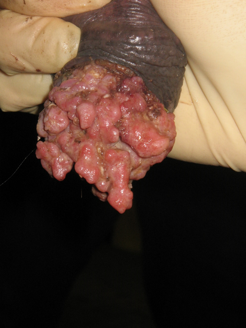
Squamous cell carcinoma
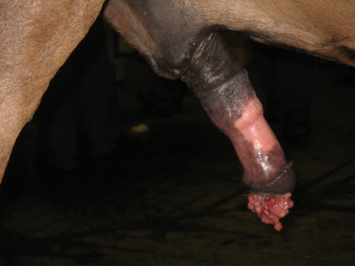
Squamous cell carcinoma
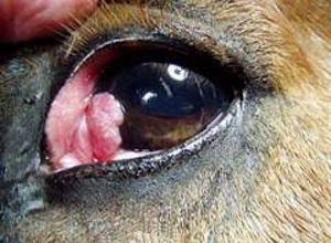
Squamous cell carcinoma
melanoma
melanoma
describe mixed sarcoid
Areas of 2 or more types of sarcoids with no distinguishable margin between them. (This one is verrucose with fibrobalstic parts).
describe malignant sarcoid
A rarer form of sarcoid which have extensive rapid spread over a wide area of skin with cords of sarcoid tissue interspersed with nodules and fibroblastic lesions.
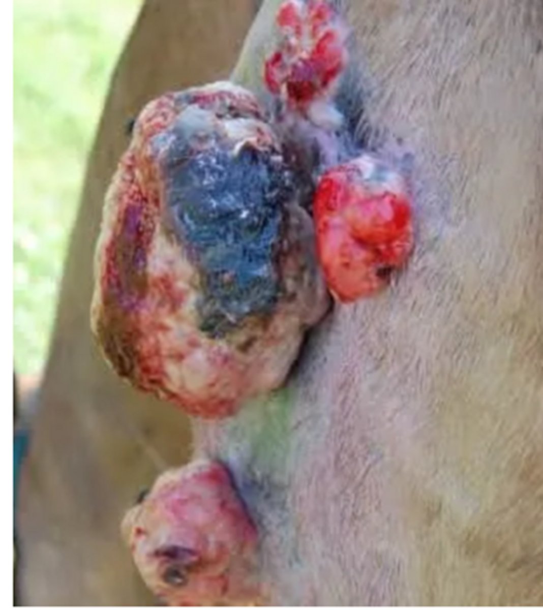
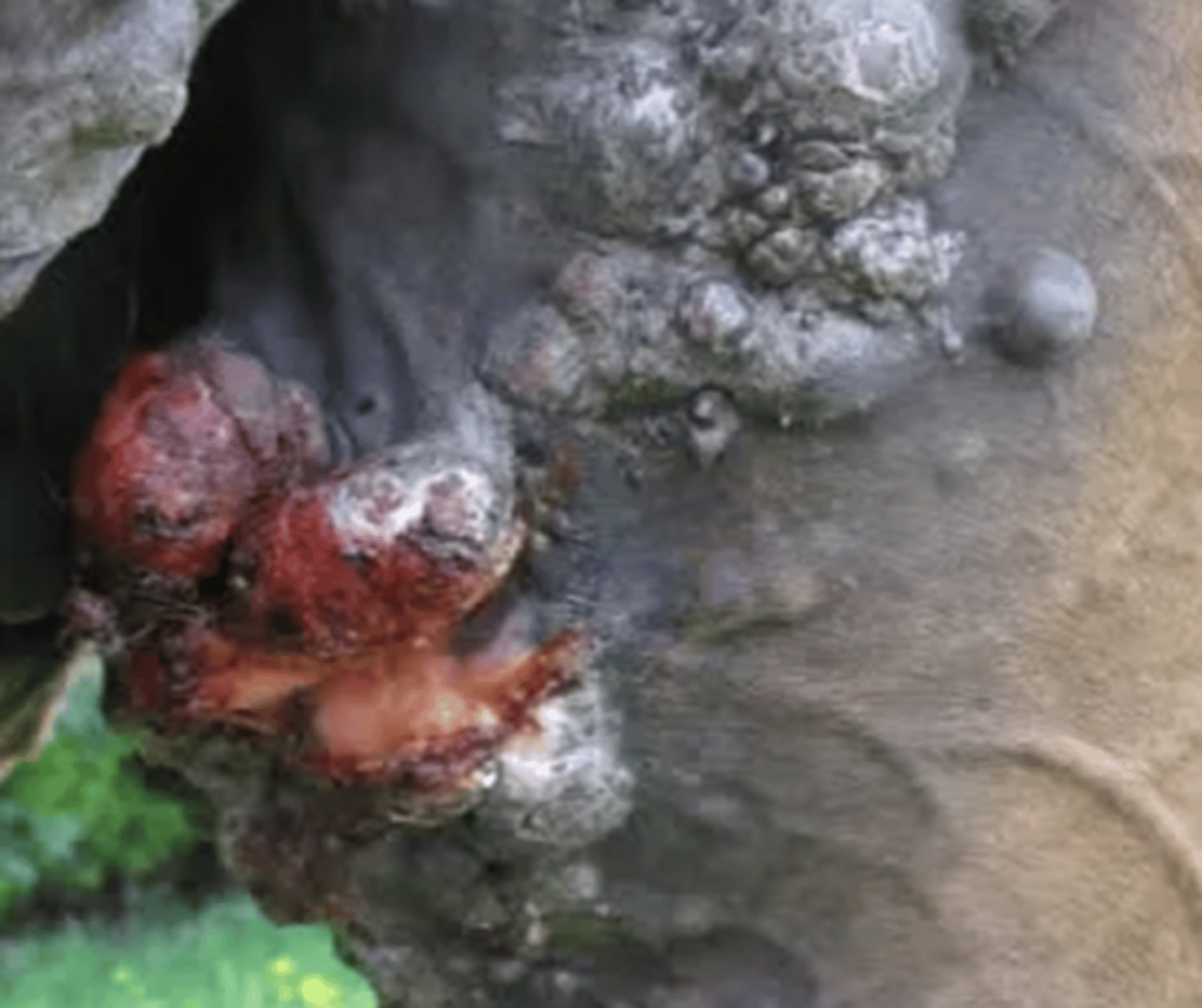
describe fibroblastic sarcoid
Ulcerative nodular masses that frequently bleed when knocked, they can appear like granulation tissue
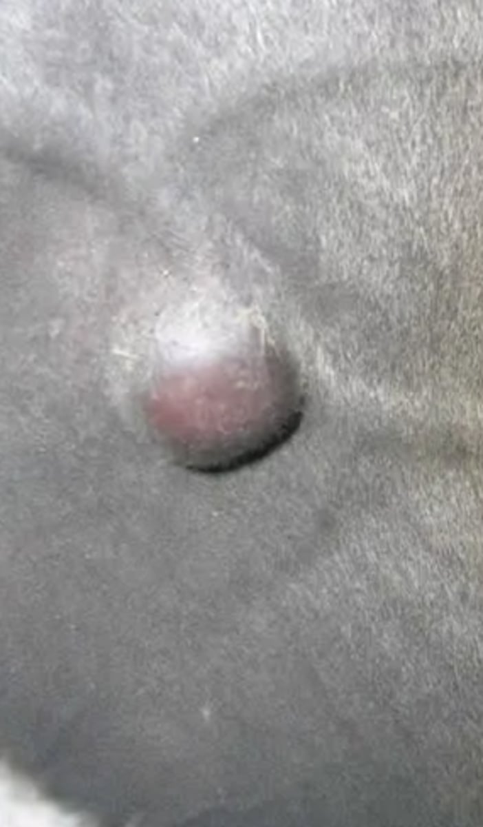
describe nodular sarcoid
Firm round nodules within the skin, usually fixed within the skin, however the skin can normally move freely over the underlying tissue
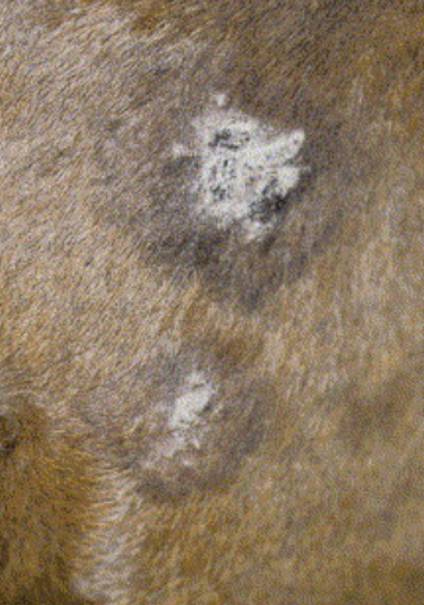
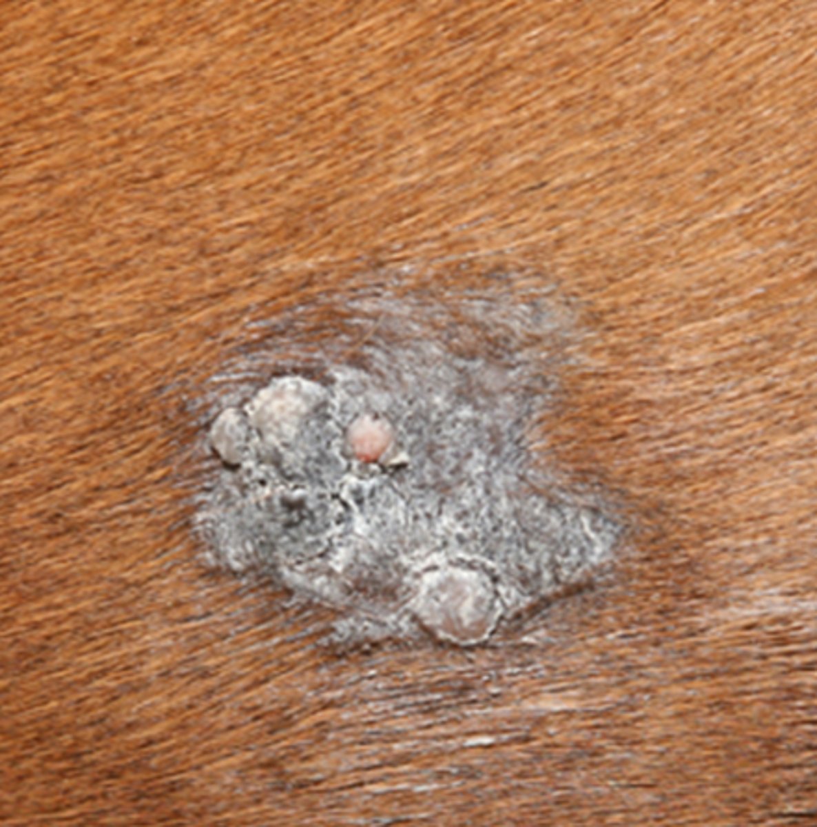
describe verrucose sarcoid
plaques of cracked flaky irregular skin, often described as wart like.
The margins of these can be difficult to determine
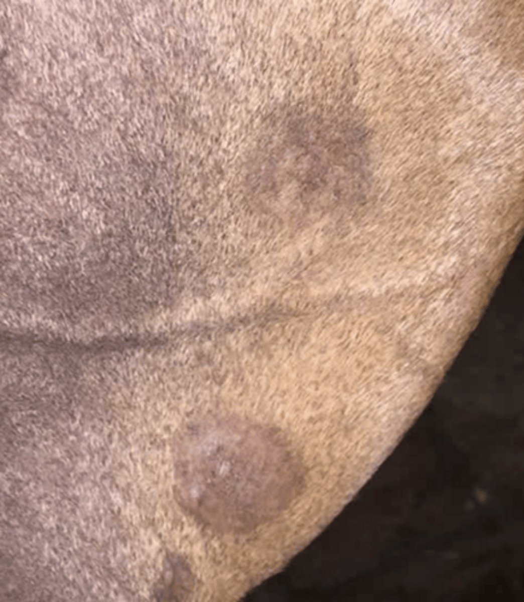
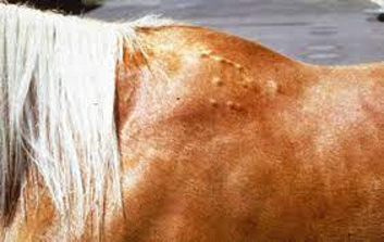
describe occult sarcoid
flat often hairless areas of skin, can be subtle and difficult to spot, may even be difficult to determine from ringworm