Erythrocyte and Morphologic Abnormalities of RBC
1/84
There's no tags or description
Looks like no tags are added yet.
Name | Mastery | Learn | Test | Matching | Spaced | Call with Kai |
|---|
No analytics yet
Send a link to your students to track their progress
85 Terms
Maturation of RBC
18 to 21 days
BFU-E →CFU-E = 7 days
CFU-E →Pronormoblast = 7 days
Pronormoblast →Mature RBC = 7 days
Morphologic Trend as Cell Matures (Erythroblast vs. More mature stage)
Cell size
N:C ratio
Nucleoli
Nuclear chromatin
Color of cytoplasm
Cell size - erythroblast larger, mature smaller
N:C ratio - erythroblast hgher, mature lower
Nucleoli - erythroblast present, mature absent
Nuclear chromatin - erythroblast fine, homogenous, mature coarse, clumped, condense
Color of cytoplasm - erythroblast blue, mature pink
Nomenclature of RBC Maturation series
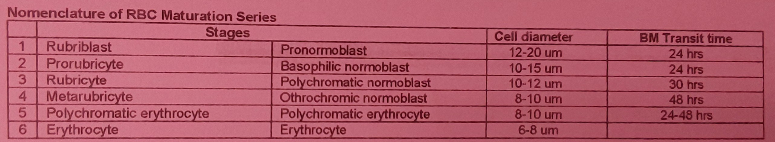
First morphologically identifiable RBC precursor
Contains 1 to 2 nucleoli
Globin production begins
Pronormoblast
Intensely basophilic (deeper blue) cytoplasm
Chromatin begins to condense (resembles a wheel with broad spokes)
Detectable hemoglobin synthesis occurs
Basophilic normoblast
First stage in which pink color (due to hemoglobin) is seen
Hemoglobin synthesis increases
Last stage capable of mitosis
Polychromatic erythroblast
Complete condensation of nucleus (pyknotic)
Hemoglobin production continues on the remaining ribosomes
Last nucleated stage (nucleus is ejected from the cell later in this stage)
Orthochromic normoblast
Completion of hemoglobin production
Residual RNA imparts a bluish tinge to the cytoplasm
Also known as diffusely basophilic erythrocyte
Polychromatic erythrocyte
Characteristics of Mature RBC
Size: _ um
Central pallor: _ of cell size
Shape: _
Primary function: _
Size: 7-8 um
Central pallor: 1/3 of cell size
Shape: biconcave/discocyte
Primary function: deliver oxygen to tissues
RBC membrane consists of:
_% carbohydrates
_% lipids
_% proteins
8% carbohydrates
40% lipids
52% proteins
Lipids in RBC membrane (2)
Phospholipids - dynamic fluidity to membrane
Cholesterol - tensile strength to the lipid bilayer
2 types of protein in RBC membrane
Integral/Transmembrane Protein
Peripheral/Cytoskeletal Protein
transport ions, water, and glucose, and anchor cell membrane receptors
also provides vertical membrane support
Integral/Transmembrane Protein
Example of Integral/Transmembrane Protein
Aquaporin-1
Glycophorins (A,B,C)
certain blood group antigens (Rh)
provides lateral or horizontal membrane stability
Peripheral protein
Example of Peripheral proteins
Alpha-spectrin
Beta-spectrin
Ankyrin
Adducin
Actin
Protein 4.1 and 4.2
Tropomodulin
Tropomyosin
Production of RBCs
Stimulus:
Source of EPO:
Stimulus: Hypoxia
Source of EPO: Peritubular interstitial cells of the kidney
EPO is a glycoprotein hormone and growth factor that has the following effects (3)
early release of reticulocytes in BM
preventing apoptosis of RBC progenitors
less time of RBC maturation in BM
EPO measurement
specimen: _
normal value: _
EDTA plasma measured by chemiluminescence
10 to 30 U/L
Destruction of RBCs
Extravascular Hemolysis
→%:
→Mechanism:
→Lab findings in excessive hemolysis:
→%: 80% to 90%
→Mechanism: RES macrophage-mediated
→Lab findings in excessive hemolysis: High serum conjugated bilirubin (B1) and urine urobilinogen
Destruction of RBCs
Extravascular Hemolysis
→%:
→Mechanism:
→Lab findings in excessive hemolysis:
→%: 10% to 20%
→Mechanism: Fragmentation of RBCs
→Lab findings in excessive hemolysis: low serum haptoglobin and hemopexin, hemoglobinemia, hemoglobinuria
Reference method used for RBC survival studies:
Chromium-51 Radioisotope Labelling
RBC Metabolic Pathways
_______
main source of ATP (90%)
glucose is catabolized into _
produces a net gain of _ molecules of ATP
important enzyme: _
EMBDEN-MEYERHOF PATHWAY
main source of ATP (90%)
glucose is catabolized into PYRUVIC ACID
produces a net gain of 2 molecules of ATP
important enzyme: HEXOKINASE, PYRUVATE KINASE, LACTATE DEHYDROGENASE
Embden-Meyerhof Pathway is also called _
anaerobic glycolysis
RBC Metabolic Pathways
_______
detour of glycolysis (5% to 10%)
generates _ to reduce glutathione
detoxifies oxidative compounds and safeguards hemoglobin
important enzyme: _
RBC Metabolic Pathways
HEXOSE MONOPHOSPHATE PATHWAY
detour of glycolysis (5% to 10%)
generates NADPH to reduce glutathione
detoxifies oxidative compounds and safeguards hemoglobin
important enzyme: G6PD
Hexose Monophosphate Pathway is also called _
Aerobic glycolysis
RBC Metabolic Pathways
_______
prevents formation of _
maintains iron in ferrous state (Fe2+)
important enzyme: _
RBC Metabolic Pathways
METHEMOGLOBIN REDUCTASE PATHWAY
prevents formation of METHEMOGLOBIN
maintains iron in ferrous state (Fe2+)
important enzyme: CYTOCHROME B5-REDUCTASE
RBC Metabolic Pathways
_______
generates _
this binds between globin chains in hemoglobin and enhances delivery of oxygen ro tissues
RBC Metabolic Pathways
RAPOPORT-LUEBERING PATHWAY
generates 2,3-DIPHOSPHOGLYCERATE
this binds between globin chains in hemoglobin and enhances delivery of oxygen ro tissues
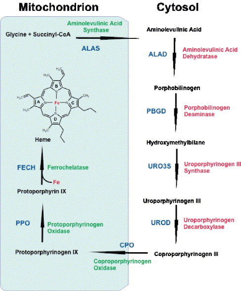
Main function is to transport O2 from the lungs to tissues and transport CO2 from the tissues to the lungs for exhalation
RBC
A molecule of hemoglobin consists of:
2 pairs of polypeptide chains
4 heme group
Each heme group is composed of a complex ring structure called _
protoporphyrin IX
Heme consists of:
1 atom of ferrous (Fe2+) iron
4 pyrrole rings
Each hemoglobin is capable of binding _ molecules of O2
1 g hemoglobin can carry _ of O2
A small percentage of HbA is glycated. The most characterized of the glycated hemoglobin is _ in which glucose attaches to the -
Each hemoglobin is capable of binding 4 molecules of O2
1 g hemoglobin can carry 1.34 mL of O2
A small percentage of HbA is glycated. The most characterized of the glycated hemoglobin is HbA1c in which glucose attaches to the N-terminal valine of the beta chain
Heme synthesis occurs in the _ and _ of bone marrow erythroid precursors
cytoplasm and mitochondria
The genes that code for the production of globin chains are located on: _
Translation of mRNA to the globin polypeptide chain occurs on the ribosomes in cytoplasm
Chromosome 16: alpha, beta
Chromosome 11: beta, epsilon, delta, gamma
Intrauterine (Embryonic) Hemoglobin
Gower I: 2 epsilon: 2 zeta
Gower II: 2 epsilon: 2 alpha
Portland: 2 zeta: 2 gamma
Newborn hemoglobin
HbF (60 to 90%)
HbA (10 to 40%)
2 years to adulthood hemoglobin
HbA (>95%)
HbA2 (<3.5%)
HbF (1 to 2%)
Describes how the affinity of hemoglobin for oxygen relates to the partial pressure of oxygen
Hb-O2 Dissociation Curve
Hb-O2 Dissociation Curve
Shape:_
PO2 of approximately _ results in 50% saturation of hemoglobin molecule (P50 value)
sigmoidal
27 mmHg
Hb-O2 Dissociation Curve
Shift to the left
HIGH oxygen affinity
LOW oxygen release to tissues
LOW PCO2
LOW temperature
LOW H+ ions/alkalosis
LOW 2,3-DPG
Hb-O2 Dissociation Curve
Shift to the right
LOW oxygen affinity
INCREASED release of oxygen to tissue
HIGH PCO2
HIGH TEMP
HIGH H+ ions / acidosis
HIGH 2,3-DPG
dysfunctional hemoglobins that cannot transport oxygen
Dyshemoglobin
Methemoglobin
Carboxyhemoglobin
Sulfhemoglobin
Methemoglobin (Hi)
Hb iron is in _
Acquired/inherited
Causes (2)
associated with _ blood color
Treatment: _
Hb iron is in FERRIC STATE (Fe3+)
Acquired/inherited
Causes CYANOSIS, DYSPNEA
associated with CHOCOLATE BROWN blood color
Treatment: METHYLENE BLUE / ASCORBIC ACID
Carboxyhemoglobin (HbCO)
_ times greater affinity to Hb than O2
Acquired (smoking, car exhaust)
Toxic effects (_% levels)
assocaited with _ colored blood
Treatment: _
240 times greater affinity to Hb than O2
Acquired (smoking, car exhaust)
Toxic effects (20-30% levels)
assocaited with CHERRY RED colored blood
Treatment: REMOVE CO SOURCE, O2 THERAPHY
Sulfhemoglobin (SHb)
sulfur atom at heme pyrrole ring
acquired (drugs/sulfur chemicals)
cannot be converted to _
Associated with _ color
Treatment: _
sulfur atom at heme pyrrole ring
acquired (drugs/sulfur chemicals)
cannot be converted to NORMA HbA
Associated with MAUVE LAVENDER/GREENISH PIGMENT color
Treatment: AVOIDANCE OF THE OFFENDING AGENT
Abnormal variation in RBC volume or diameter
Anisocytosis
Normocytic: 7 to 8 um
Microcytic: <6 um
Macrocytic: >8 um
Variation in hemoglobin content of RBCs
Anisochromia
Normochromic: 1/3 central pallor, MCHC = 32 to 36 g/dL
Hypochromic: <1/3 central pallor, MCHC<32 g/dL
Hyperchromic: >1/3 central pallor, MCHC>36 g/dL
Abnormal variation in RBC shape
Poikilocytosis
RBC Morphologic Abnormalities
deep staining large oval red cell
Macroovalocyte
RBC Morphologic Abnormalities
Macroovalocyte
Megaloblastic anemia
RBC Morphologic Abnormalities
small,dense RBC
no central pallor
low surface area:volume ratio
Spherocyte
RBC Morphologic Abnormalities
Spherocyte
Heriditary spherocytosis
AIHA
Severe burns
RBC Morphologic Abnormalities
Few irregularly spaced projections of varying length and width
Acanthocyte/ Thorn cell
RBC Morphologic Abnormalities
Acanthocyte/Thorn cell
severe liver disease
abetalipoproteinemia
McLeod syndrome
RBC Morphologic Abnormalities
RBC with blunt or pointed, short projections that are evenly spaced
Burr cells / Echinocyte
RBC Morphologic Abnormalities
Burr cell / Echinocyte
Uremia
Pyruvate kinase deficiency
RBC Morphologic Abnormalities
Hb concentrated in the center and around the periphery
Target cell / codocyte
RBC Morphologic Abnormalities
Target cell / Codocyte
Liver disease
Obstructive jaundice
Thalassemia
Hemoglobin C Disease
RBC Morphologic Abnormalities
slit like (mouth like) area of central pallor
Hereditary stomatocytosis
Acute alcoholism
Rh null syndrome
RBC Morphologic Abnormalities
narrow oval cells
egg-shaped or cigar shaped
Elliptocyte
RBC Morphologic Abnormalities
Elliptocyte
Hereditary elliptocytosis
IDA
Thalassemia major
RBC Morphologic Abnormalities
Fragmented RBCs caused by rupture in peripheral circulation
Schistocyte
RBC Morphologic Abnormalities
Schistocyte
Disseminated Intravascular Coagulation
Hemolytic Uremic Syndrome
Microangiopathic Hemolytic Anemia
Thrombotic Thrombocytopenic Purpura
RBC Morphologic Abnormalities
has single pointed extension resembling tear drop or pear
Dacryocyte
RBC Morphologic Abnormalities
Dacryocyte
Primary Myelofibrosis
Thalassemia
Myelopthisic Anemia
Megaloblastic anemia
RBC Morphologic Abnormalities
Crescent-shaped cell
Caused by crystalization of HbS due to low oxygen tension
Sickle cell / Drepanocyte
RBC Morphologic Abnormalities
Sickle cell/Drepanocyte
Sickle cell anemia
Sickle cell-beta thalassemia
RBC inclusions
Bluish tinge throughout cytoplasm
Also called polychromasia
Contect: RNA
Diffuse basophilia
RBC inclusions
Diffuse basophilia
Hemolytic anemia
After treatment for iron, vitamin B12 or folate deficiency
RBC inclusions
dark blue-purple, punctuate granules
Content: precipitated RNA
Coarse basophilic stippling
RBC Inclusions
Coarse Basophili Stippling
Lead poisoning
Megaloblastic anemia
Thalassemia
Hemoglobinopathies
Myelodysplastic Syndrome
RBC Inclusions
Dark blue-purple round granule
Content: DNA remnants
Howell-Jolly bodies
RBC Inclusions
Howell-Jolly bodies
Post-splenectomy
Megaloblastic anemia
Thalassemia
Myelodysplastic syndrome
RBC Inclusions
Not visible in Wight's staine
In supravital stain: dark blue-pyrple granule near RBC membrane
Content: Denatured hemoglobin
Heinz bodies
RBC Inclusions
Heinz bodies
G6PD deficiency
Unstable hemoglobin
Oxidant drugs/chemicals
RBC Inclusions
Irregular clusters of light to dark blue granules near periphery
Content: Non-heme iron
Pappenheimer bodies
RBC Inclusions
Blue rings or figure-8 inclusion
Content: mitotic spindle remnant
Megaloblastic anemia
Myelodysplastic syndrome
RBC Inclusions
Not visible in wright's stain
Supravital stain: Fine, evenly dispersed dark blue granules
“golf-ball RBCs”, “raspberry”
Content: precipitated beta-globin chains
Hemoglobin H inclusion
RBC Inclusions
Hemoglobin H inclusion
Hemoglobin H disease
RBC Inclusions
hexagonal, rod-shaped crystals
bar of gold appearance
Hemoglobin C crystal
RBC Inclusions
finger-like or quartz like crystal of dense hemolgobin protruidng from RBC membrane
Hemoglobin SC crystals
RBC Inclusions
Hemoglobin SC crystals
Hemoglobin SC disease
RBC Inclusions
NRBCs in bone marrow with dots surrounding th nucleus
Content: Non-heme iron
Ringed sideroblast
RBC Inclusions
Ringed sideroblast
Sideroblastic anemia
Myelodysplastic syndrome