Connective Tissue
1/33
Name | Mastery | Learn | Test | Matching | Spaced | Call with Kai |
|---|
No study sessions yet.
34 Terms
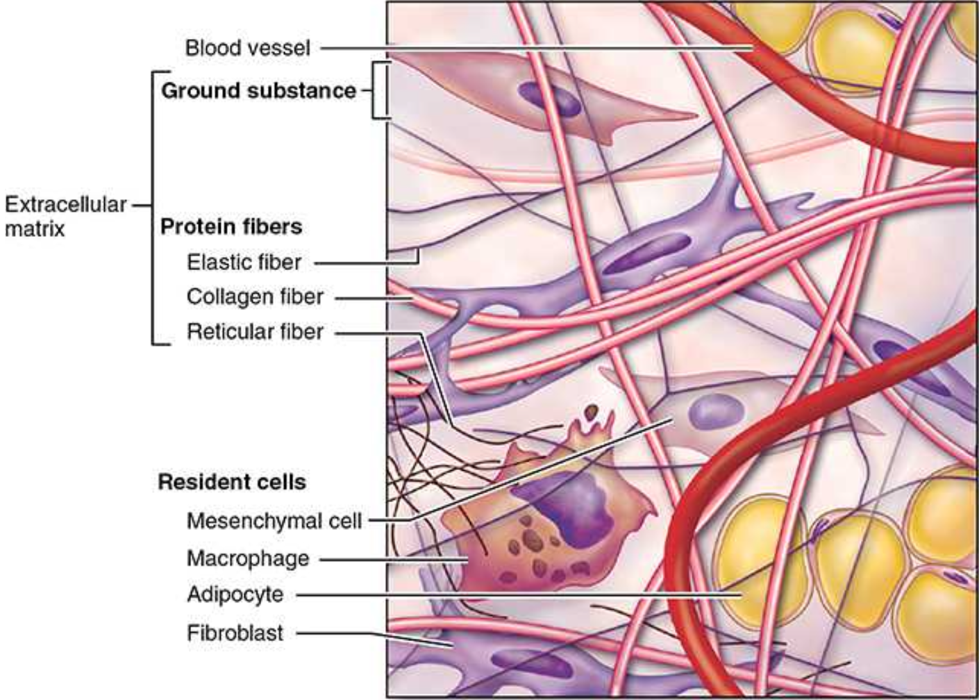
What is the ECM made out of?
The ___ contain different combinations of long protein fibers (collagen and elastic fibers) and ground substance, a complex of anionic hydrophilic proteoglycans, glycosaminoglycans (GAGs), and multiadhesive glycoproteins (laminin, fibronectin, and others).
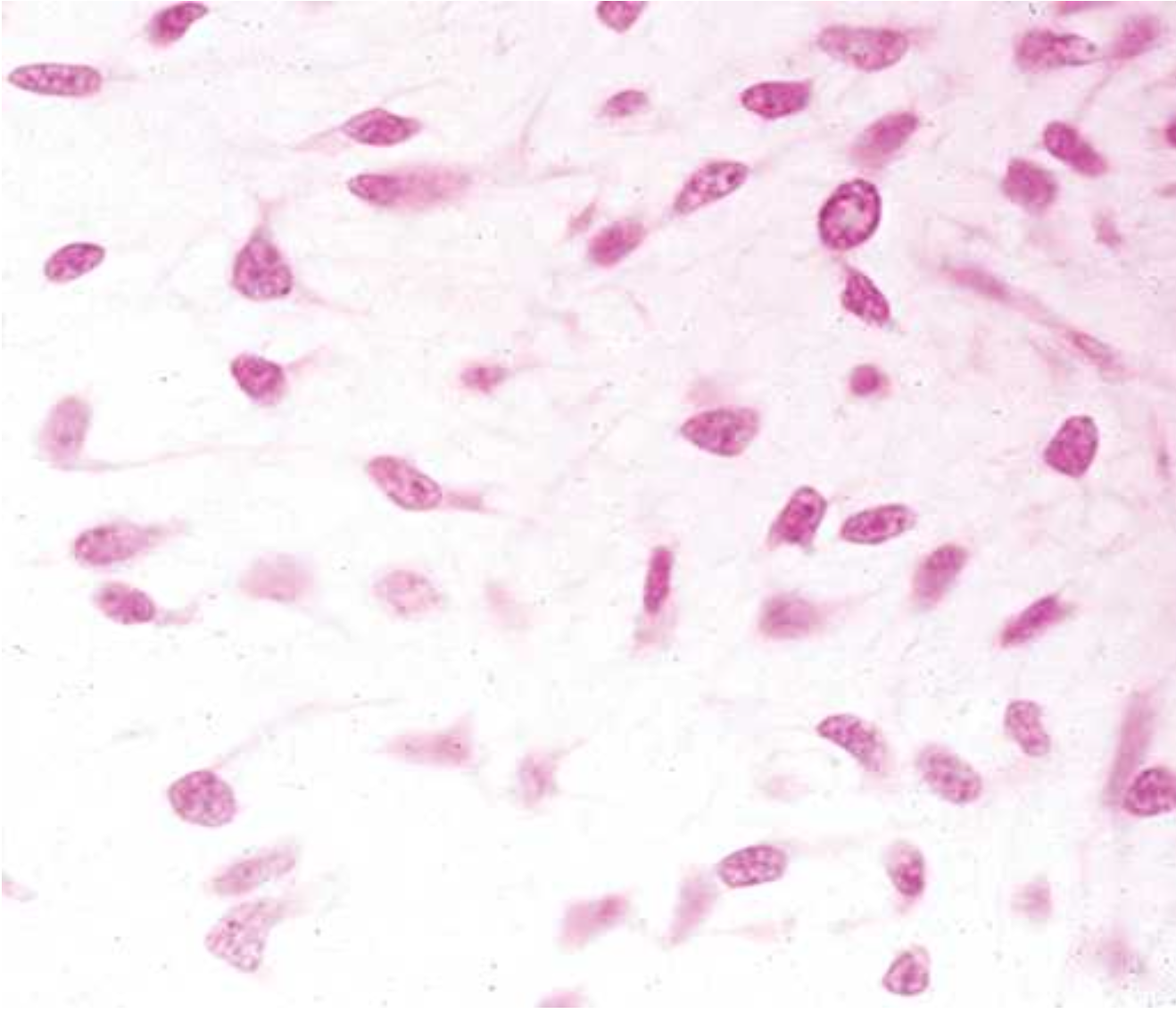
What is mesenchyme?
a tissue developing mainly from the middle layer of the embryo, the ___derm. _______ consists largely of viscous ground substance with few collagen fibers.

Mesenchymal cells have _____ nuclei with ____ chromatin and ____ nucleoli.
large, fine, prominent
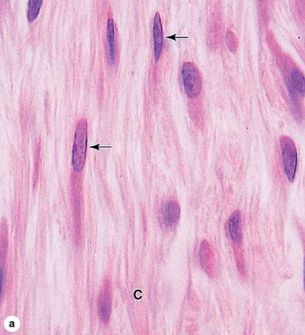
What are fibroblasts?
They are key cells in connective tissue proper.

What do fibroblasts do?
They synthesize and secrete collagen (the most abundant protein of the body) and elastin, which both form large fibers, as well as the GAGs, proteoglycans, and multiadhesive glycoproteins comprising the ground substance.

What do plasma cells do?
They make antibodies- y-shaped proteins that target antigens (toxins or bacteria).
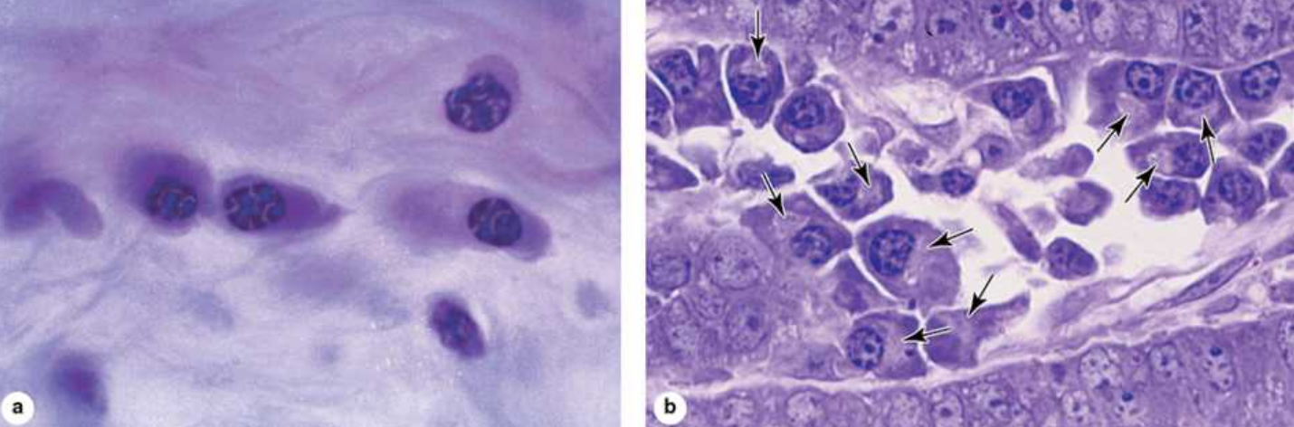
What do eosinophilic leukocytes do?
They modulate allergic/vasoactive reactions and defend against parasites.
What do neutrophilic leukocytes do?
Phagocytosis of bactera.

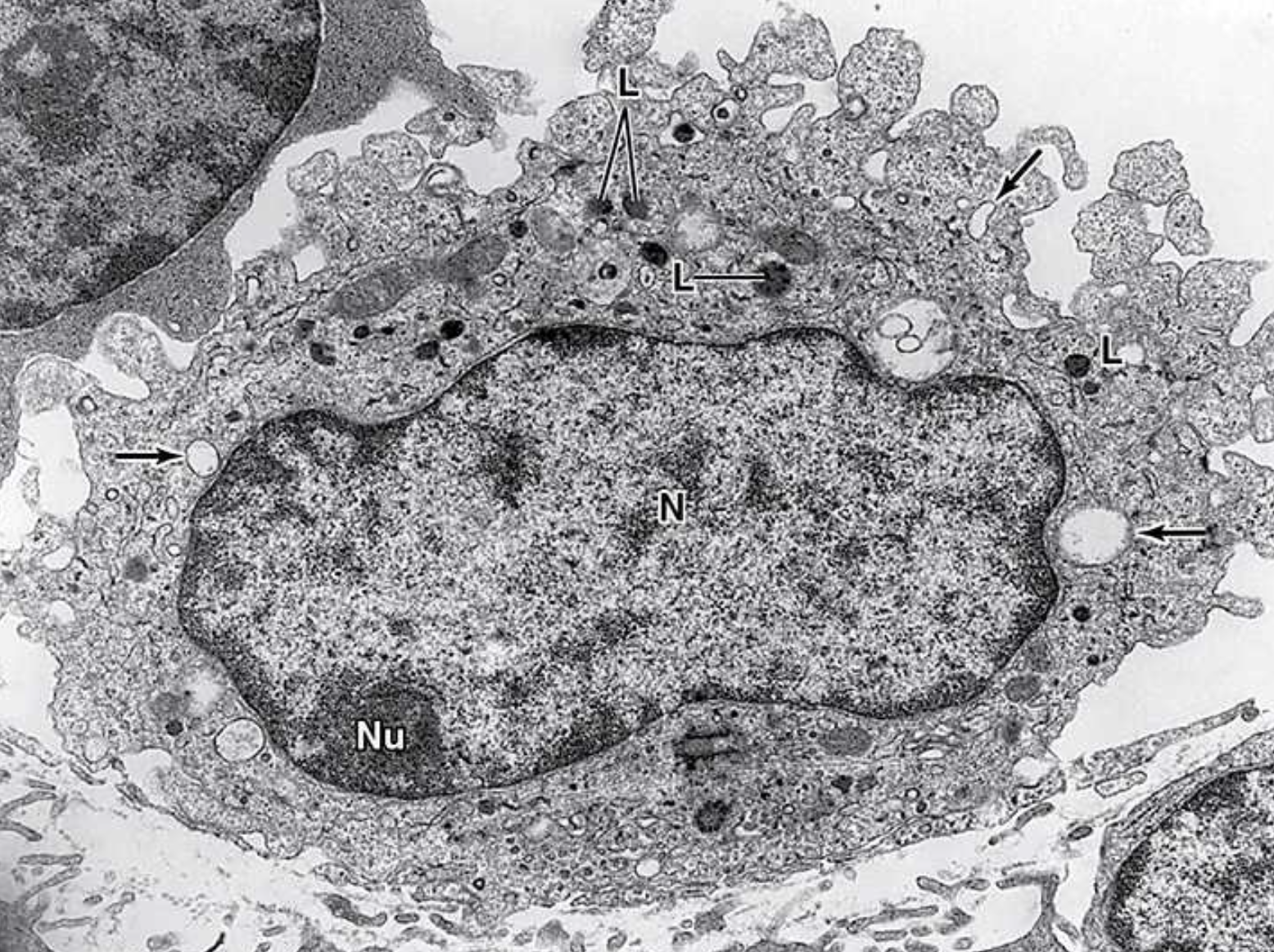
What do macrophages do?
Phagocytosis of ECM debris, antigen presentation and processing, secretion of growth facotrs, cytokines (chemical messengers made of proteins), etc.
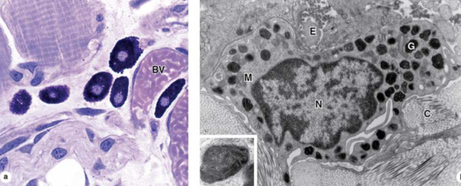
What do mast cells and basophilic leukocytes do?
They make pharmacologically active molecules like histamines.

What do adipocytes do?
They store neutral fats.
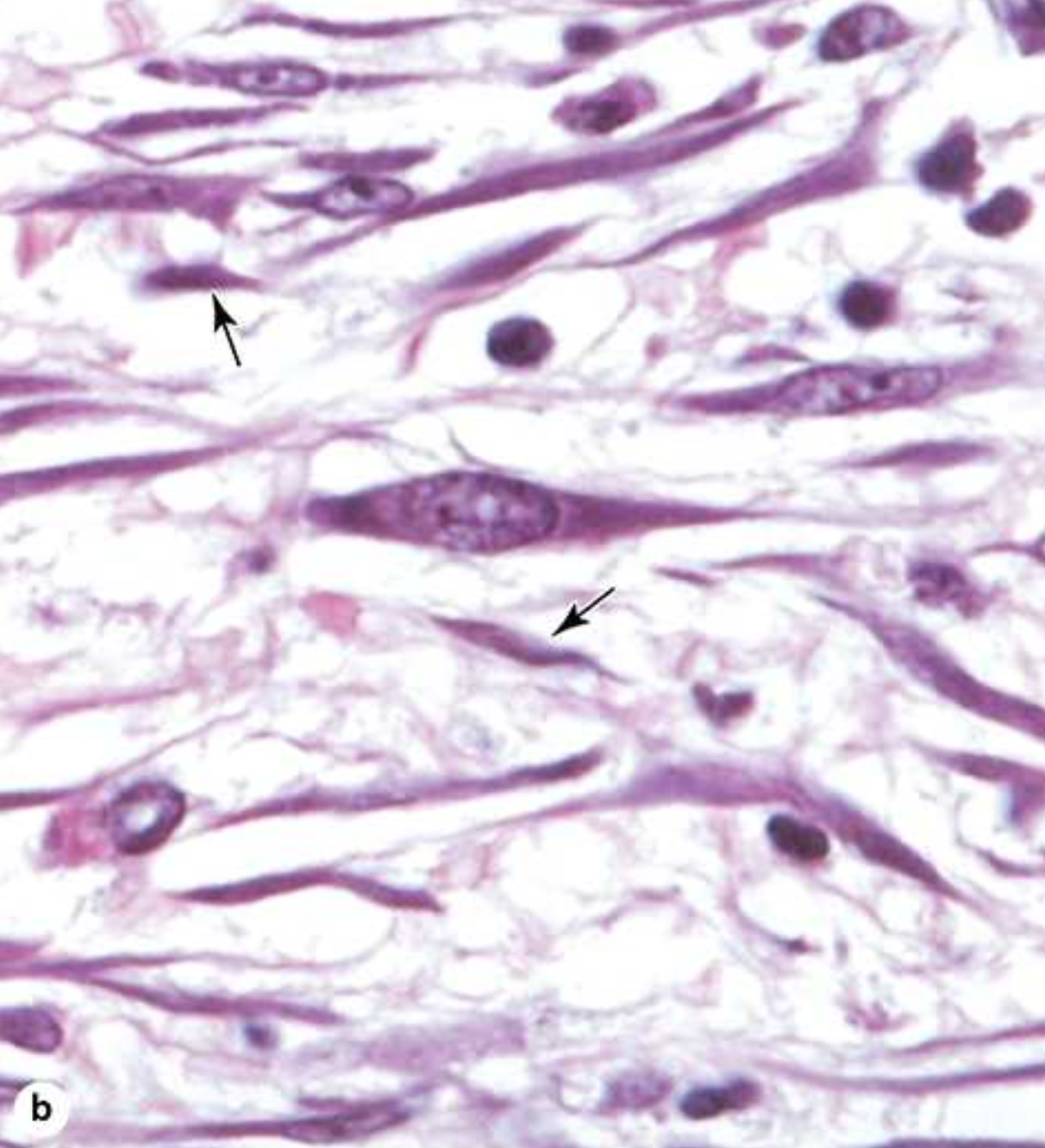
What is the difference between fibroblasts and fibrocytes?
Fibroblasts are highly active with high metabolic activity while fibrocytes are relatively inactive and “mature”.

Active fibroblasts have more _____ and irregularly _____ cytoplasm. They have ____ rough endoplasmic reticulum and a ___-_______ golgi apparatus.
abundant, branched, much, well-developed.
What are growth factors?
They are a family of proteins which influence cell-growth and differentiation.
Myofibroblasts are involved in ___ ________. They have a form of ____ found in smooth muscle cells.
wound healing, actin
What is the purpose of adipose tissue?
It serves to cushion and insulate the skin and other organs. Made of cells specialized to store lipid as neutral fats or less commonly, to produce heat
What is a monocyte?
A precuros cell that circulates the blood. They cross the epithelial wall of small vessels called venules to enter connective tissue, where they mature, and acquire the morphologic features of macrophages
Where are macrophages located?
in the connective tissue, lymphoid organs, lungs, bone marrow, pleural and peritoneal cavities.
What are the three main kinds of fibers?
Collagen, reticular, and elastic
Collagen 1
The most abundant and widely distributed collagen, forms large, eosinophilic bundles usually called collagen fibers. These often densely fill the connective tissue, forming structures such as tendons, organ capsules, and dermis.

Collagen 4
Network or sheet-forming collagen
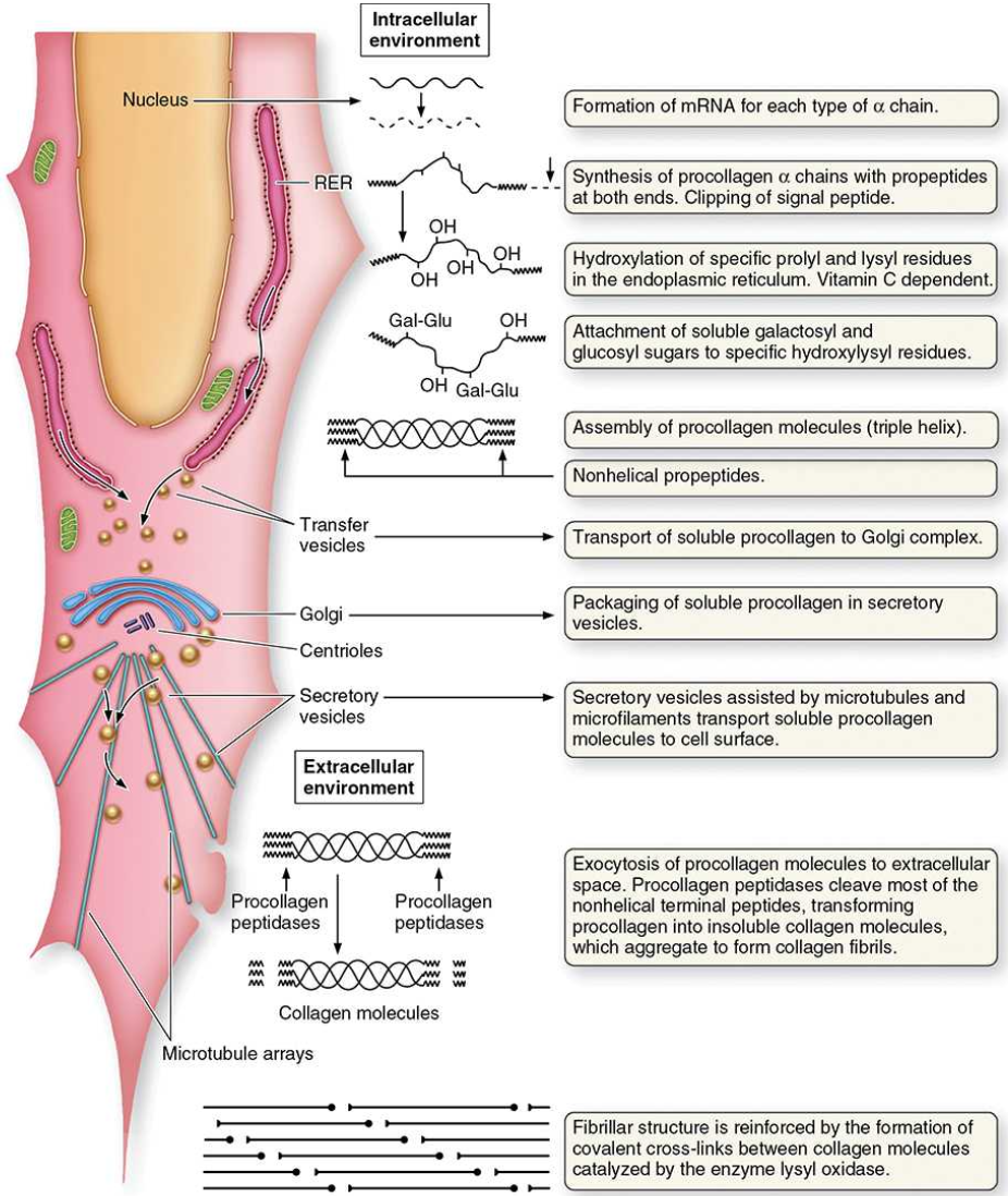
Synthesis of type 1 collagen
α‑chain production: Procollagen α chains are synthesized on RER ribosomes and enter the RER, where their Gly‑X‑Y–rich sequences form the basis of the future triple helix.
Post‑translational modifications: In the RER, selected proline and lysine residues are hydroxylated (requiring O₂, Fe²⁺, and vitamin C), and some hydroxylysines are glycosylated.
Triple‑helix formation: The non‑helical terminal regions help align three α chains. Disulfide bonds at the C‑terminus stabilize the assembly, allowing the triple helix to form. Procollagen is then sent through the Golgi and secreted.
Extracellular processing: Procollagen peptidases remove terminal peptides, converting procollagen into collagen, which spontaneously assembles into fibrils near the cell surface.
Fibril maturation: Proteoglycans and accessory collagens (e.g., types V and XII) organize and stabilize fibrils, which then aggregate into larger fibers.
Cross‑linking: Lysyl oxidase catalyzes covalent cross‑links between collagen molecules, strengthening fibrils and preventing their breakdown.
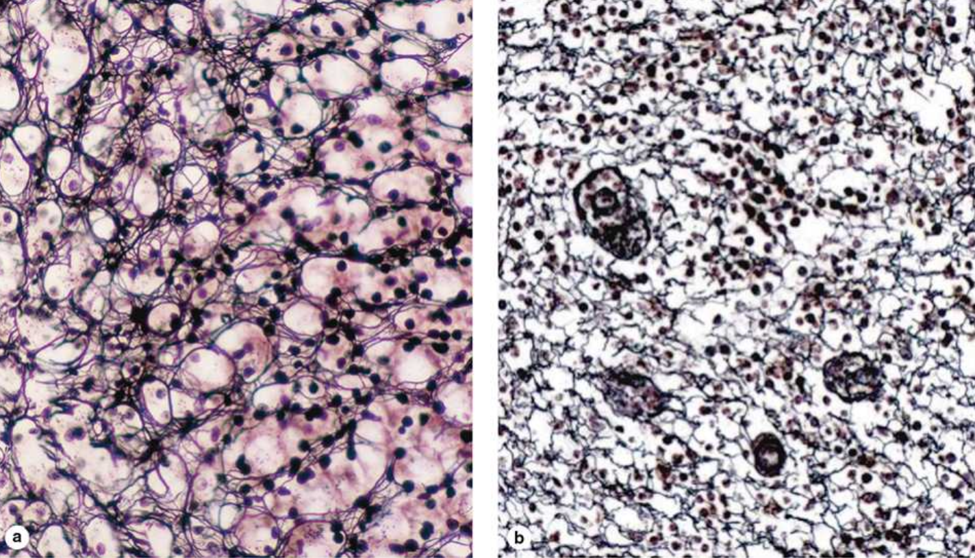
Reticular fiber
Found in delicate connective tissue of many organs, notably in the immune system, ____ ___consist mainly of collagen type III, which forms an extensive network (reticulum) of thin (diameter 0.5–2 μm) fibers for the support of many different cells. These fibers serve as a supportive stroma in most lymphoid and hematopoietic organs and many endocrine glands. The fibers consist of type III collagen that is heavily glycosylated, producing the black argyrophilia, an affinity for stains containing silver salts.

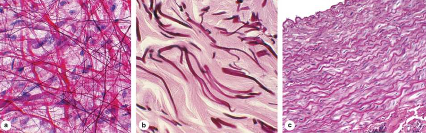
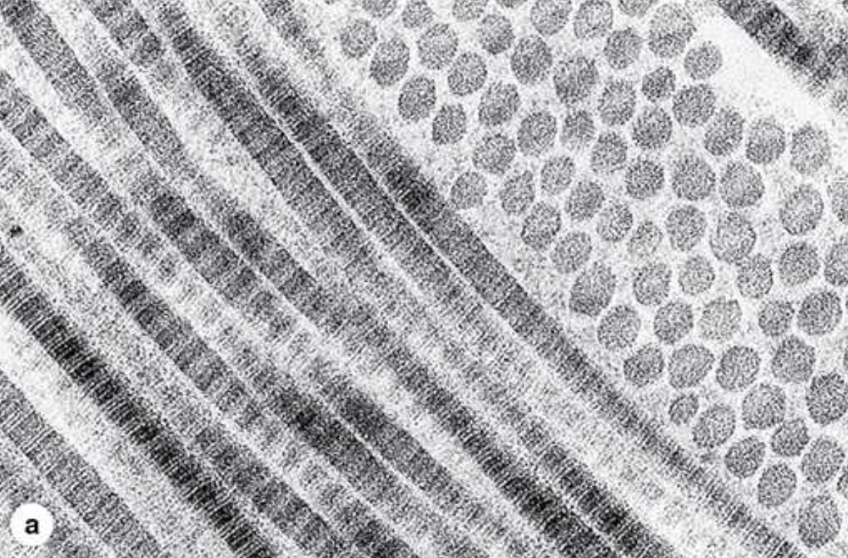
Elastic fibers
form sparse networks interspersed with collagen bundles in many organs, particularly those subject to regular stretching or bending. As the name implies, ______ ____ have rubberlike properties that allow tissue containing these fibers, such as the stroma of the lungs, to be stretched or distended and return to their original shape.


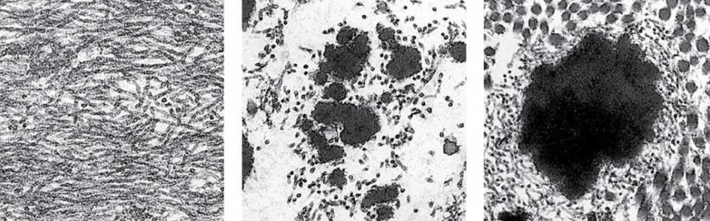
Making elastic fiber
They are composed of fibrillin (350 kDa), which forms a network of microfibrils, embedded in a larger mass of cross- linked elastin (60 kDa). Both proteins emerge by secretion from fibroblasts (and smooth muscle cells in vascular walls) and give rise to elastic fibers in a stepwise manner as shown in Figure 5–14. Initially, microfibrils with diameters of 10 nm develop from fibrillin and various glycoproteins. The microfibrils act as scaffolding upon which elastin is then deposited. Elastin accumulates around the microfibrils, eventually making up most of the elastic fiber, and conferring the rubberlike property.
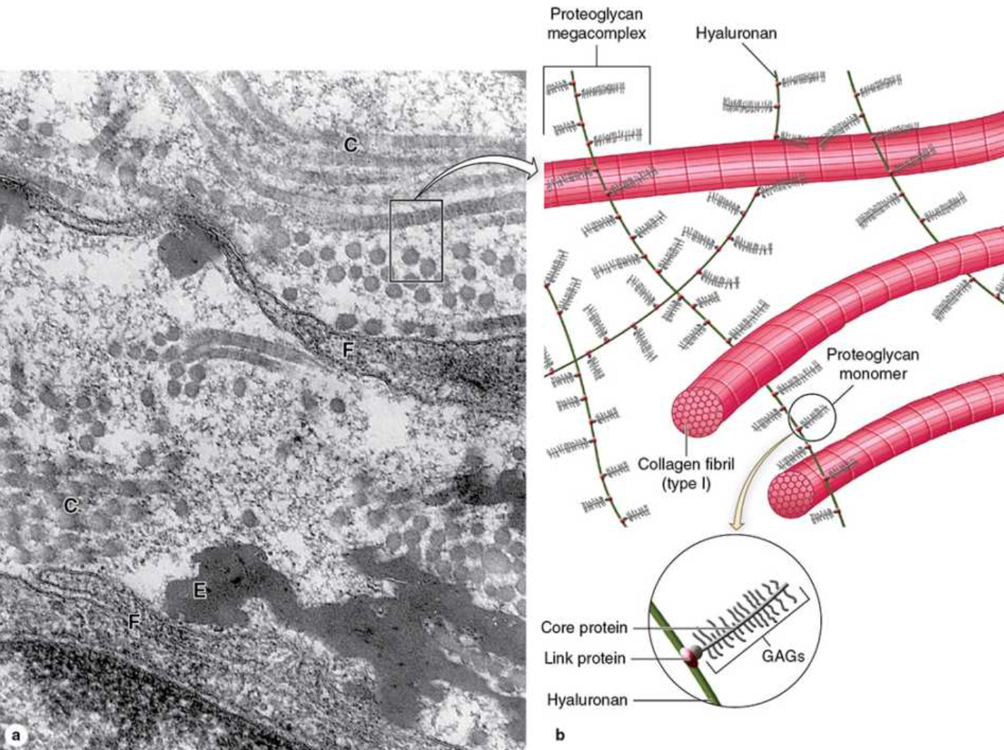
Ground Substance
The _____ _______ of the ECM is a highly hydrated (with much bound water), transparent, complex mixture of three major kinds of macromolecules: glycosaminoglycans (GAGs), proteoglycans, and multiadhesive glycoproteins. Filling the space between cells and fibers in connective tissue, ______ _________ allows diffusion of small molecules and, due to its viscosity, acts as both a lubricant and a barrier to the penetration of invaders.

What are the two main forces that act on water in the capillaries?
hydrostatic pressure and osmotic pressure

What is hydrostatic pressure?
the blood caused by the pumping action of the heart, which forces water out across the capillary wall
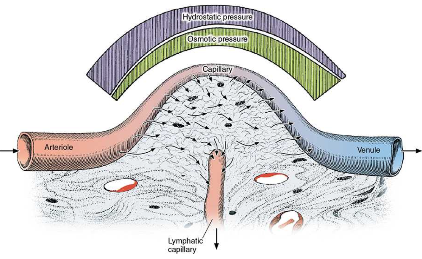
What is osmotic pressure?
produced by plasma proteins such as albumin, which draws water back into the capillaries


What is loose connective tissue?
occurs widely, forming a layer beneath the epithelial lining of many organs and filling internal spaces of muscle and nerve- also called areolar tissue. it typically contains cells, fibers, and ground substance in roughly equal parts. Fibroblasts predominate among the cells, but the other types of connective tissue cells also normally occur, along with nerves and small blood vessels.


What is dense irregular tissue?
similar to loose but with fewer cells, almost all fibroblasts, and a clear predominance of bundled type I collagen fibers over ground substance. The abundance of collagen here protects organs and strengthens them structurally. In _____ _______ _____, bundles of collagen fibers appear randomly interwoven, with no definite orientation. The tough three-dimensional collagen network provides considerable resistance to stress from all directions.
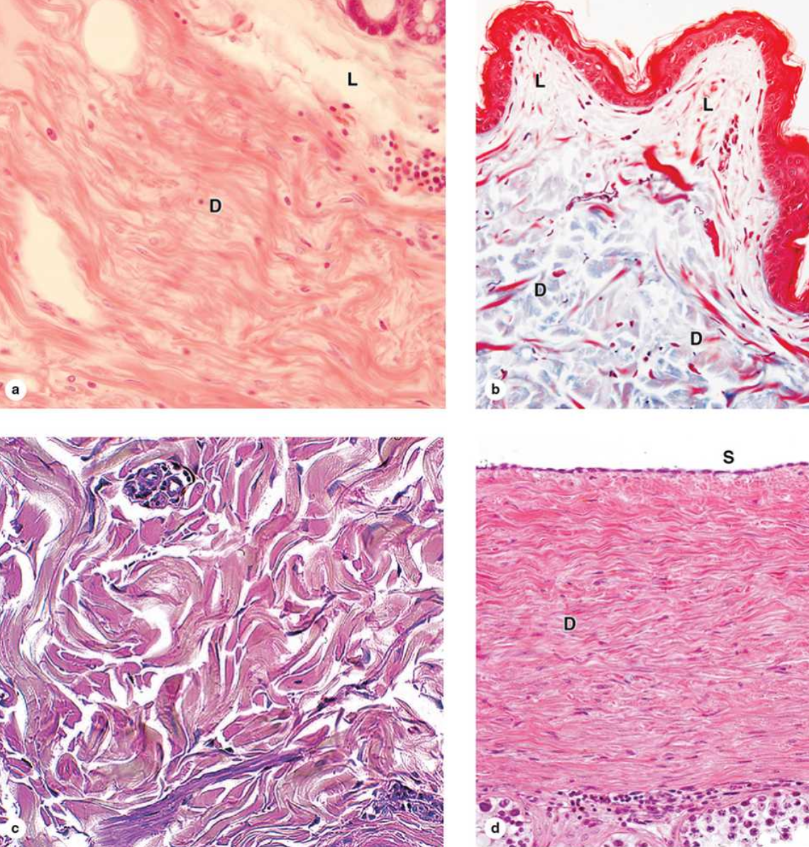

What is dense regular connective tissue?
consists mostly of type I collagen bundles and fibroblasts aligned in parallel for great resistance to prolonged or repeated stresses from the same direction.
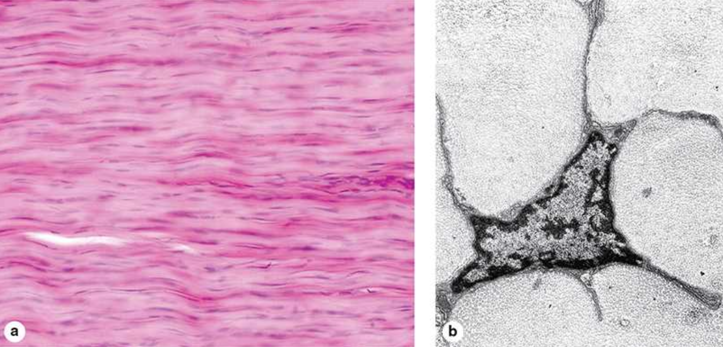
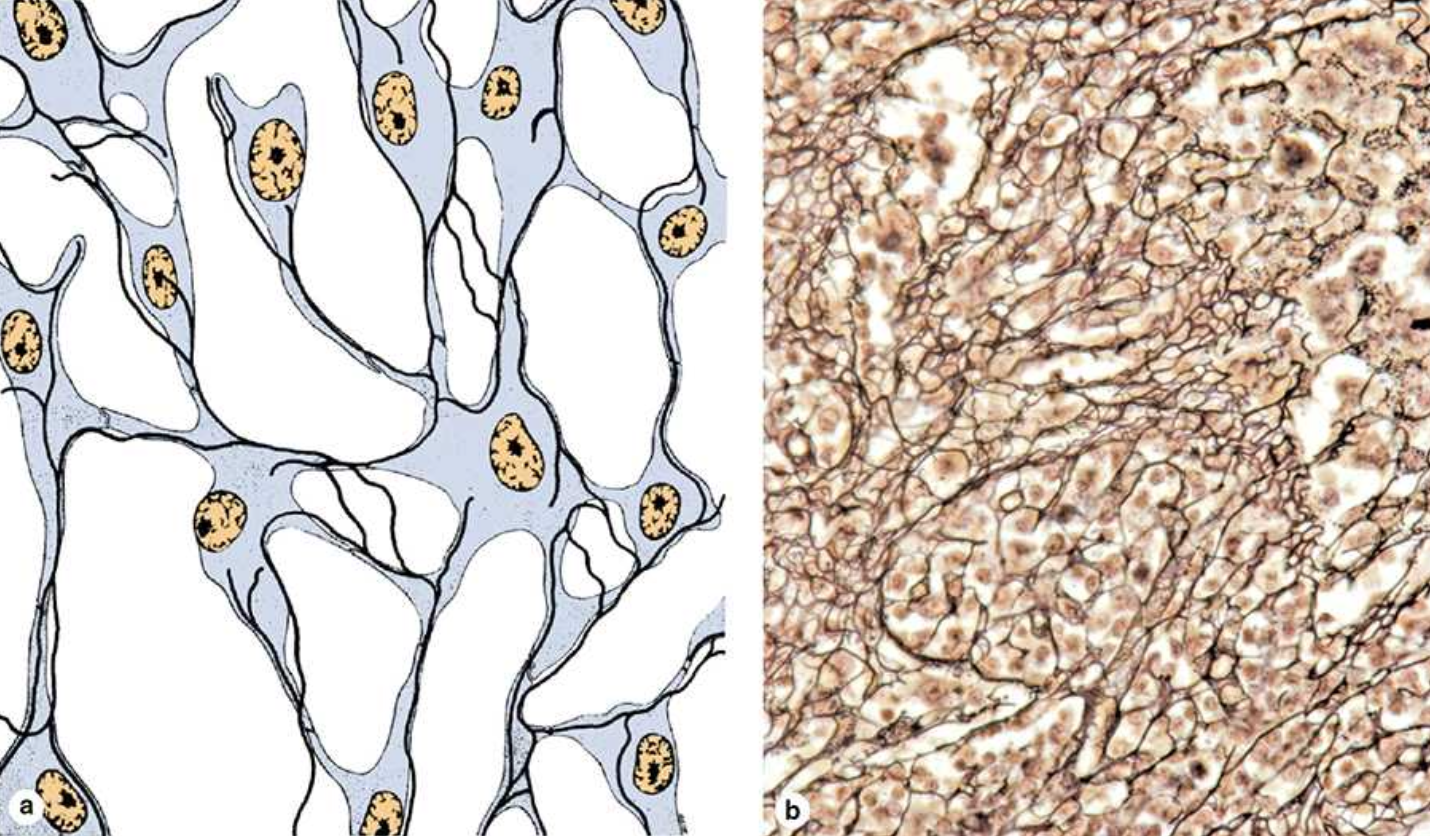
Reticular fibers
characterized by abundant fibers of type III collagen (Figure 5–12), forms a delicate network supporting various types of cells. Also known as reticulin, type III collagen is produced by modified fibroblasts also often called reticular cells, which remain associated with and partially cover the collagen fibers


Mucoid (mucous) tissue
represents the principal component of the fetal umbilical cord, where it is referred to as Wharton’s jelly. With abundant ground substance composed chiefly of hyaluronan, mucoid tissue is gelatinous, with sparse collagen fibers and scattered fibroblasts
