10. Surgical diseases of the salivary glands and ducts. Symptoms, diagnosis and therapy.

0.0(0)
studied byStudied by 0 people
learnLearn
examPractice Test
spaced repetitionSpaced Repetition
heart puzzleMatch
flashcardsFlashcards
Card Sorting

1/88

encourage image

There's no tags or description

Looks like no tags are added yet.

Study Analytics
Name
Mastery
Learn
Test
Matching
Spaced

No study sessions yet.

89 Terms

1
New cards
What are the four major salivary glands in dogs and cats?
Parotid, mandibular, sublingual, and zygomatic
2
New cards
Where is the parotid gland located?
Below the ear, overlying the masseter muscle
3
New cards
Where does the parotid duct open?
Into the oral cavity at the level of the upper fourth premolar (maxillary P4) tooth
4
New cards
Where is the zygomatic gland located?
Near the eye, below the zygomatic arch, and deep to the orbit
5
New cards
Where does the zygomatic duct open?
Into the oral cavity opposite the last upper molar (maxillary M2) tooth
6
New cards
Where is the mandibular gland located?
Caudal to the angle of the mandible, near the bifurcation of the external jugular vein
7
New cards
Where does the mandibular duct open?
At the sublingual caruncle, located on the floor of the mouth, near the base of the tongue
8
New cards
What are the two parts of the sublingual salivary gland?
Monostomatic and polystomatic
9
New cards
Where is the monostomatic part of the sublingual gland located?
Close to the mandibular gland
10
New cards
Where is the polystomatic part of the sublingual gland located?
Along the ventral floor of the mouth
11
New cards
Where do the ducts of the sublingual gland open?
  1. The monostomatic duct opens with the mandibular duct at the sublingual caruncle.

  2. The polystomatic ducts have multiple small openings along the sublingual mucosa.

12
New cards
Which minor salivary gland is present in cats but not dogs?
Molar salivary gland
13
New cards
Where is the molar salivary gland located in cats?
Near the lower molar teeth
14
New cards
Where does the molar salivary gland duct open in cats?
Directly into the oral cavity on the floor of the mouth near the lower molars
15
New cards
Where are the labial glands located?
In the lips
16
New cards
Where do the labial gland ducts open?
Into the oral vestibule (area between the lips/cheeks and teeth)
17
New cards
Where are the buccal glands located?
Within the mucosa of the cheeks
18
New cards
Where do the buccal gland ducts open?
Into the cheek mucosa and the oral vestibule
19
New cards
Where are the lingual glands located?
In the tongue, especially near the base
20
New cards
Where do the lingual gland ducts open?
Directly into the mouth at various locations on the tongue
21
New cards
Where are the palatine glands located?
In the soft palate
22
New cards
Where do the palatine gland ducts open?
Into the mucosa of the soft palate
23
New cards
What are the functions of saliva in dogs and cats?
Lubricating food, aiding swallowing, cooling, cleansing the mouth, buffering acids, reducing bacterial growth, and protecting oral tissues. It plays little role in carbohydrate digestion.
24
New cards
What are the two types of saliva produced by the major salivary glands?
Serous (watery) and mucus-rich
25
New cards
Which glands produce serous saliva?
Parotid and mandibular
26
New cards
Which glands produce mucus-rich saliva?
Sublingual and zygomatic
27
New cards
How is the final composition of saliva determined?
Initial sodium-rich saliva is modified as it flows through ducts, with sodium reabsorption and bicarbonate and potassium secretion.
28
New cards
What controls salivary gland function?
The autonomic nervous system
29
New cards

What effect does parasympathetic stimulation have on saliva production?

Increases saliva production.

30
New cards
What nerves mediate parasympathetic stimulation of salivary glands?
Facial and mandibular nerves
31
New cards
What effect does sympathetic stimulation have on saliva production?
Causes minor inhibition after an initial increase due to myoepithelial cell contraction
32
New cards

What are examples of surgical diseases of the salivary glands and ducts?

  1. Sialadenosis

  2. Sialadenitis and necrotising sialometaplasia

  3. Sialocele

  4. Sialoliths

  5. Rupture or fistula of salivary ducts

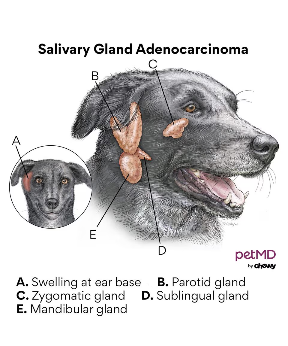
  6. Salivary gland neoplasia

  7. Salivary production disorders

33
New cards
What is sialadenosis?
Non-inflammatory, non-neoplastic, bilateral swelling of salivary glands, most commonly affecting the mandibular glands
34
New cards

What is the aetiology of sialadenosis?

Unknown

35
New cards
What are the clinical signs of sialadenosis?
Retching, gulping, lip smacking, hypersalivation, and weight loss. Oesophageal abnormalities may be present.
36
New cards
How is sialadenosis diagnosed?
Usually by excluding other causes
37
New cards
How is sialadenosis treated?

Phenobarbital (1-2 mg/kg PO every 12 hours). Surgery alone is ineffective.

38
New cards
What is sialadenitis and necrotising sialometaplasia?
Inflammation of the salivary glands that may progress to glandular necrosis and ductal metaplasia
39
New cards
How do the clinical signs of sialadenitis and necrotising sialometaplasia differ from sialadenosis?
They are similar, but with the addition of pain on palpation of the glands and vomiting.
40
New cards
What is a possible cause of sialadenitis and necrotising sialometaplasia?
Concurrent gastrointestinal or oesophageal disease and hyperstimulation of the vagal nerve
41
New cards

What are clinical signs of sialadenitis and necrotising sialometaplasia?

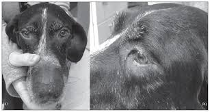
Fever, depression, painful and swollen SG on palpation, vomiting.

Rupture of abscessed gland discharges pus into the surrounding tissue or the mouth. Rupture through the skin may form a salivary fistula.

42
New cards

How is sialadenitis diagnosed?

Clinical exam, histopathology

43
New cards
How is sialadenitis and necrotising sialometaplasia treated?
Same as for sialadenosis (phenobarbital).
44
New cards
What is a sialocele (salivary mucocele)?
A collection of saliva within subcutaneous tissue, lined by inflammatory connective tissue. Not a true cyst.
45
New cards
What are some causes of sialoceles?
Trauma (surgical and non-surgical), sialoliths, foreign bodies, and neoplasia
46
New cards
Which breeds are predisposed to sialoceles?
Poodles, German Shepherds, Australian Silky Terriers, and Dachshunds
47
New cards
What are the different types of sialoceles?
  1. Cervical mucocele

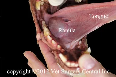
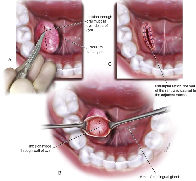
  2. Ranula (sublingual mucocele)

  3. Zygomatic mucocele

  4. Pharyngeal mucocele

48
New cards
What are the clinical signs of a cervical mucocele?
Acute onset of non-painful swelling in the intermandibular or cranioventral cervical region
49
New cards
What are the clinical signs of a ranula?
Medial deviation of the tongue, difficulty prehending, chewing, and swallowing
50
New cards
What are the clinical signs of a zygomatic mucocele?
Exophthalmos, protrusion of the third eyelid, or painless orbital swelling
51
New cards
What are the clinical signs of a pharyngeal mucocele?
Caudal dorsal or lateral pharyngeal swelling, rostral to the epiglottis. Laboured breathing or stridor.
52
New cards
How are sialoceles diagnosed?

Aspiration of saliva, cytology, sialography, biopsy, USG, MRI, and CT

53
New cards
How are sialoceles treated?
  1. Sialadenectomy of the affected gland

  2. Removal of saliva by incision or aspiration (often recurs)

  3. Ranulas are treated by marsupialisation.

<ol><li><p>Sialadenectomy of the affected gland</p></li><li><p>Removal of saliva by incision or aspiration (often recurs)</p></li><li><p>Ranulas are treated by marsupialisation.</p></li></ol><p></p>
54
New cards
What are sialoliths?

Calcified masses (salivary stones) that form in salivary glands (calcium, oxalate, phosphate, etc.)

55
New cards
Where do sialoliths most commonly form?
Parotid duct, but also in the mandibular-sublingual duct complex
56
New cards
What are the clinical signs of parotid sialoliths?
Swelling on the lateral face, which can be painful and recurring
57
New cards
How are sialoliths diagnosed?

Palpation, radiography, USG, and CT scan

58
New cards
How are sialoliths treated?

Depends on location, infection, and recurrence risk. Options include:

  1. Surgical removal of the affected duct-gland complex if easily accessible.

  2. For stones near the oral papilla, mucosal incision and removal, healing by second intention.

  3. Duct ligation, resection and anastomosis, or primary repair for complex cases.

  4. Marsupialization into the oral cavity for cases with duct dilation

59
New cards
What causes rupture or fistula of salivary ducts?

Trauma to the mandibular, zygomatic, or sublingual salivary glands.

Injury such as bite wound, abscess drainage, or prior surgery causes iatrogenic rupture

60
New cards
What must duct rupture/fistula be differentiated from?

Draining sinuses caused by foreign bodies or endodontic disease of a mandibular tooth.

61
New cards
Which salivary gland is most commonly affected by rupture/fistula?
Parotid gland
62
New cards
How are ruptured/fistulated salivary ducts treated?
Surgical ligation of the duct or complete excision of the gland.
63
New cards
Is salivary gland neoplasia common?
No, it is uncommon.
64
New cards
What type of neoplasia is most common in salivary glands?
Epithelial origin, mostly adenocarcinoma or acinic carcinoma.
65
New cards
Which salivary glands are most commonly affected by neoplasia?
Mandibular and parotid glands
66
New cards
What are the clinical signs of salivary gland neoplasia?
Unilateral, non-painful, firm swelling in the upper neck, base of the ear, upper muzzle or maxilla, halitosis, exophthalmos, and dysphagia
67
New cards
How is salivary gland neoplasia diagnosed?

Mass and lymph node sampling (biopsy), three-view thoracic radiographs for metastases, and CT/MRI for surgical planning.

68
New cards
How is salivary gland neoplasia treated?

Sialadenectomy (removal of the affected gland with tumour). Radiotherapy.

69
New cards

What are examples of salivary production disorders?

  1. Ptyalism

  2. Hypoptyalism

70
New cards
What is ptyalism?
Drooling of saliva, caused by hypersialosis (hypersecretion) or pseudoptyalism (conformational or swallowing abnormalities).
71
New cards
What are some causes of ptyalism?
  1. Drugs, toxins or poisons (organophosphates)

  2. Local irritation or inflammation associated with stomatitis, glossitis (especially in cat), oral foreign bodies, neoplasms, injuries or mucosal defects

  3. Infectious diseases (Rabies), nervous form of distemper, or other convulsive disorders

  4. Motion sickness, fear, nervousness, excitement

  5. Reluctance to swallow or interference of swallowing (esophageal irritation, obstruction)

  6. Sublingual lesions

  7. Tonsillitis

  8. Administration of medicine

  9. Conformational defects

  10. Metabolic disorders (hepatic encephalopathy)

  11. Abscess or other inflammatory blockage conditions of the salivary gland

72
New cards

What is important before examining an animal with ptyalism?

Eliminate possibility of rabies (vaccine, history of dog/fox bites)

73
New cards
How is ptyalism treated?

Treating the underlying cause.

Glycopyrrolate reduces production of saliva

74
New cards
What is hypoptyalism?
Decreased saliva secretion, resulting in dry mouth (xerostomia).
75
New cards
Is hypoptyalism common in dogs and cats?
No, it is uncommon.
76
New cards
What are some causes of hypoptyalism?
Radiation treatment, atropine, extreme dehydration, pyrexia, and anaesthesia
77
New cards
How is hypoptyalism treated?

Treating the underlying cause.

Pilocarpine stimulates saliva production. Encourage drinking water.

78
New cards

What are examples of surgeries of the salivary glands?

  1. Sialadenectomy (extirpation)

  2. Marsulialisation

79
New cards
What is the most common surgery performed on the salivary glands?
Sialadenectomy
80
New cards
What are the two main surgical approaches for sublingual and mandibular sialadenectomy?
Lateral and ventral approaches
81
New cards
What is the positioning for the lateral approach to sialadenectomy?
Lateral recumbency with the affected side up and neck extended
82
New cards
What is the incision for the lateral approach to sialadenectomy?
Horizontal cut over the mandibular gland, between the linguofacial and maxillary veins
83
New cards
What are the steps of the lateral approach to sialadenectomy?
  1. Blunt and sharp dissection to expose and decompress sialocele.

  2. Identify and dissect glands from surrounding tissues.

  3. Ligate blood vessels and continue blunt dissection rostrally along ducts.

  4. Ligate or carefully dissect ducts, avoiding the lingual branch of the trigeminal nerve (V → V3 n. mandibularis → n. lingualis).

  5. Transect ducts and glands near the oral cavity if necessary.

84
New cards
What is the positioning for the ventral approach to sialadenectomy?
Dorsal recumbency, prepared from the midcervical region to the ventral mandible
85
New cards
What is the incision for the ventral approach to sialadenectomy?
From 4-5 cm caudal to the mandibular ramus, extending toward the mandibular symphysis
86
New cards
What are the steps of the ventral approach to sialadenectomy?
  1. Incise platysma to expose external jugular bifurcation.

  2. Blunt dissection to expose salivary glands (differentiate from mandibular lymph nodes).

  3. Incise capsule, ligate vessels, and retract glands caudally.

  4. Bluntly dissect sublingual gland under the digastricus muscle, clamp ducts, and excise glands.

  5. If ranula is present, extend dissection past lingual nerve to sublingual caruncle, ligate and transect duct.

  6. Closure: Use absorbable monofilament to close tissues, optional drainage for sialocele.

87
New cards
How is the surgical site closed after sialadenectomy?
With absorbable monofilament sutures, with optional drainage for sialocele.
88
New cards

What is the procedure for marsupialisation?

  1. Drain mucocele (usually ranulas) by excising an elliptical, full-thickness section of the mucocele wall.

  2. Suture the granulation tissue lining to the sublingual mucosa to encourage draining for several days.

  3. Marsupialised ranulas contract and heal quickly by secondary intention

89
New cards

What type of tumour can be seen in the salivary glands?

Adenoma/adenocarcinoma