AP Biology Unit 4
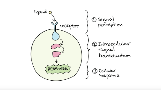
topics 1,2,3, and 4:
cell communication
-critical for the function and survival of cells
-responsible for the growth and development of multicellular organisms
how do cells communicate?
direct contact
local signaling
long distance signaling
direct contact
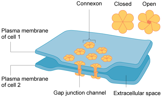
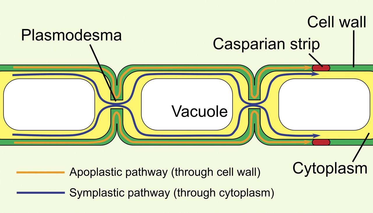
-communication through cell junctions
-signaling substances and other material dissolved in the cytoplasm can pass freely between adjacent (next to each other) cells.
animal cells: gap junctions
plant cells: plasmodesmata
-key to remember the difference: plant and plasmodesmata both start with P
-an example of direct contact are immune cells
Local Regulators
-a secreting cell will release chemical messages (local regulators/ligands) that travel a SHORT distance through the extra cellular fluid
-the chemical messages will cause a response in a target cell
paracrine
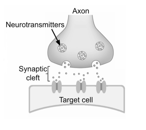
synaptic
-paracrine: secretory cells release local regulators (growth factors) via exocytosis to an adjacent cell
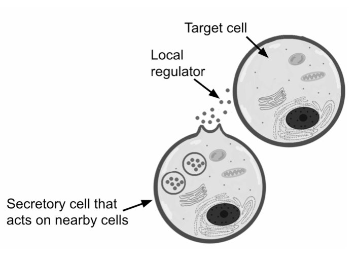
-synaptic: occurs in animal nervous systems, neurons secrete neurotransmitters and diffuse across the synaptic clef-space between the nerve cell and target cell
Long Distance Signaling
-animals and plants use hormones for long distance signaling
-plants: release hormones that travel in plant vascular tissue (xylem and phloem) or through the air to reach target tissues
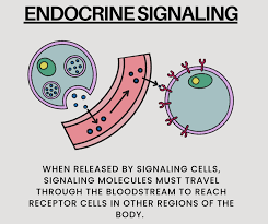
-animals: use endocrine signaling, specialized cells release hormones into the circulatory system where they reach target cells
example: insulin
-insulin is released by the pancreas into the bloodstream where it circulates through the body and binds to target cells
Cell Signaling
three stages:
reception
-ligand binds to receptor
transduction
-signal is converted
response
-a cell process is altered
stage one: reception
the detection and receiving of a ligand by a receptor in the target cell
receptor: macromolecule that binds to a signal molecule (ligand)
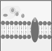
-receptors can be located in either the plasma membrane or intracellular
all receptors have an area that interacts with the ligand and an area that transmits a signal to another protein
binding between ligand and receptors is highly specific
-when the ligand binds to the receptor, the receptor activates (via a conformational change)
-allows the receptor to interact with other cellular molecules
-initiates transduction signals
plasma membrane receptors vs intracellular receptors
plasma membrane:
-most common type of receptor
-binds to ligands that are polar, water-soluble, and large
examples:
ligand gated ion channels
G protein coupled receptors (GPCRs)
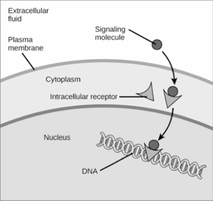
intracellular receptors
-found in the cytoplasm or nucleus of target cell
-binds to ligands that can pass through the plasma membrane
examples:
hydrophobic molecules
steroid and thyroid hormones
gasses like nitric oxide
stage two: transduction
-transduction: a ligand-receptor binding converts the signal into a form that triggers a response in the target cell
-requires a signal transduction pathway
signal transduction pathway:
transforms the binding of a ligand to its receptor into a specific cellular response
-regulates protein activity through phosphorylation by kinase, relaying a signal inside the cell, dephosphorylation by phosphatase, and shutting off the pathways.
dephosphorylation: removal of a phosphate
phosphorylation: addition of a phosphate
kinase: an enzyme that catalyzes phosphorylation
phosphatase: an enzyme that catalyzes dephosphorylation
-during transduction, the signal is amplified
second messengers: small, non protein molecules and ions help relay the message and amplifed the response; cyclic AMP (cAMP) is a common second messenger
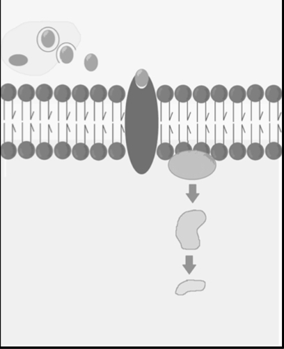
stage three: response
response: the final molecule in the signaling pathway converts the signal to a response that will alter a cellular process
examples
protein that can alter membrane permability
enzyme that will change a metabolic process
protein that turns genes on or off
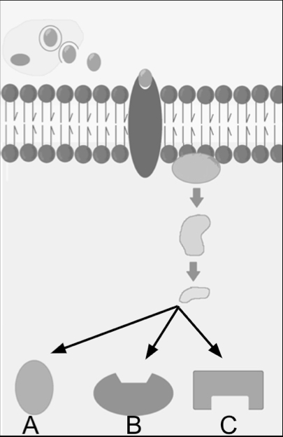
signal transduction pathways
-influence how a cell responds to its environment
-they can result in changes in gene expression and cell function
-can alter phenotypes or result in cell death
changes in signal transduction pathways
-mutations to receptor proteins or to any component of the signaling pathway will result in change to the transduction of the signal
important receptors:
-G protein coupled receptors (GPCRs)
-Ion Channels
GPCRs
largest category of cell surface receptors
important in animal sensory systems
binds to a G protein that can bind to GTP (similar to ATP)
the GPCR, enzyme, and G protein are inactive until ligand binding to GPCR on the extracellular side
ligand binding causes cytoplasmic side to change shape
allows for the G protein to bind to GPCR
activates the GPCR and G protein
GDP becomes GTP
part of the activated G protein can then bind to the enzyme
activates enzyme
amplifies signal and leads to a cellular response
ion channels
ligand gated ion channels
-located:
in the plasma membrane
-important in the:
nervous system
receptors that act as a gate for ions
when a ligand binds to the receptor, the “gate” opens/closes allowing the diffusion of specific ions
initiates a series of events that lead to a cellular response
quorum sensing
-controlled through population density
-quorum sensing could be used to solve antibiotic resistance through pathogency
topic five
homeostasis- the state of relatively stable internal conditions
feedback loops
negative
reduces the effect of the stimulus
the most common feedback
sweat, blood sugar, heart rate
positive
increases the effect of the stimulus
child labor, blood clotting, fruit ripening
the difference summarized: negative feedback reduces a stimulus’s effect to maintain homeostasis, while positive feedback amplifies it.
-stimulus: a variable that will cause a response
-receptor/sensor: sensory organs that detect a stimulus (this is sent to the brain)
-effector: muscle or a gland that will respond
-response: changes that either increase or decrease the effect of the stimulus
homeostatic imbalances
-disease: when the body is unable to maintain homeostasis
cell signaling as a means of homeostasis
-in order to maintain homeostasis, the cells in a multicellular organism must be able to communicate, which occurs through the signal transduction pathways
topics six and seven
cell cycle
allows for the reproduction of cells, growth of cells, and tissue repair
the cell cycle is the life of a cell from its formation until it divides
organization of DNA
-dna associates with and wraps around proteins known as histones to form nucleosomes
-strings of nucleosomes form chromatin
-when a cell is not activately dividing, chromatin is in a non-condensed form
-after DNA replication, chromatin condenses to form a chromosome
-since the DNA was replicated, each chromosome has a duplicated copy
-those copies join together to form sister chromatids
centromere: the region on each sister chromatid where they are most closely attached (the center)
kinetochore: proteins attached to the centromere that link each sister chromatid to the mitotic spindle
genome
a genome is all of a cells genetic information (DNA)
prokaryotic: singular, circle DNA
eukaryotic: one or more linear chromosomes
-every eukaryotic has a specific number of chromosomes
-homologous chromsomes: two chromsomes that are the same length, same centromere position, and carry genes with the same characteristic
somatic cells vs gametes
somatic cells:
-body cells
-diploid: two sets of chromosomes
-divide by mitosis
gametes:
-reproductive cells
-haploid: one set of chromosomes
-divide by meiosis
the cell cycle
interphase:
-G1: the cell grows and carries out normal functions
-S “synthesis” phase: DNA replication and chromsome duplication occurs
-G2 phase: final growth and preparation for mitosis
M phase:
mitosis: nucleus divides
results in 2 identical diploid daughter cells
cytokenesis: cytoplasm divides
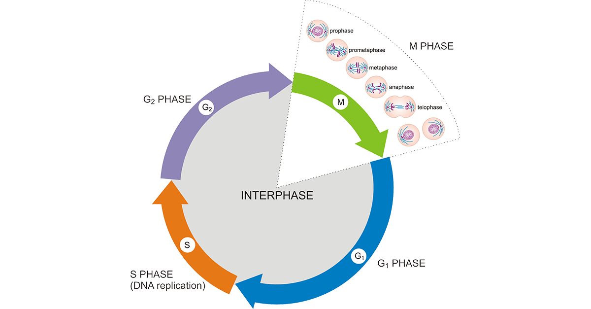
phases of mitosis
prophase
prometaphase
metaphase
anaphase
telophase
phase one: prophase
chromatin condenses into visible chromosomes, each with two sister chromatids joined at the centromere
the nuclear envelope starts to break down, facilitating the attachment of spindle fibers
the mitotic spindle forms and extends from centrosomes that move to opposite poles of the cell, preparing for chromsome movement and alignment
phase two: prometaphase
nuclear envelope fragements
microtubules enter nuclear area and some attach to kintechores
phase three: metaphase
centrosomes are at opposite poles
chromosomes line up at the metaphase plate
microtubules are attached to each kinetochore
phase four: anaphase
sister chromatids seperate and move to opposite ends of the cell due to microtubules shortening
cell elongates
phase five: telophase
two daughter nuclei form
nucleoli reappear
chromosomes become less condensed
cytokenesis occurs
animals: a cleavage furrow appears due to a contractile ring of actin filaments
plants: vesicles produced by the golgi travel to the middle of the cell and form a cell plate
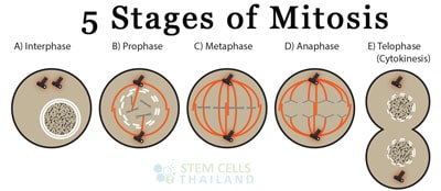
review: chromosomes
-cohesins are responsible for holding the sister chromatids together
-DNA is copied by synthesis, which forms sister chromatids because it results in two identical copies of each chromosomes
-gametes are reproductive cells
-somatic cells are anything other than reproductive cells
-eukaryotic cells undergoes mitosis
-a haploid contains one set of chromosomes (haploid number for humans is 23)
-a diploid contains two sets of chromosomes (diploid number for humans is 46)
regulation of the cell cycle
-throughout the cell there are checkpoints
G1 checkpoint:
most important
checks for cell size, growth factors, and DNA damage
stop/go signals:
-go: completes the whole cell cycle
-stop: cell enters a non dividing state known as the G0 phase
G0:
some cells stay in the G0 forever (muscle or nerve cells)
some cells can be called back into the cell cycle
G2:
checks for the completion of DNA replication and DNA damage
stop/go signals:
-go: cell proceeds to mitosis
-stop: cell cycle crops and the cell will attempt to repair damage
if the damage cannot be repaired the cell will undergo apoptosis, which is programed cell death
M (spindle) checkpoint:
checks for microtubule attachment to chromosomes at the kinetichores at metaphase
stop/go signals:
-go: cell proceeds to anaphase and completes mitosis
-stop: cell will pause mitosis to allow for spindles to finish attaching to chromosomes
internal cell cycle regulators
regulation of the cell cycle involves an internal control system that consists of:
proteins known as cycling (synthesized and degraded at specific stages of the cell cycle)
enzymes known as cyclin-dependent kinases (CKDs)
concentration remains constant through each phase of the cell cycle
active ONLY when its specific cyclin is presented
each CDK has a specific regulator effect
active CDK complexes phosphorylation target proteins, which help regulate key events in the cell cycle
external cell cycle regulators
growth factors:
hormones released by cells that stimulate cell growth
signal transduction pathway is initiated
CDKs are activated leading to progression through the cell cycle
contact (or density) inhibition:
cell surface receptors recognize contact with other cells
initiates signal transduction pathway that stops the cell cycle in the G1 phase
anchorage dependence:
cells rely on attachement to other cells or the extracellular matrix to divide
cancer: evasion of the cell cycle
normal cells become cancerous through: DNA mutations
-DNA mutation: change in DNA
normal cells vs cancer cells
normal cells:
follows checkpoints
divide on average 20-50 times in culture
go through apoptosis when there are errors
cancer cells:
don’t follow checkpoints
divide infinitely when in culture
considered to be immortal
evade apoptosis and continue to divide when there are errors
cancer cells
-uncontrollable growth of cancer cells can lead to a tumor (a mass of tissue formed by abnormal cells)
benign tumor: cells are abnormal, but aren’t cancerous
-the cells are unable to spread elsewhere in the body
malignant tumor: cancerous cells
-cells are able to spread somewhere else in the body
metastasis: cells seperate from the tumor and spread elsewhere in the body (malignant tumor)