2. rad lung patterns

studied byStudied by 0 people
0.0(0)
Get a hint
Hint

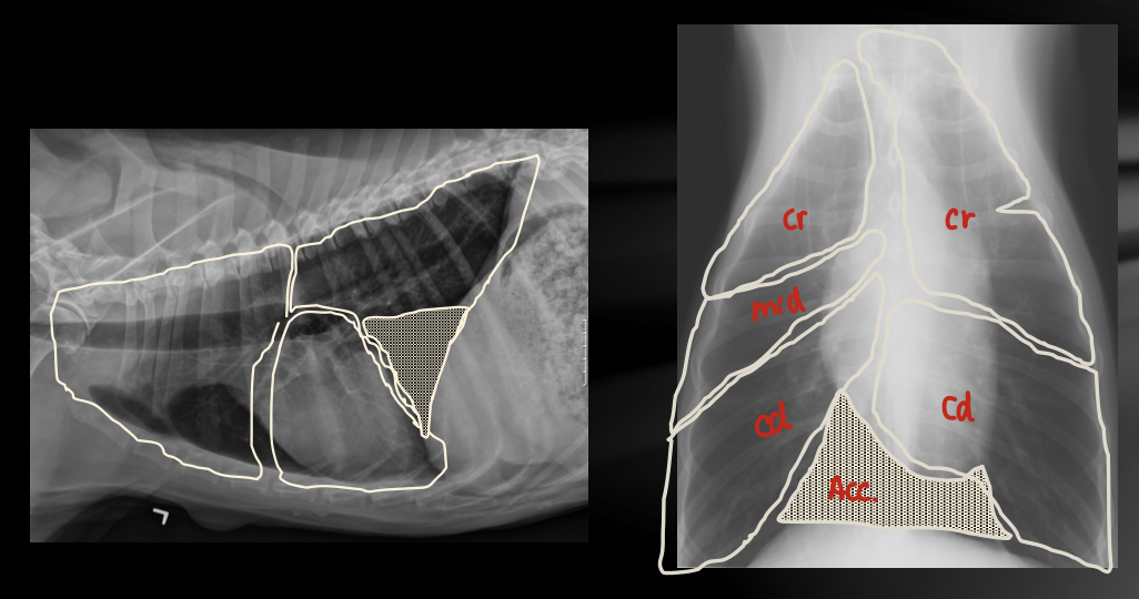
Lung lobe refresher on rads

1 / 12

encourage image

There's no tags or description

Looks like no one added any tags here yet for you.

13 Terms

1

Lung lobe refresher on rads

New cards
2

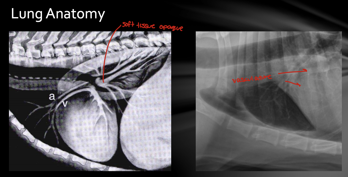
Normal lung is what opacity

radiolucent

New cards
3

how to pulmonary diseases alter lung opacity

pulmonary diseases result in increased pulmonary opacity

New cards
4

What 3 things could decrease pulmonary opacity (hyperlucency)

  • decreased vascularity of a pulmonary segment (thromboembolism)

  • hyperinflation (emphysema)

  • cavity lung lesion

New cards
5

lung lesions can be described in 2 different ways

focal or diffuse/generalized

New cards
6

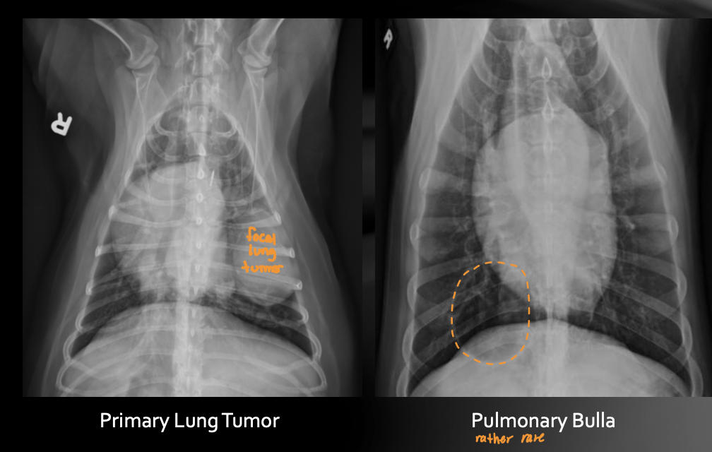
focal opaque vs focal lucent lesion

opaque - tumor

lucent - bulla

New cards
7

4 lung patterns

  1. bronchial

  2. interstitial

    1. structured - nodules

    2. unstructured - diffuse + busy (blurry vessel)

  3. alveolar - all soft tissue opaque

  4. (vascular)

<ol><li><p>bronchial</p></li><li><p>interstitial</p><ol><li><p>structured - nodules</p></li><li><p>unstructured - diffuse + busy (blurry vessel)</p></li></ol></li><li><p>alveolar - all soft tissue opaque</p></li><li><p>(vascular)</p></li></ol><p></p>
New cards
8

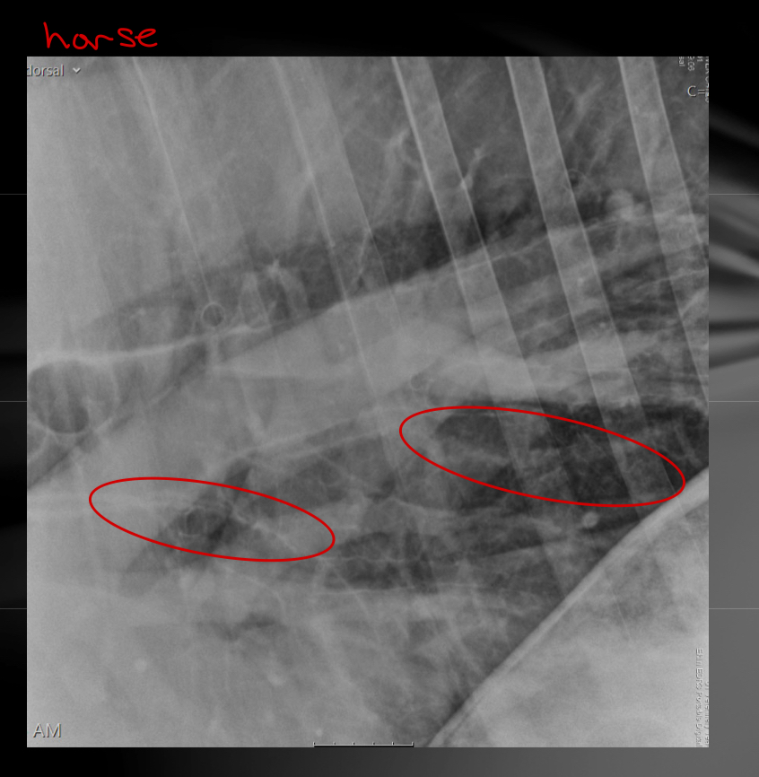
bronchial pattern

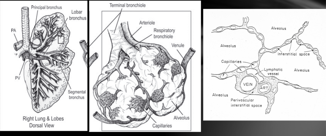
  • Increased visibility, conspicuity and thickening of bronchial walls

    • Doughnuts 

    • Tramlines

Differentials

  • See with “lower airway disease of infectious, parasitic or allergic etiology (bronchitis, lungworms, asthma)

  • recurrent airway obstruction in horse

  • Some bronchial wall mineralization is common in older dogs
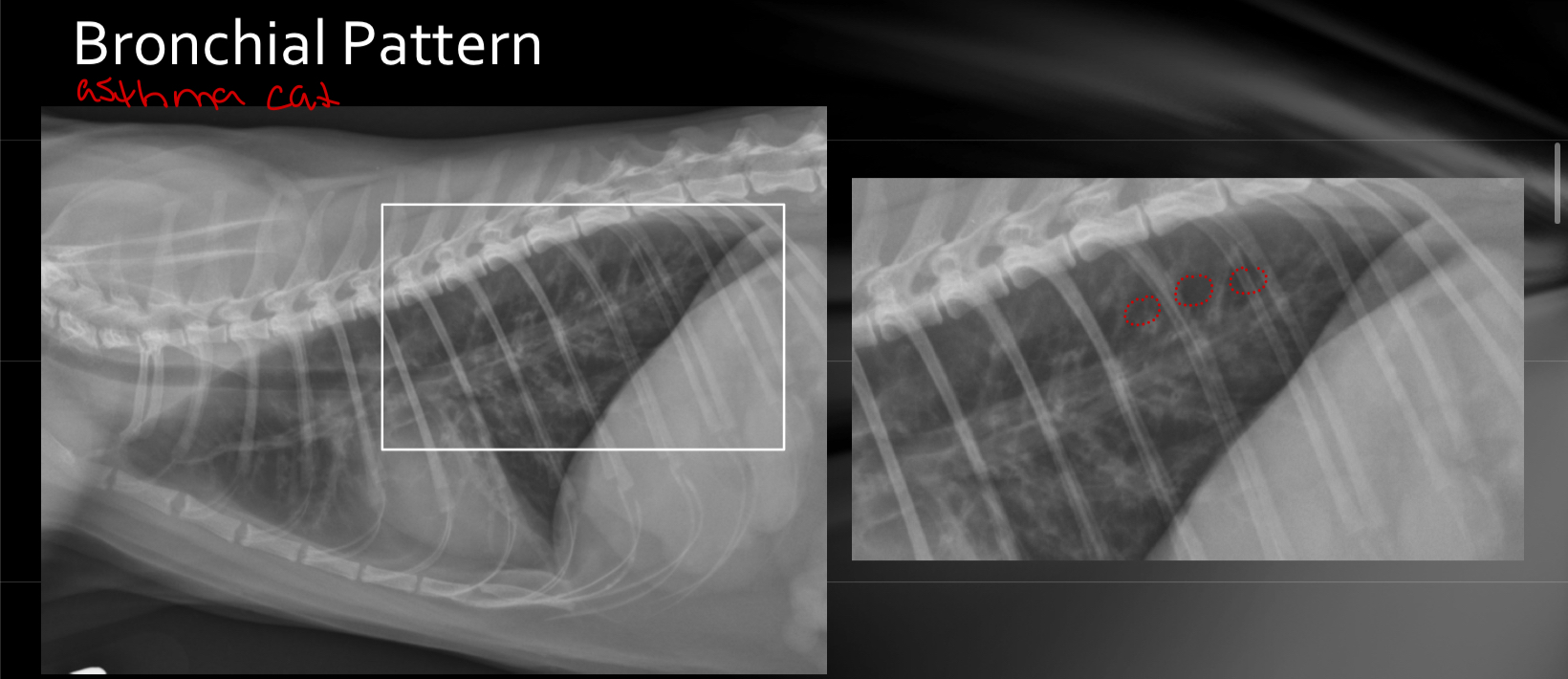
<ul><li><p><span>Increased visibility, conspicuity and thickening of bronchial walls</span></p><ul><li><p><span>Doughnuts&nbsp;</span></p></li><li><p><span>Tramlines</span></p></li></ul></li></ul><p>Differentials</p><ul><li><p><span>See with “lower airway disease of infectious, parasitic or allergic etiology (bronchitis, lungworms, asthma)</span></p></li><li><p><span>recurrent airway obstruction in horse</span></p></li><li><p><span>Some bronchial wall mineralization is common in older dogs</span></p></li></ul><img src="https://knowt-user-attachments.s3.amazonaws.com/67007514-64ca-4d9e-8ab6-a9b8d5f0bf67.jpg" data-width="100%" data-align="center"><img src="https://knowt-user-attachments.s3.amazonaws.com/355fc6e9-d1ae-449c-abb1-a5e113f41ffd.jpg" data-width="100%" data-align="center"><p></p>
New cards
9

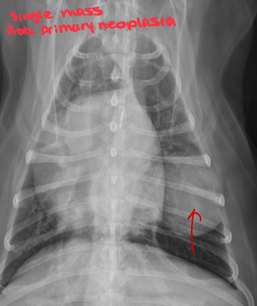
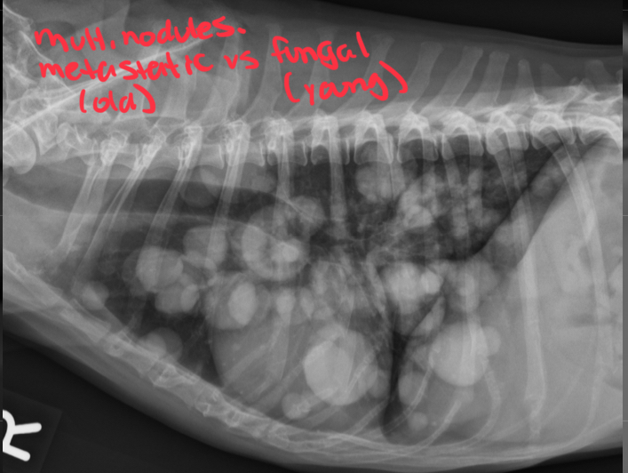
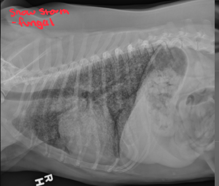
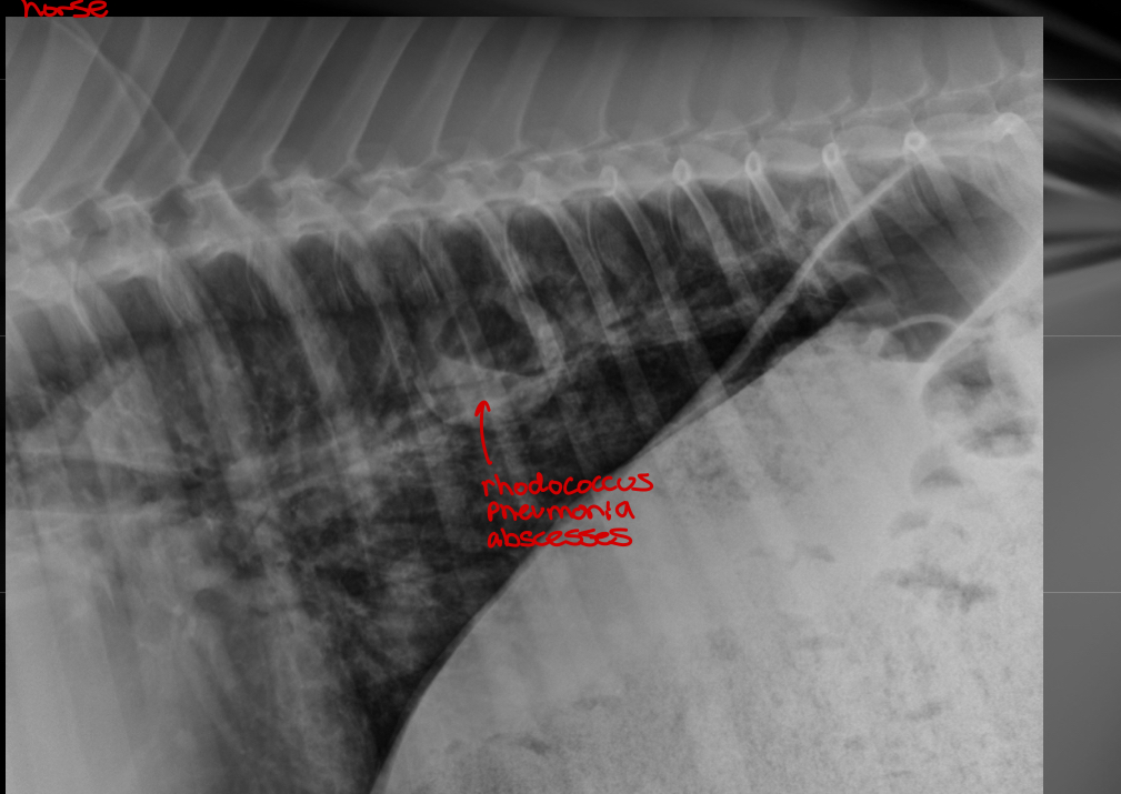
interstitial structured

  • Single mass 

  • Multiple nodules

  • TNTC nodules - miliary pattern or snowstorm lungs

Differential dx: 

  • Lung neoplasia (primary or metastatic)

  • Fungal disease

  • Rhodococcus pneumonia abscesses

  • other

New cards
10

Interstitial unstructured

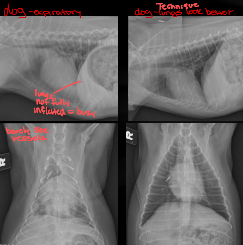
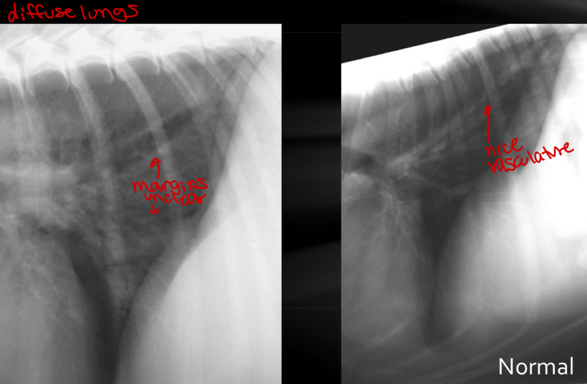
  • Lung is diffusely increased in opacity, but vessels remain visible

differentials (incidental to fatal)

  • Pulmonary edema

  • Interstitial pneumonia

  • Interstitial hemorrhage

  • Pulmonary fibrosis

  • Lymphoma

  • Early granulomatous (fungal) disease

  • Normal variant (age, expiration, obese patient)

incidental - inspiratory vs expiratory rads
significant - pulmonary lymphoma
New cards
11

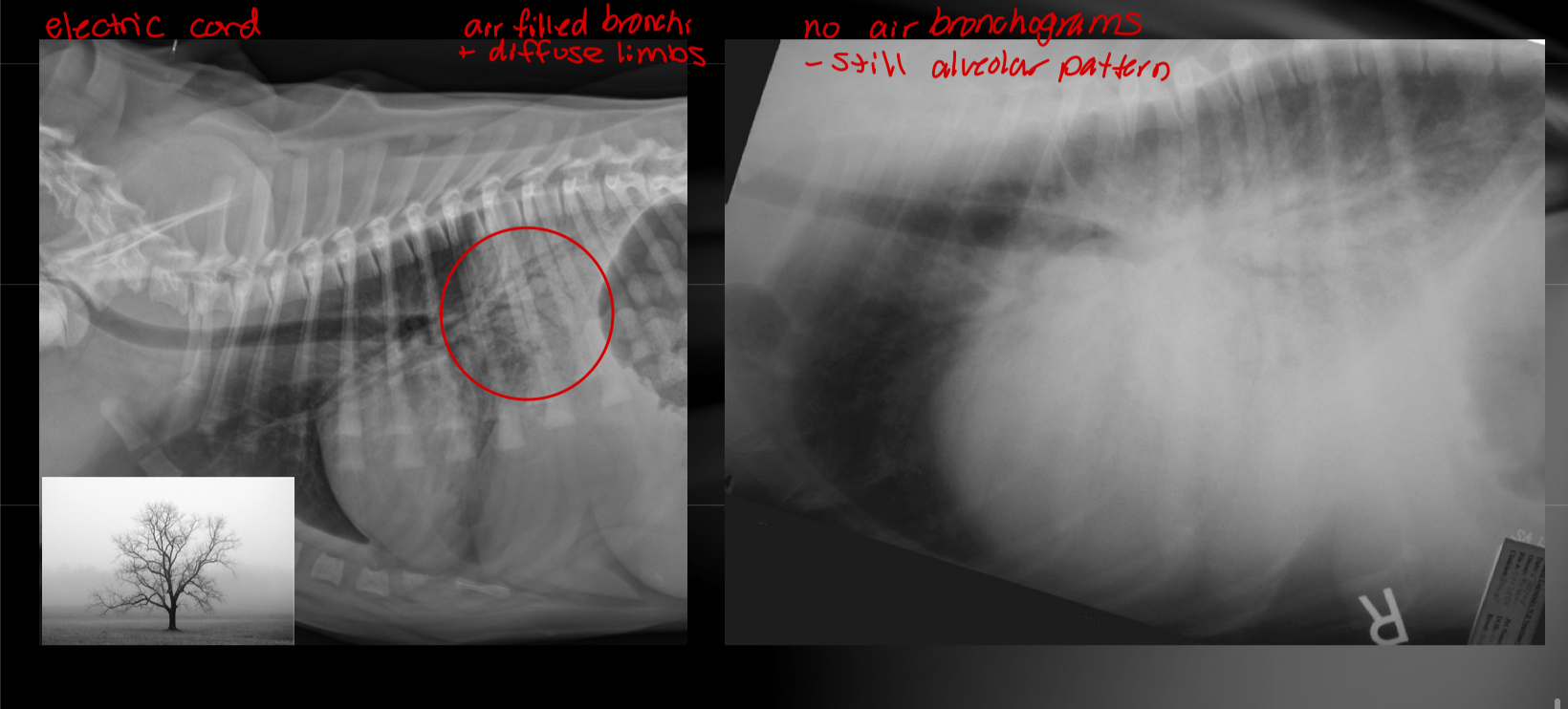
alveolar pattern

  • Increased pulmonary opacity to the point where vessels CANNOT be identified

  • Caused by decreased volume of air in lungs

    • Lungs filled with crap (consolidation) - blood, pus, water, cells

    • Lung is collapsed (atelectasis)

    • pulmonary edema → interstitial pattern early → alveolar pattern when bad

  • Air bronchograms - trees in the fog

    • Could be alveolar even if you don’t see the “trees”

    • gas filled bronchi contrasting with soft tissue opaque lungs

    • go away when soft tissue or fluid fills bronchial lumen as well

Differentials

  • NOT possible to determine what kind of fluid/cells makes increased density on radiographs

  • Consider

    • Location + distribution

    • Other radiographic abnormalities (heart, mediastinum, ribs)

  • Possible causes

    • Pulmonary edema (cardiogenic and noncardiogenic like electric cord)

    • Pneumonia

    • Hemorrhage

    • Atelectasis - no air/collapse

    • other

<ul><li><p><span>Increased pulmonary opacity to the point where <strong>vessels CANNOT</strong> <strong>be identified</strong></span></p></li><li><p><span>Caused by decreased volume of air in lungs</span></p><ul><li><p><span>Lungs filled with crap (consolidation) - blood, pus, water, cells</span></p></li><li><p><span>Lung is collapsed (atelectasis)</span></p></li><li><p>pulmonary edema → interstitial pattern early → alveolar pattern when bad</p></li></ul></li><li><p><span>Air bronchograms - trees in the fog</span></p><ul><li><p><span>Could be alveolar even if you don’t see the “trees”</span></p></li><li><p><span>gas filled bronchi contrasting with soft tissue opaque lungs</span></p></li><li><p><span>go away when soft tissue or fluid fills bronchial lumen as well</span></p></li></ul></li></ul><p>Differentials</p><ul><li><p><span>NOT possible to determine what kind of fluid/cells makes increased density on radiographs</span></p></li><li><p><span>Consider</span></p><ul><li><p><span>Location + distribution</span></p></li><li><p><span>Other radiographic abnormalities (heart, mediastinum, ribs)</span></p></li></ul></li><li><p><span>Possible causes</span></p><ul><li><p><span>Pulmonary edema (cardiogenic and noncardiogenic like electric cord)</span></p></li><li><p><span>Pneumonia</span></p></li><li><p><span>Hemorrhage</span></p></li><li><p><span>Atelectasis - no air/collapse</span></p></li><li><p><span>other</span></p></li></ul></li></ul><img src="https://knowt-user-attachments.s3.amazonaws.com/603691f6-b8ca-4946-b7ea-7c169c629e5c.jpg" data-width="100%" data-align="center"><p></p>
New cards
12

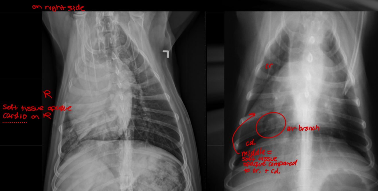
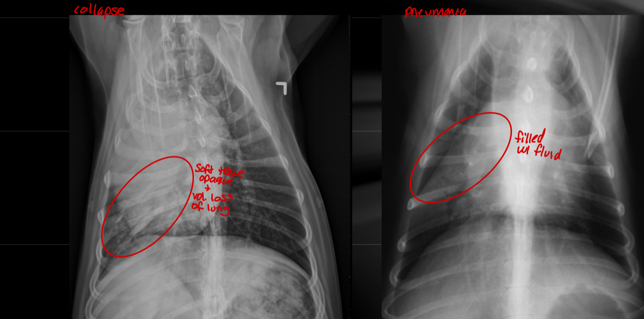
atelectasis vs true alveolar pattern

Atelectasis

  • Structures are shifted to one side

  • Soft tissue opaque structures

  • Volume loss of lung

True alveolar pattern

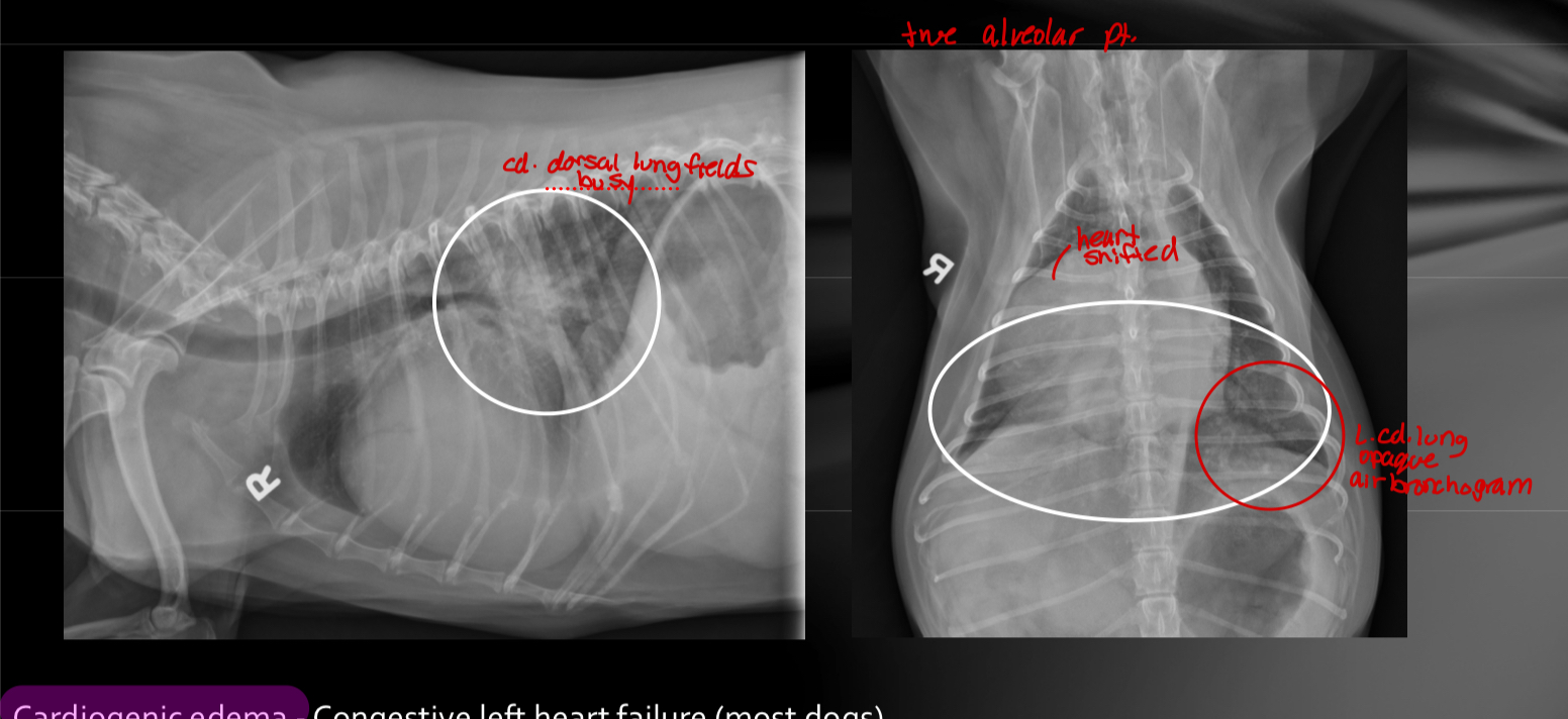
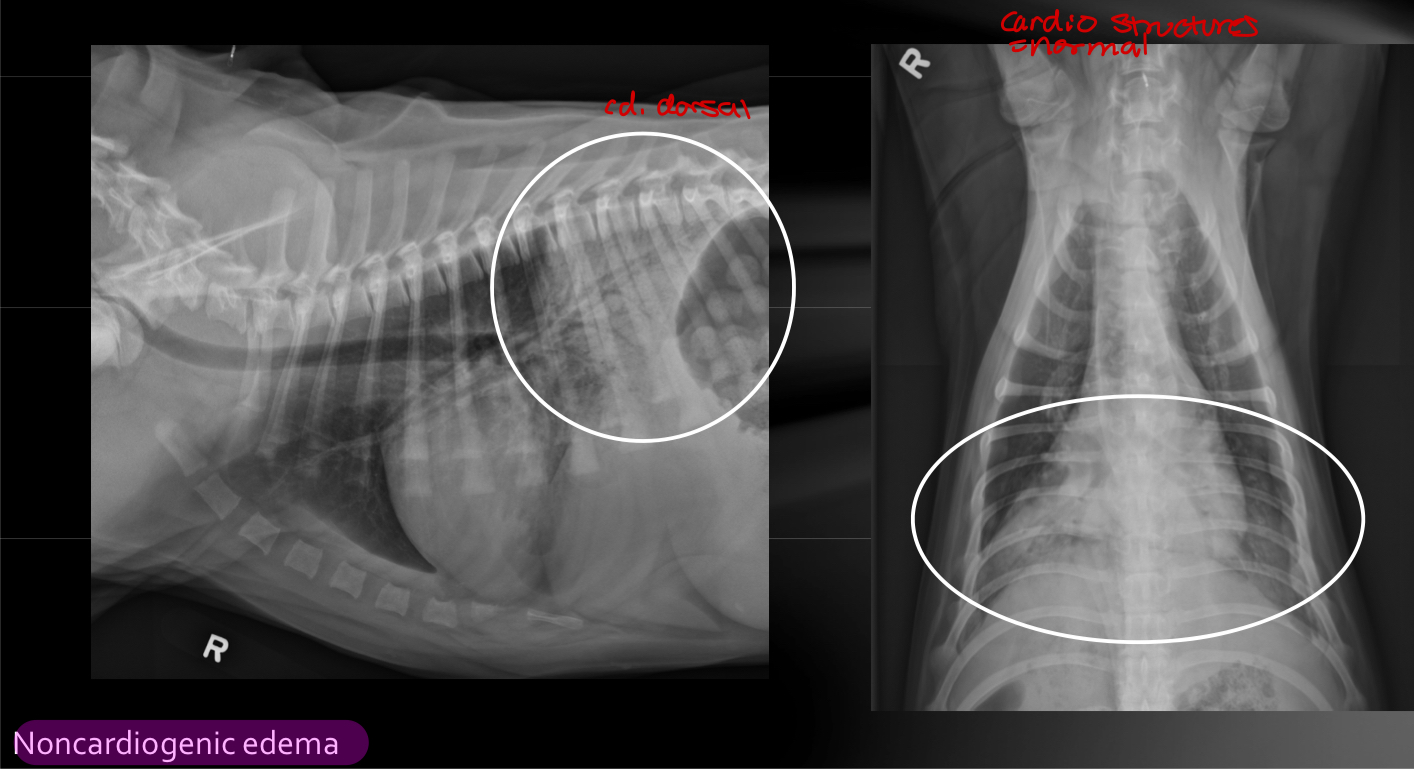
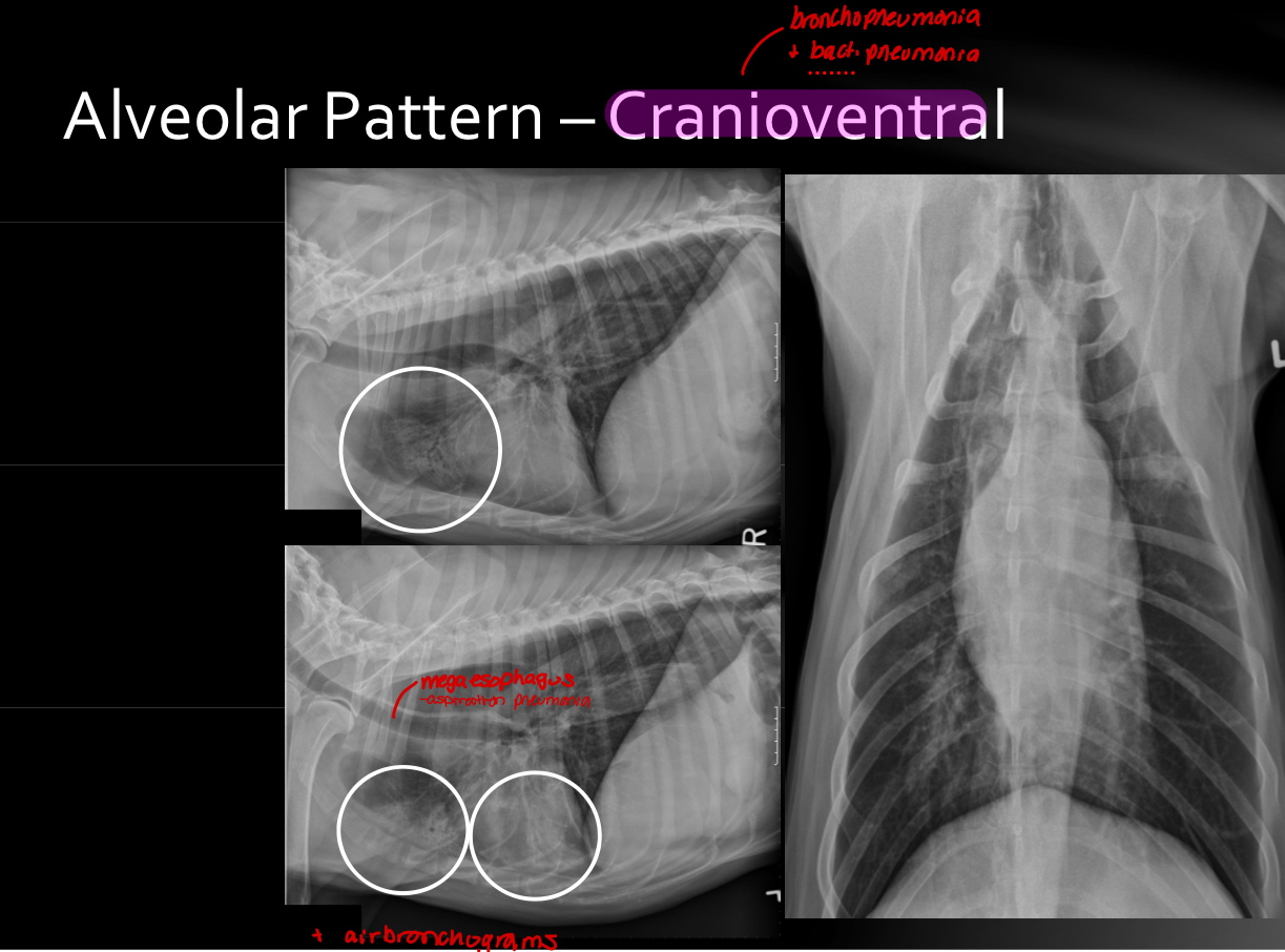
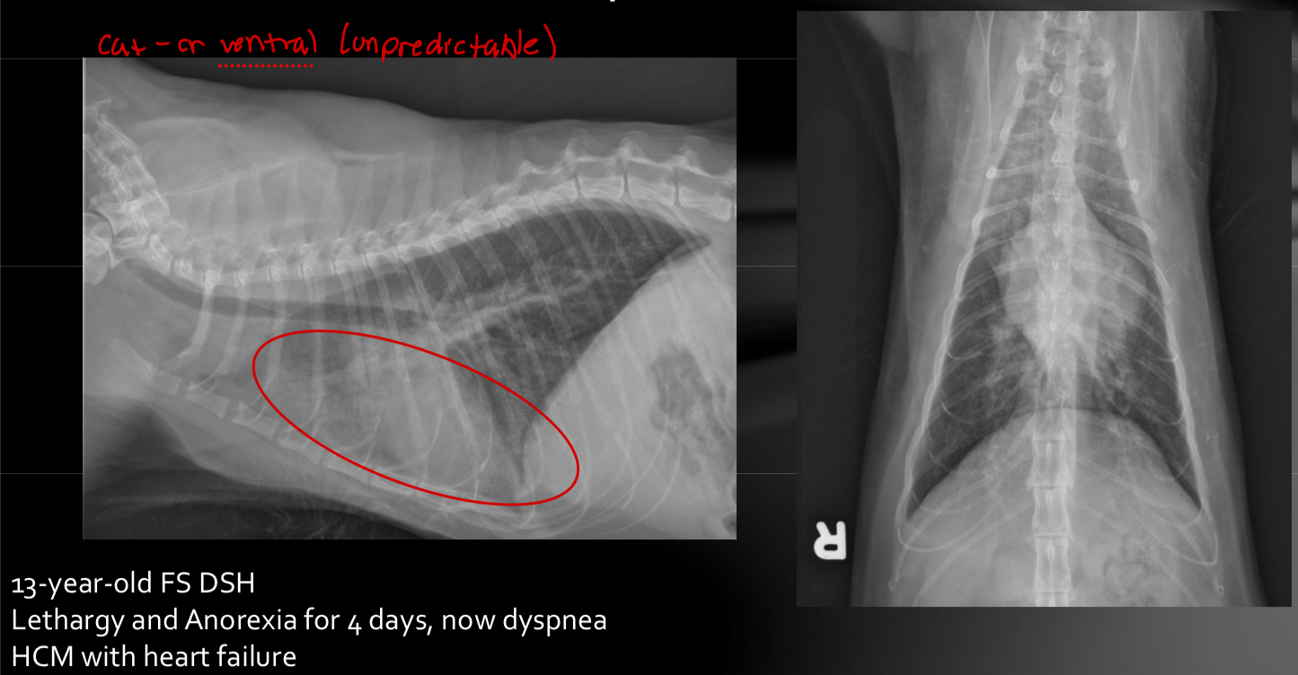
  • middle lung lobe opacities are like soft tissue opaque compared to cr. and cd. Lung lobes (filled with fluid)

  • Pneumonia

  • Cardiogenic edema - congestive heart failure in dogs is caudodorsal

  • Noncardiogenic edema - has the same caudodorsal opacities with normal cardiac structures

  • bronchopneumonia /bacterial pneumonia - cranioventral opacities (aspiration pneumonia)

    • Cranioventral in horses, ruminants, pigs

  • alveolar pattern can be anywhere

    • Trauma (contusions, hemorrhage)

    • Hemorrhage for other reasons

    • Heart failure in cats

    • Dobermans and dogs with acute chordae tendinae rupture

    • other

cardiogenic edemanoncardiogenic edema

New cards
13

hierarchy of lung patterns?

who knows BUT

  1. structured interstitial and alveolar (consolidation)

  2. bronchial (with cough is significant and could just be old animal)

  3. unstructured interstitial (± importance)

New cards

Explore top notes

note Note
studied byStudied by 635 people
... ago
4.8(5)
note Note
studied byStudied by 11 people
... ago
5.0(1)
note Note
studied byStudied by 1 person
... ago
5.0(1)
note Note
studied byStudied by 11 people
... ago
4.5(2)
note Note
studied byStudied by 21 people
... ago
5.0(1)
note Note
studied byStudied by 93 people
... ago
5.0(1)
note Note
studied byStudied by 7 people
... ago
5.0(1)
note Note
studied byStudied by 13 people
... ago
5.0(1)

Explore top flashcards

flashcards Flashcard (32)
studied byStudied by 47 people
... ago
4.0(1)
flashcards Flashcard (22)
studied byStudied by 6 people
... ago
5.0(1)
flashcards Flashcard (500)
studied byStudied by 24 people
... ago
5.0(1)
flashcards Flashcard (90)
studied byStudied by 2 people
... ago
4.0(1)
flashcards Flashcard (118)
studied byStudied by 9 people
... ago
4.0(1)
flashcards Flashcard (25)
studied byStudied by 2 people
... ago
4.0(1)
flashcards Flashcard (30)
studied byStudied by 10 people
... ago
5.0(1)
flashcards Flashcard (88)
studied byStudied by 559 people
... ago
5.0(1)
robot