Tietze Chapter 5 Diagnostics (General, Cardio)
1/31
There's no tags or description
Looks like no tags are added yet.
Name | Mastery | Learn | Test | Matching | Spaced | Call with Kai |
|---|
No analytics yet
Send a link to your students to track their progress
32 Terms
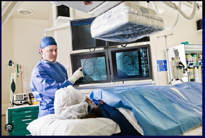
Angiography
It is a radiographic test used to evaluate blood vessels and the circulation (Figure 5-1). Radiopaque material is injected through a catheter inserted in the blood vessel, and images are recorded using standard radiographic techniques

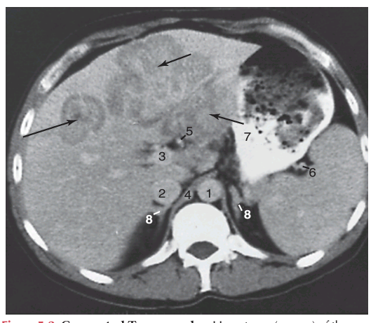
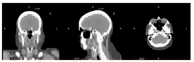
Computed Tomography (CT Scan)
It uses a computerized x-ray system to produce detailed sectional x-ray images (Figure 5-2). The system is very sensitive to differences in tissue density and produces detailed two-dimensional planar images; the use of contrast agents increases attenuation. In spiral or helical CT scanning pictures are taken continuously, which decreases the time needed to obtain images.
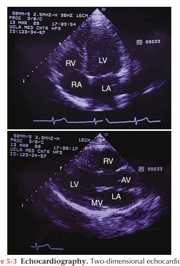
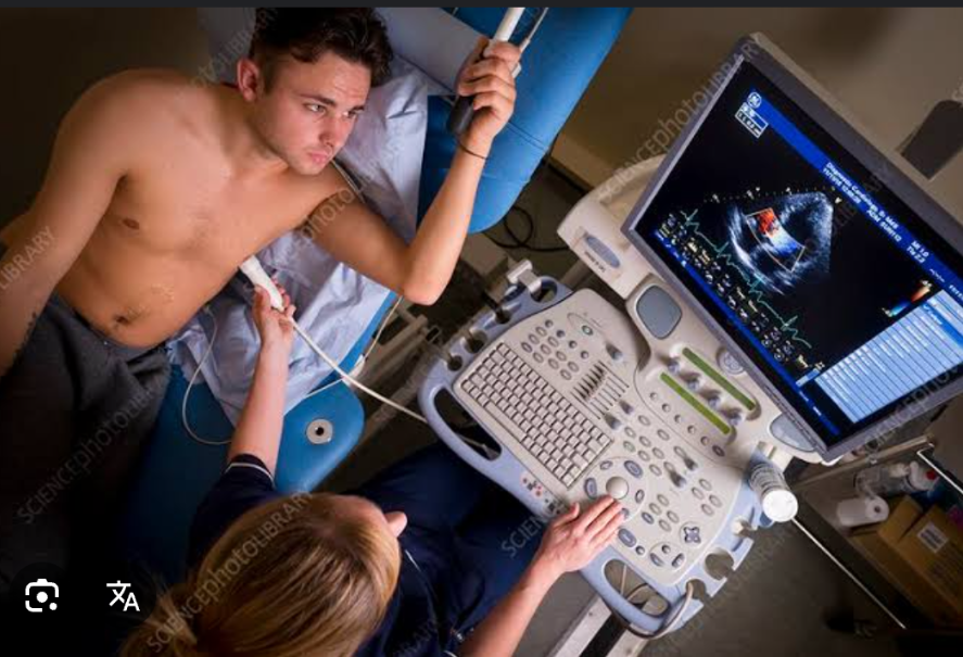
Doppler Echography (Echocardiograph)
It uses ultrasound technology to measure shifts in frequency caused by object movement. For example, it is used to evaluate blood flow velocity and turbulence in the heart and peripheral circulation.
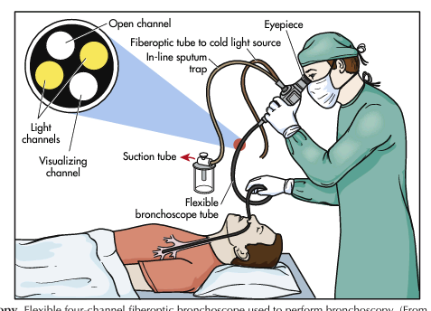
Endoscopy
It is used to examine the interior of a hollow viscus (e.g., digestive, respiratory, and urogenital organs and the endocrine system) or canal (e.g., bile ducts, pancreas). The instrument, a flexible or inflexible tube with a camera and light source, is inserted into a body orifice (Figure 5-4). Still and/or video images are recorded and biopsy specimens are obtained for tissue examination or other laboratory diagnostic tests
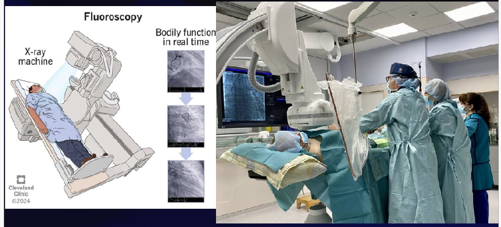
Fluroscopy
It makes the shadows of x-rays visible, to provide real-time visualization of procedures. It exposes a patient to more radiation than routine radiography but often is used to guide needle biopsy procedures and nasogastric tube advanceme
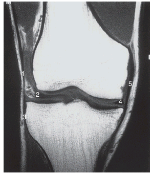
Magnetic Resonance Imaging
It uses an externally applied magnetic field to align the axis of nuclear spin of cellular nuclei (Figure 5-9). The patient is surrounded by the magnetic field. Brief radiofrequency pulses are applied to displace the alignment. The energy emitted when the displacement ends is detected, which allows finely detailed planar and three-dimensional images to be produced; the use of contrast agents increases the attenuation (Figure 5-10).
Molecular imaging
It is an investigational technology that assesses biologic processes at the cellular and subcellular level in living tissue. Areas of interest include the biology of cancer and cardiovascular disease.
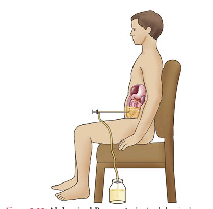
Abdominal Paracentesis
Removal of fluid in abdomen
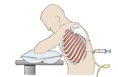
Thoracentesis
Removal of fluid in chest
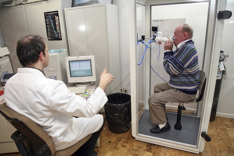
Plethysomnography
It assesses changes in the size of vessels and hollow organs by measuring displacement of air or fluid from a containment system. It is used to assess pulmonary function
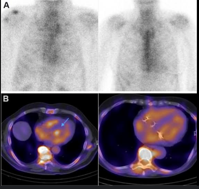
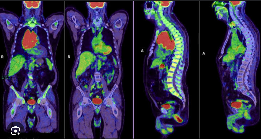
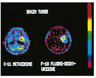
Positron Emission Tomography
It uses positron-emitting radionuclides (e.g., fluorodeoxyglucose F 18) to visualize organs and tissues of the body. The radionuclides decay, producing positrons that collide with electrons. A special camera detects photons released when the positrons and electrons collide.
Provides quantitative information regarding the structure and function of organs and tissues. It is commonly used to detect and monitor malignancies.
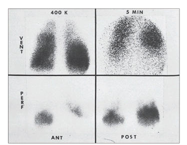
Radionuclide Studies
It involve the administration of oral, parenteral, or inhaled radioactive chemicals or pharmaceuticals. X-ray images, usually serial, record the collection and dispersion of the radioactive material
Single photon emission computed tomography SPCET
It is similar to PET but involves the administration of radionuclides that emit gamma rays. It is less expensive than PET but provides more limited image resolution. It is commonly used to assess the coronary arteries, bone, brain, prostate, and thyroid.
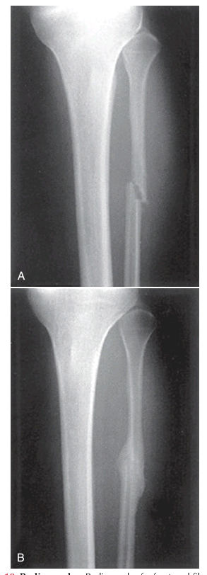
Standard Radiography
images are produced on photographic plates by passing x-rays (roentgen rays) through the body. Difficult to interpret because the three dimensionality is lost on the planar images.

Ultrasonography (Echography)
It uses ultrasound (high-frequency sound waves imperceptible to the human ear) to create images of organs and vessel
Cardiac catheterization
— is used to evaluate cardiac function. A catheter is passed into the right or left side of the heart. Transducers on the tip of the catheter record pressures in the vessels and chambers of the heart. Ports in the catheter provide access for obtaining blood samples for the determination of oxygen content and cardiac output.
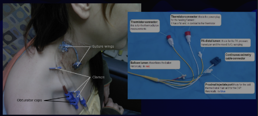
Central Line Placement with hemodynamic monitoring
a catheter is placed into the central venous system and advanced into the right side of the heart. The right atrial, right ventricular, pulmonary artery, and pulmonary artery occlusion (formerly known as pulmonary capillary wedge) pressures are measured, and cardiac output is calculated. These parameters are used to monitor the hemodynamic status of the patient and to calculate the pulmonary and peripheral vascular resistances.
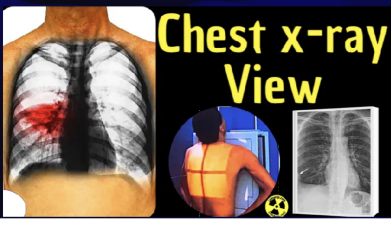
Chest radiograph
used to diagnose cardiac disease and monitor patient response to drug and nondrug therapy. It is used to determine the size and shape of the atria and ventricles, to calculate the cardiothoracic ratio, and to detect abnormalities in the lung fields and pleural spaces.
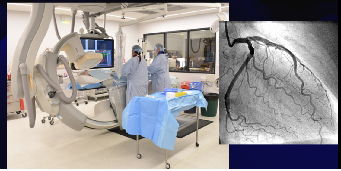
Coronary Angiography
the cardiac vessels are visualized by injecting the vessels with a contrast agent.
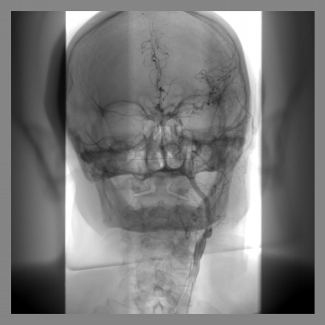
Digital Subtraction Angiography
background images are obtained before the contrast agent is injected. The background images are then “subtracted” from the images obtained after the injection of the contrast agent. This technique improves image resolution.
Contrast echocardiography
Visualization of the rightsided chambers of the heart is enhanced by the injection of contrast agents.
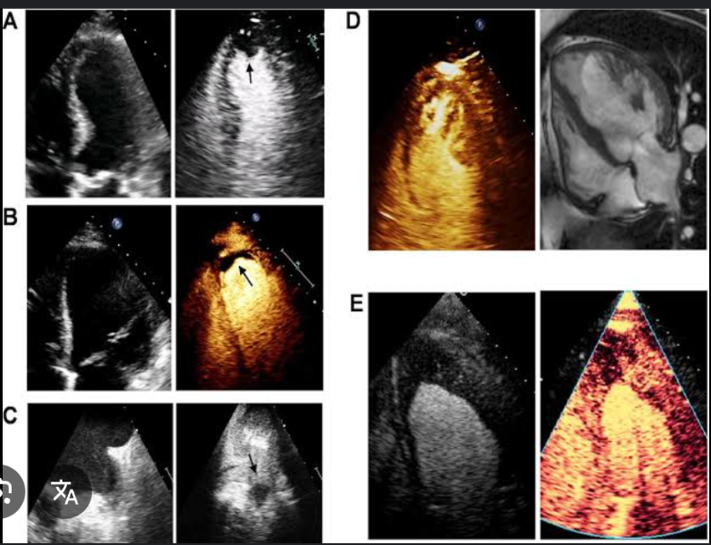
Doppler echocardiography
Doppler imaging and echocardiography techniques are combined to evaluate cardiac blood flow patterns.
Exercise echocardiography
compares echocardiograms obtained before and during exercise.
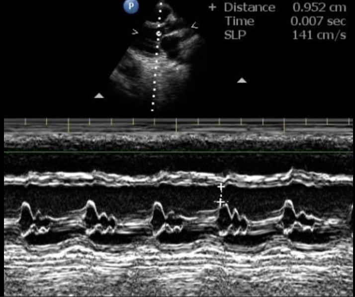
M-Mode echocardiography
records the motion of the heart over time. It is used to evaluate the structures of the heart throughout the cardiac cycle
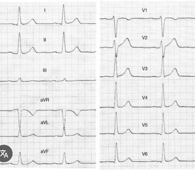
Electrocardiography
records the electrical activity of the heart. The ECG is used to diagnose cardiac disease, monitor patient response to drug therapy, and monitor for adverse drug effects.
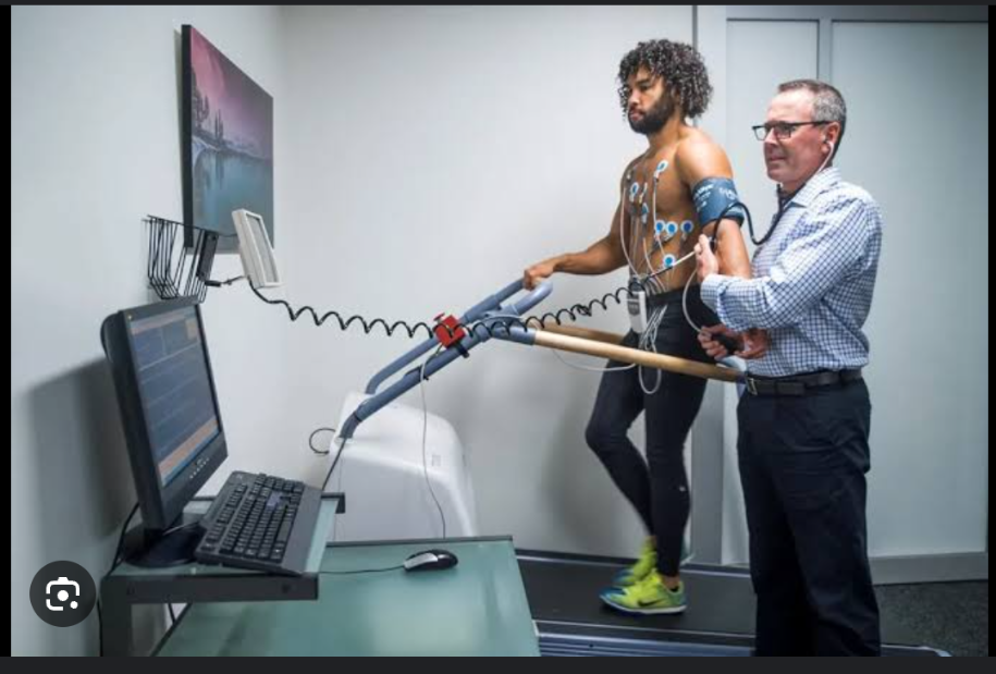
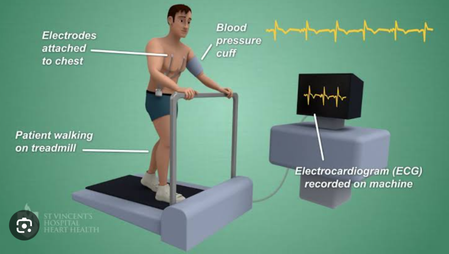
Electrocardiogram with stress
In the stress test, the ECG is recorded during a standardized exercise protocol with gradually increasing levels of exercise or with the patient at rest after the administration of dobutamine or dipyridamole;
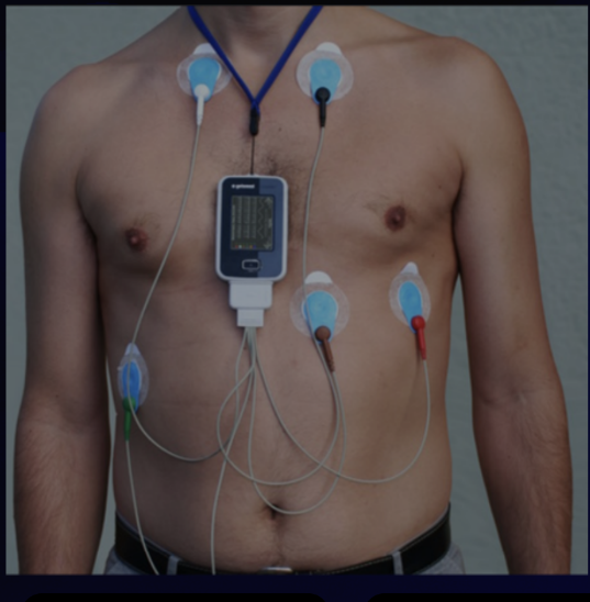
Holter monitoring (ambulatory electrocardiogram)
is a portable recorder used to record the ECG continuously throughout the patient’s usual activities
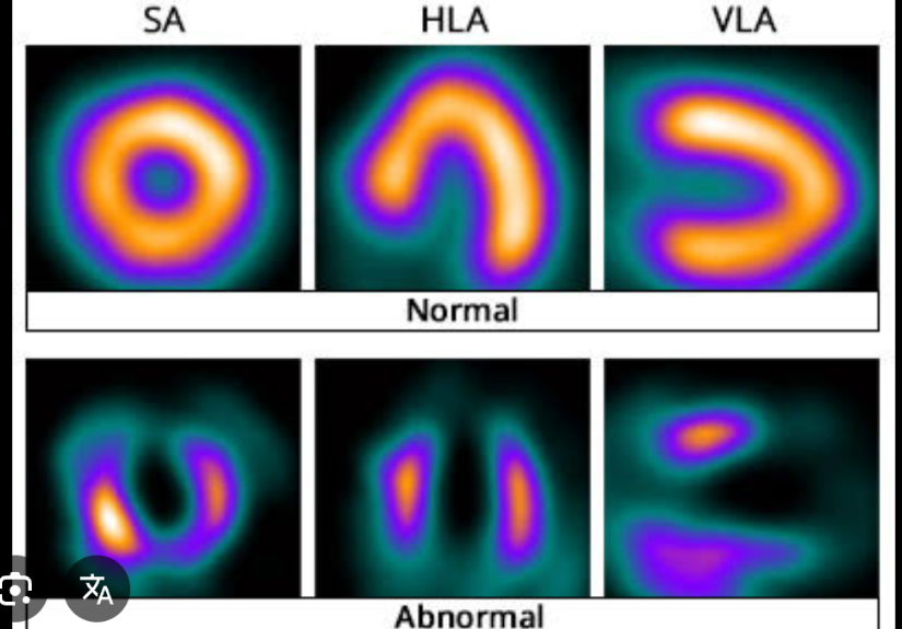
Thalluium stress test
combines the parenteral administration of thallium Tl 201, a radionuclide taken up by healthy myocardial tissue, and the stress test (either exercise or pharmacologic). A gamma camera is used to record serial images of the myocardium
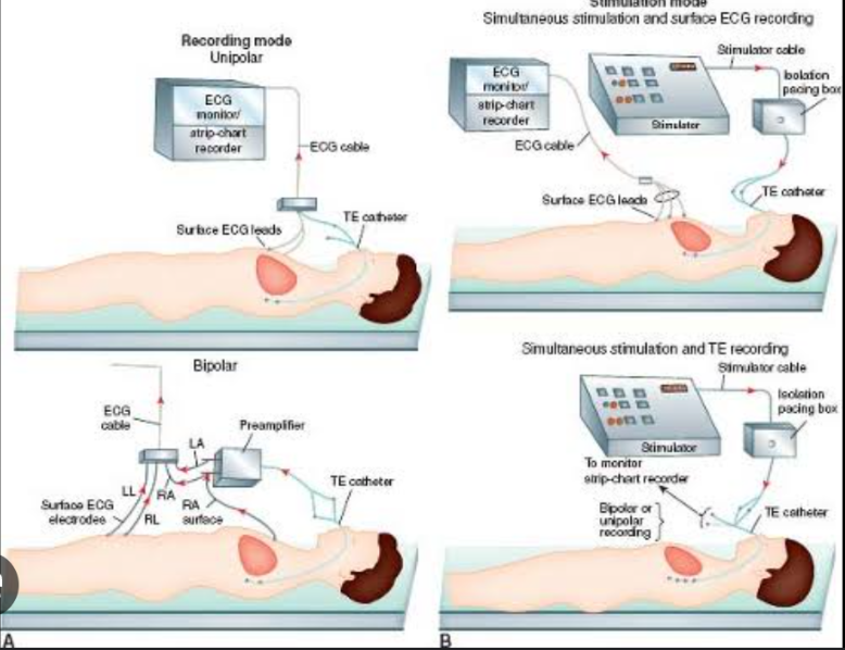
Intracardiac electrophysiologic studies
are tests in which special catheters with electrodes are used to stimulate the cardiac tissue to assess the nature and origin of cardiac arrhythmias and the response to antiarrhythmic drug therapy
Lymphoscintigraphy
evaluates the patency and anatomy of peripheral lymph vessels by placing a radioactive agent in the tissue drained by the lymph system being evaluated. The test is used to assess lymphedema and tumor involvement or regional lymph nodes that cannot be visualized with other imaging procedures
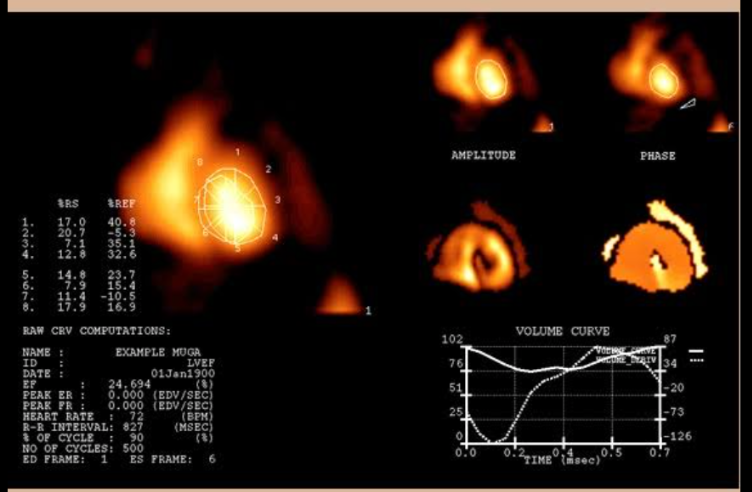
Multiple gated acquisition scan (radionuclide angiocardiography)
evaluates ventricular function, cardiac wall motion, ejection fraction, and cardiac output after the injection of albumin or red blood cells (RBCs) labeled with a radionuclide (technetium Tc 99m).
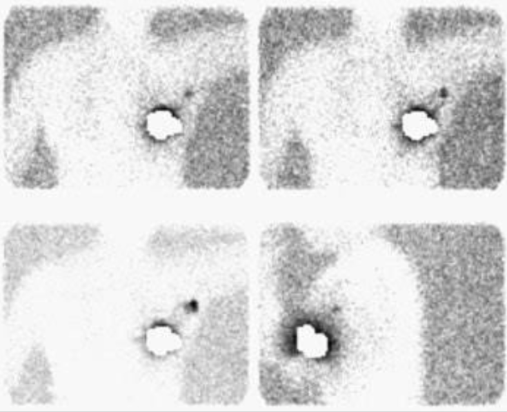
Technetium Tc 99m Pyrophosphate Uptake
Infarcted myocardial tissue shows an increased uptake of technetium Tc 99m compared with healthy tissue. The isotope is injected parenterally, and serial images of the heart are obtained to evaluate the location and extent of the myocardial damage.