Unit 3b - Special Senses
1/36
There's no tags or description
Looks like no tags are added yet.
Name | Mastery | Learn | Test | Matching | Spaced | Call with Kai |
|---|
No analytics yet
Send a link to your students to track their progress
37 Terms
Olfaction
the only sensory modality that does NOT go to thalamus first and does NOT cross the midline
the only special sense where the sensory cell is the neuron itself that carries information to the CNS
way more important as a sense in other species since it’s closely linked with taste, emotion, memory
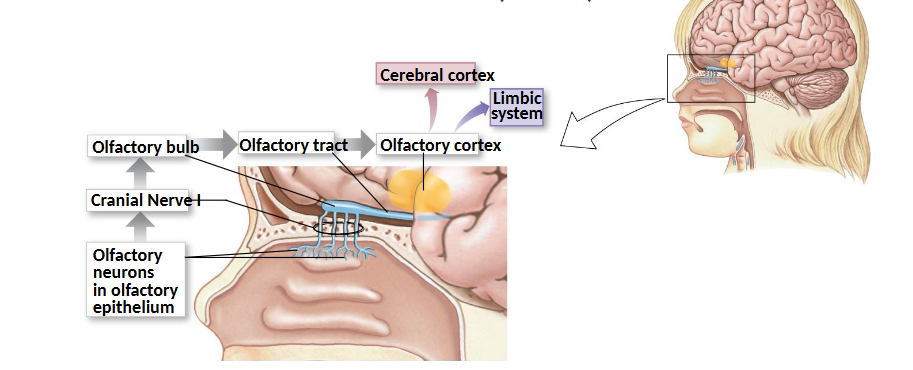
Olfactory Receptor Neurons
bipolar neurons that are replaced about 60 days (neurons are never replaced nor divide except for these)
dendrites end in non-motile cilia expressing odorant receptor proteins
axons go through gaps in cribiform plate; synapse on 2nd order neurons
odorant receptor proteins are GPCRs, one of the largest gene families in vertebrates
Olfactory Quality
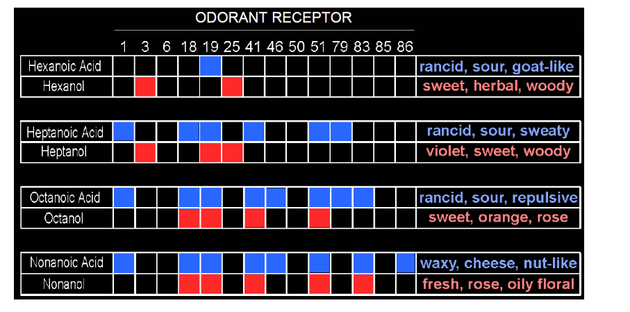
each olfactory receptor neuron expresses only one type of odorant receptor protein (GPCR) but each receptor can recognize more than one odorant AND each odorant can stimulate more than one receptor
subsequent processing en route to olfactory cortex in combination with 100s of other olfactory neurons is then interpreted as a particular odour
Receptor Codes and Perception
octanol smells fruity and floral but octanoic acid is rancid → changing a functional group changes a lot about how we perceive the smell
Linda Buck nobel for the receptor perception chart
Gustation (Taste)
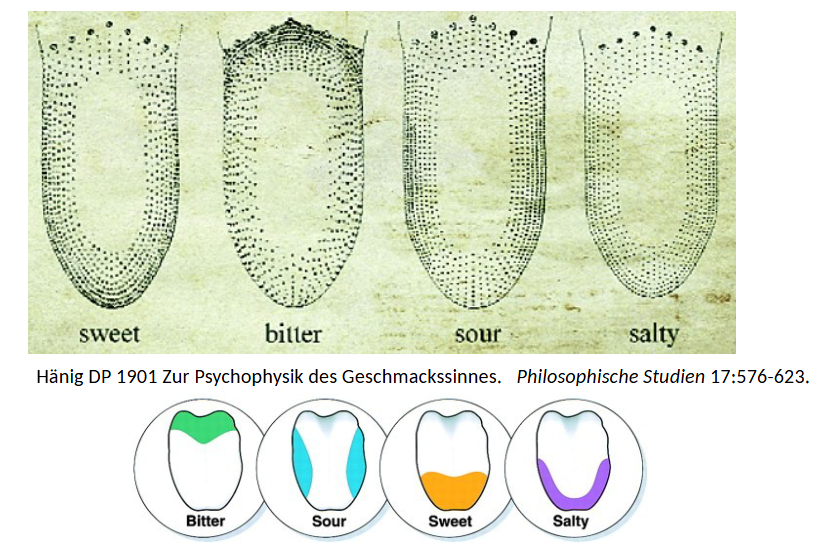
combination of 5 basic tastes: sweet, sour, salty, bitter, umami
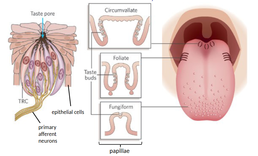
taste receptor cells are non-neural epithelial cells that release signal onto primary afferent neuron and frequently come into contact with noxious chemicals
the map of regions of tongue for different tastes is a myth
Taste Buds
number per papillae depends on type
each taste bud has 50-150 taste receptor cells
supporting epithelial cells secrete fluid into lumen of taste pore
How many receptor cell types are there for each taste?
1; 4/5 tastes only express one but bitter receptors are generic with lots of GPCRs to detect the range of bitter flavours
Taste Transduction
sweet, umami, and bitter ligands bind to GPCRs and ultimately release ATP as the signalling molecule
ligands activate taste receptor cell
various intracellular pathways activated
Ca2+ signal triggers exocytosis or ATP formation
neurotransmitter or ATP released
primary sensory neuron fires to send APs to brain
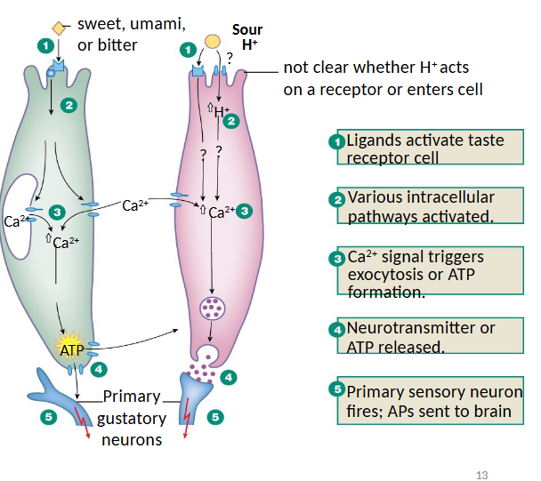
Transduction of these two tastes is poorly understood.
sour and salty
Taste Pathway to Brain
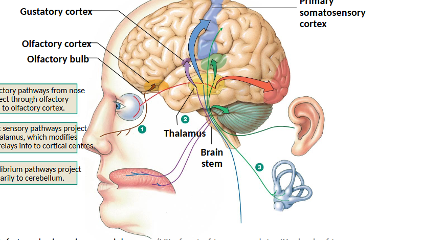
travels via various cranial nerves to medulla → thalamus → gustatory cortex → integrated with smell
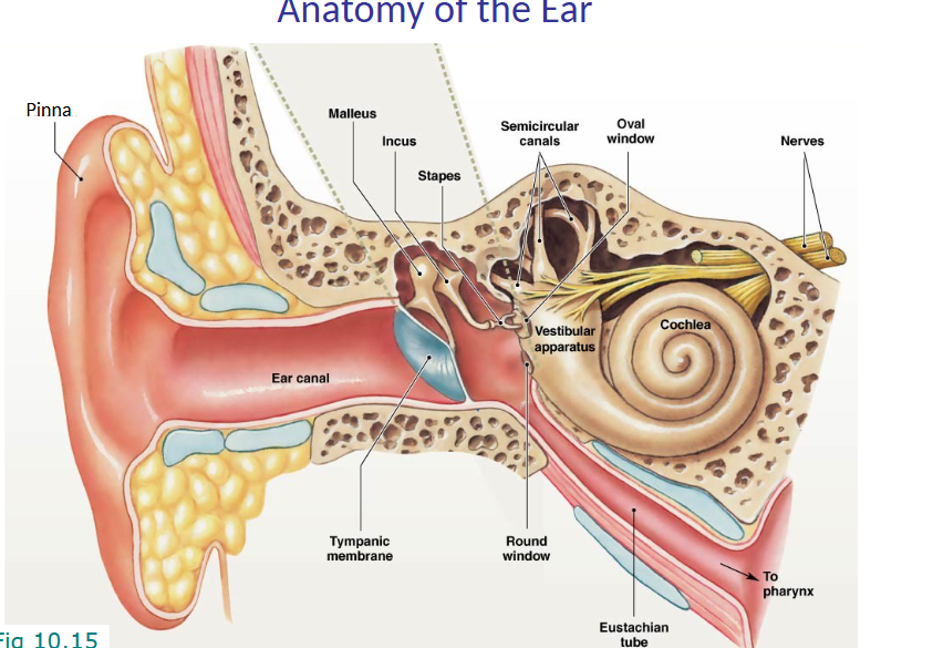
Anatomy of the Ear
external ear canal
tympanic membrane aka eardrum vibrates
sound is transduced by malleus, incus, and stapes
oval window
vestibular apparatus does equilibrium
cochlea is the sensory structure
A longer wavelength of a sound wave means what?
lower frequency and therefore lower pitch since less wavelength/time
Sound Transmission Through the Ear
sound wave reaches tympanic membrane from air-filled external canal
vibrates bony ossicles
transduced through oval window
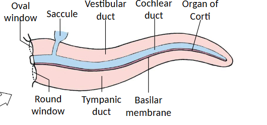
fluid-filled compartment (vestibular duct filled with perilymph) vibrates to make fluid wave
cochlear nerve
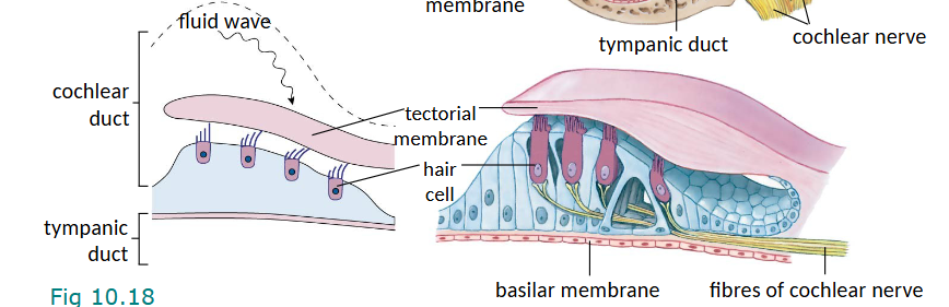
Cochlea
perilymph found in vestibular duct and tympanic duct is similar to plasms (high sodium low potassium)
endolymph in cochlear duct is similar to intracellular fluid (low sodium high potassium)
cochlear duct contains Organ of Corti with sensory hair cells and support cells
The tips of hair cells are stuck in the ____ _____ to increase the chance of being bent.
tectorial membrane so that pressure wave of fluid is felt
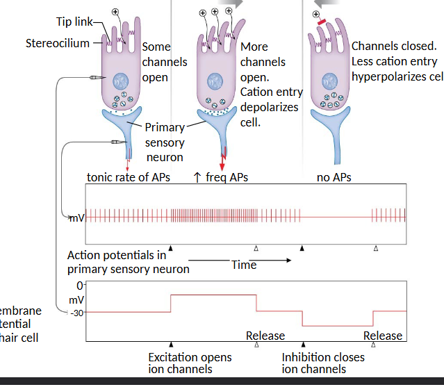
Do hair cells for equilibrium have directionality?
yes; when fluid pushes in one direction, more channels open and cation entry depolarizes cell to increase AP frequency
when hairs bend in the opposite direction, channels are closed and less cation entry hyperpolarizes the cell for no APs
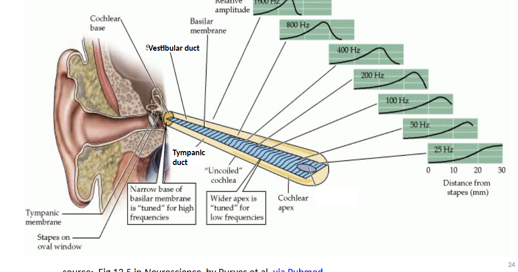
Place Code Hypothesis
sound waves trigger activity at different places along the cochlea’s basilar membrane and are perceived as different pitches - tonotopic map
wave travels along cochlea, hair cells in the area that bend the most at a given frequency encode that pitch
basically the labelled line based on which hair cells are stimulated
based on anatomy of ear
Temporal Coding Hypothesis
frequency of sound wave determines frequency of APs travelling along auditory nerve, perceived as pitch
e.g. low frequency sound → slow waves along basilar membrane → low firing rate of primary afferent neurons → perceived as low pitch sound
problem with this idea is we can hear sounds up to 20k Hz but no neuron can transmit this fast
Current Hypothesis of Coding for Pitch
multiple neurons with staggered firing rates carry the temporal code - pooled neural response interpreted as pitch (convergence)
place coding (hair cells along basilar membrane are stimulated) also plays a role
relative importance of place and temporal coding depends on pitch
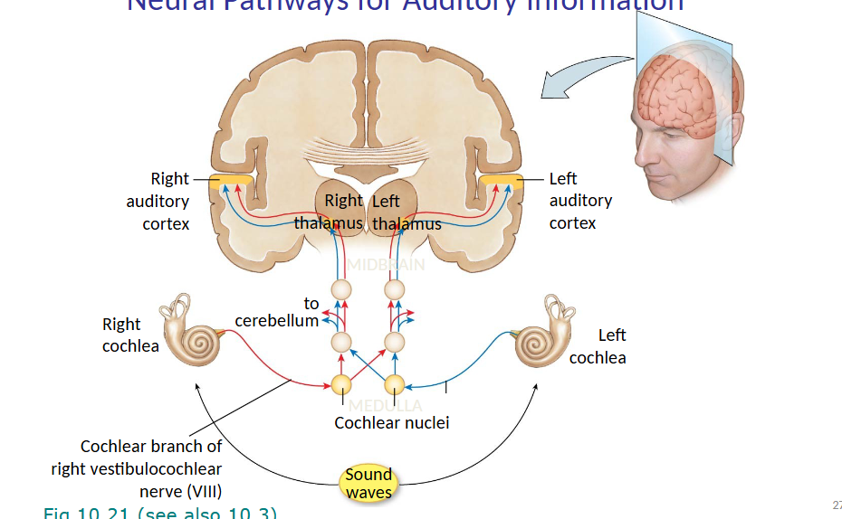
Neural Pathways for Auditory Information
sound waves reach both cochleas
travel to cochlear nuclei via cochlear branch of vestibulocochlear nerve
cerebellum
right and left thalamus
right and left auditory cortices
Conductive Hearing Loss
no transmission through either external or middle ear due to issues with earwax or fluid in middle ear
can usually be repaired
Central Hearing Loss
damage to neural pathway between ear and cerebral cortex or damage to the cortex itself
uncommon but occurs in stroke
Sensorineural Hearing Loss
damage to structures of inner ear like death of hair cells due to loud noises
common in both young and elderly; hair cells can be replaced in other vertebrate groups but not mammals
Equilibrium - Vestibular Apparatus
dynamic component - movement of body through space
static component - position of head
integrated with information from other sensory systems like muscle and joint proprioceptors and visual information
detected by hair cells lining fluid-filled chambers (otolith organs - utricle and saccule for linear acceleration and head position; semicircular canals for rotational acceleration)
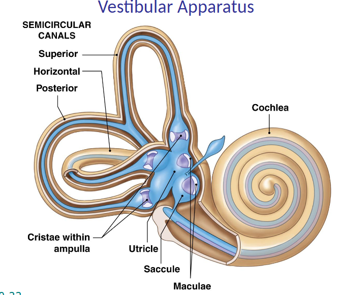
Cristae
hair cells grouped here, within ampulla of canals, detect spin (rotational acceleration) for equilibrium

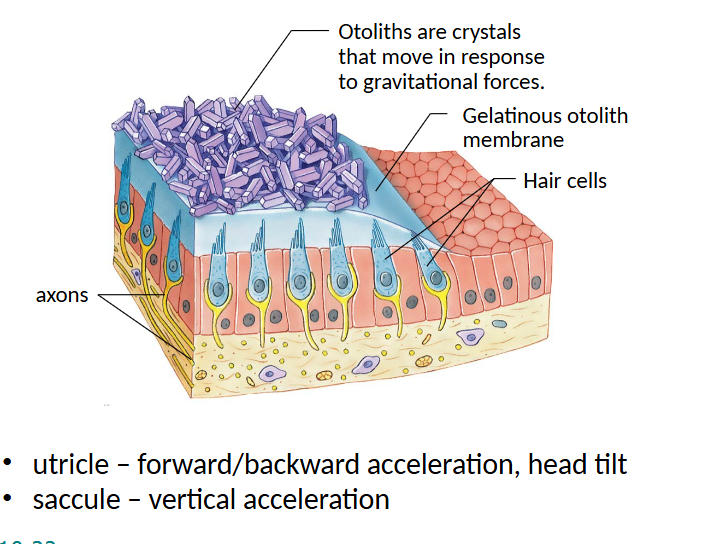
Maculae
in utricles/saccules
otoliths are crystals that move in response to gravitational forces (sit on top of mass to increase the chance of it bending)
utricle detects forward/backward acceleration, saccule for vertical acceleration
Neural Pathways for Equilibrium
goes from vestibular branch of vestibulocochlear nerve to cerebellum, NOT thalamus
cerebellum is the primary center for equilibrium
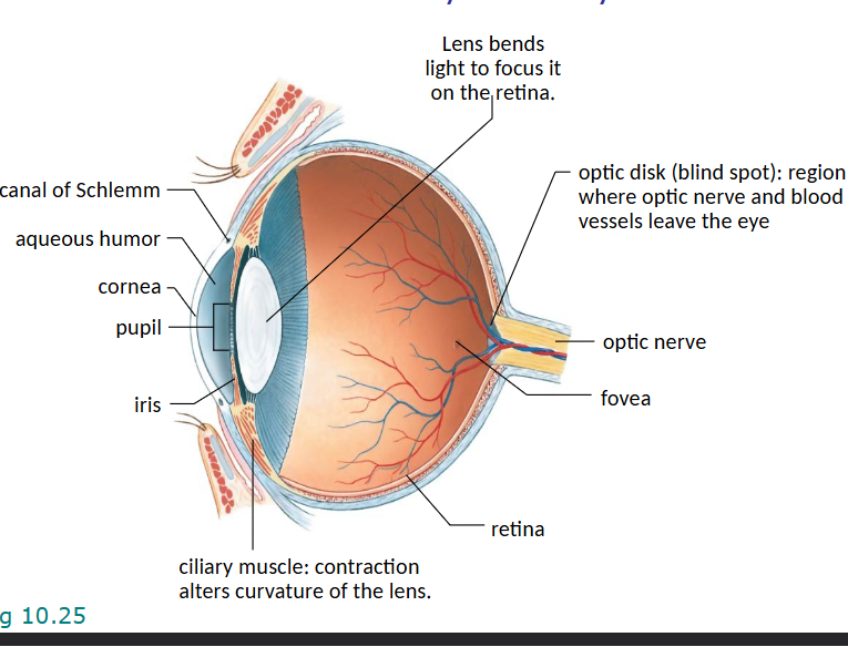
Gross Anatomy of the Eye
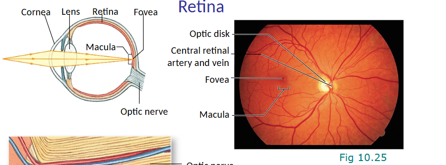
canal of Schlemm is similar to CSF and catches fluid in aqueous humour
optic disk is the blind spot where the optic nerve and blood vessels leave the eye
optic nerve is where afferent info goes
lens bends light to focus on the retina
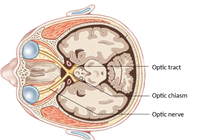
Neural Pathways for Vision - Dorsal
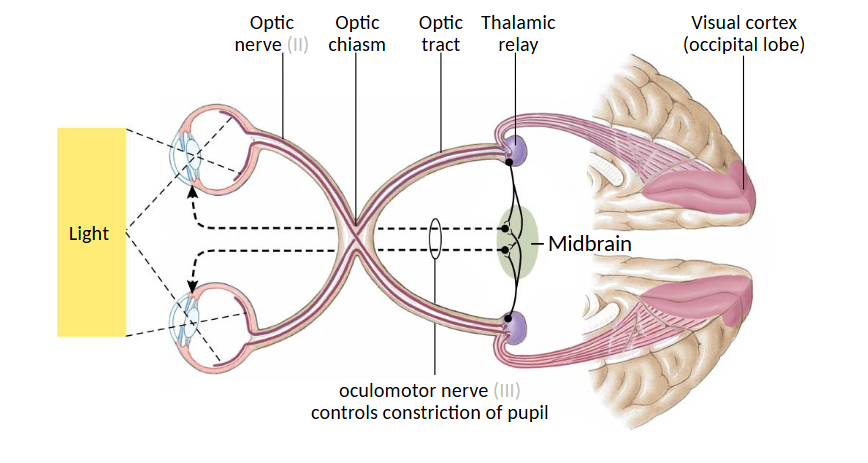
optic tract: bundle of axons in CNS
optic chiasm: crossing over point
optic nerve: bundle of axons in PNS
Pupillary Light Reflex
control of pupil diameter according to intensity of light
non-visual, autonomic reflex where:
detector = photoreceptors in retina
afferent = afferent neurons travelling in optic nerve
integrating center = thalamus/brainstem (midbrain)
efferent = motor neurons travelling in occulomotor nerve
effectors = smooth muscles regulating pupil diameter (sphincter constriction or radial dilation)
light in eyes → optic nerve → optic chiasm → optic tract → thalamic relay → midbrain
Phototransduction
conversion of light into changes in membrane potential by photoreceptor cells in retina (rods or cones)
special population of ganglion cells that mediate pupillary light reflex, circadian/seasonal rhythms
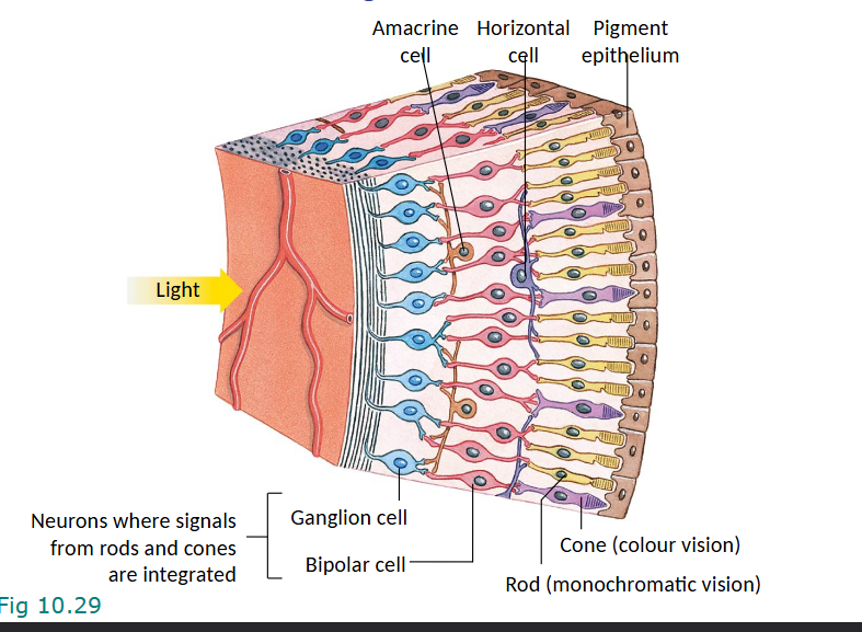
Cellular Organization of Retina
all vertebrates have inverted retina
light goes through ganglion cells, then bipolar cells (both are neurons where signals from rods and cones are integrated) and then rods and cones
photoreceptors (rods and cones) don’t make APs even though they’re neurons; only ganglion cells do
Fovea
is a dent in the retina for better access to photoreceptors
made of almost all cones
very low convergence; super sensitive spot where pigment epithelium absorbs excess light
outside of fovea there is convergence with a high chance of AP but low resolution
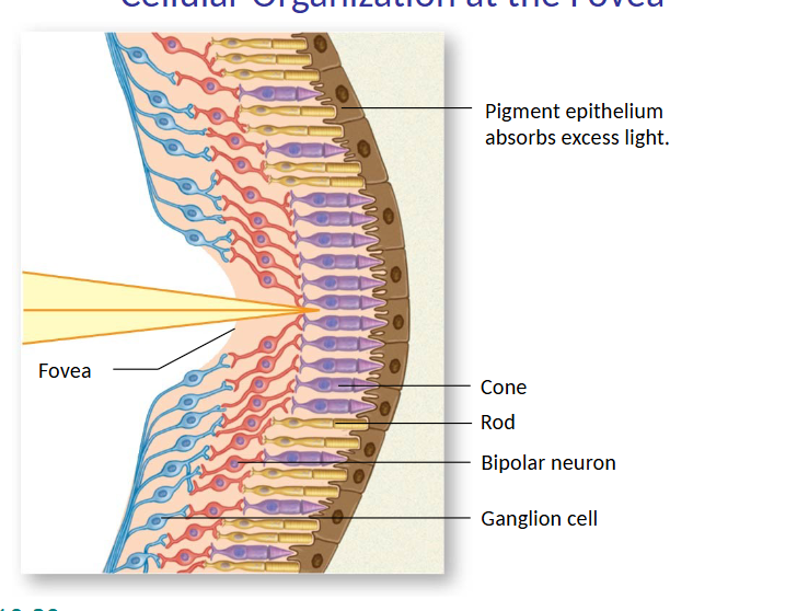
Peripheral Retina vs. Fovea
peripheral retina has mostly rods, monochrome, high convergence, high sensitivity, low resolution
fovea: all cones, low convergence, small receptive fields, low sensitivity, high resolution
Pendred Syndrome
autosomal recessive mutation in gene that codes for pendrin protein which is a transmembrane anion exchanger in ear, thyroid, kidney
loss of this in rodents causes abnormal composition of endolymph, deterioration of stria vascularis, abnormalities in inner ear structure, hearing loss
Retinal
rhodopsin is GPCR where retinal absorbs a photon to change its shape, dissociate, and activate GPCR
in resonse to light 11-cis retinal goes to all trans retinal
in the dark: cGMP is high, CNG channels are opened by cGMP, there is tonic release of glutamate
in light: retinal absorbs and dissociates, GPCR is active, cGMP is low so CNG channels close, K+ leaves still so cell hyperpolarizes and less glutamate is released
Voluntary vs. Reflexive Movement
voluntary movement is planned and intentional, initiated by decision making in brain, integrated in motor cortex, integrated lots in cerebral cortex, activates somatic MNs, activates proprioceptors, definitely informs cerebellum
reflexive movement is hard wired, initiated by external stimulus, integrated in the spinal cord, not usually integrated in cerebral cortex, activates somatic MNs, doesn’t activate proprioceptors