Unit 6 Nervous system
1/74
There's no tags or description
Looks like no tags are added yet.
Name | Mastery | Learn | Test | Matching | Spaced | Call with Kai |
|---|
No analytics yet
Send a link to your students to track their progress
75 Terms
What are the functions of the Nervous system
receives & processes sensory info
coordinated actions of skeletal muscles
controls all internal organ system
thinking & memory
What is part of the CNS
Brain & spinal cord
What is part of the PNS (peripheral NS)
autonomic nervous system
peripheral nerves
enteric nervous system
Order information processing
Nervous (neural) tissue consists of two cell types
Neurons and neuroglia (glial cells)
What are neurons
nerve cells responsible for transferring and processing info in nervous system
What are neuroglia (glial cells)
supporting cells and protect neurons
What do neurons do
for rapid communication, receive and transmit info with electrochemical impulses called action potentials
Neuron characteristics
excitability
conductivity
secretion
longevity
amitotic
What is excitability (neuron characterstic)
ability to respond to stimuli
What is conductivity
transmit signals between places
What is secretion
neurotransmitter release signaling molecules to communicate to other cells
What is longevity (neuron characteristics)
keep them until we die
What is amitotic
few neurons divide post birth
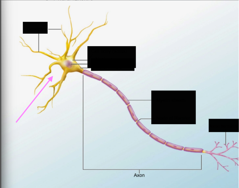
Neuron structure- what is it pointing at and what does it do?
soma
cell body or nucleus
info processing can happen
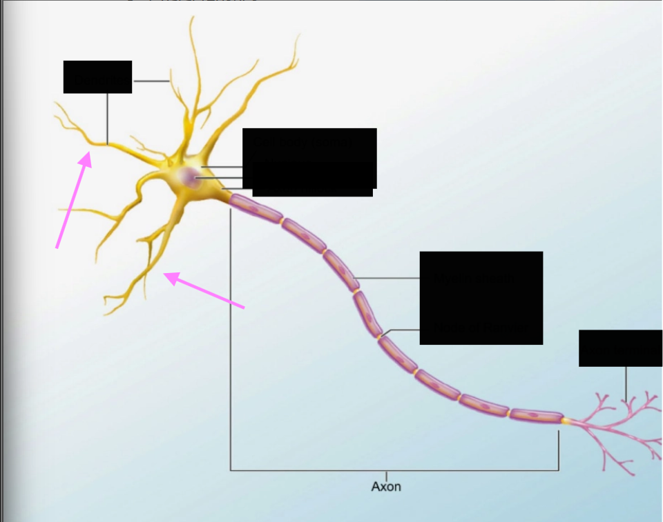
Neuron structure- what part of the neuron is it pointing at & what does it do?
dendrites
neuronal processes
inputs
how neurons bring in info
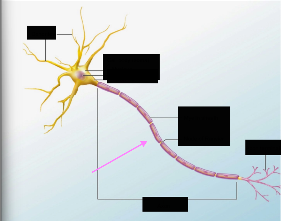
Neuronal structure - What is this & what does it do
Axon
neuronal process
outputs
where action potentials starts
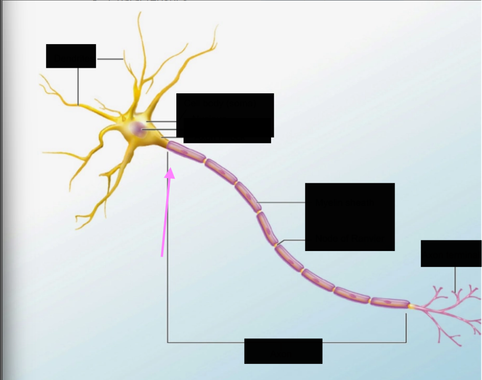
What part of the axon is this?
axon hillock
junction between soma and axon
origin site of action potentials

What is a myelin sheath?
transfers info (signal) from cell body down to axon terminal

Neuronal shapes: typically single axon & multiple dendrites
most common type in CNS
in brain & spinal cord
multipolar

Neuronal shapes: cell body is between dendrite & axon
axons are not myelinated
typically in PNS sensory branches (afferent side)
Bipolar
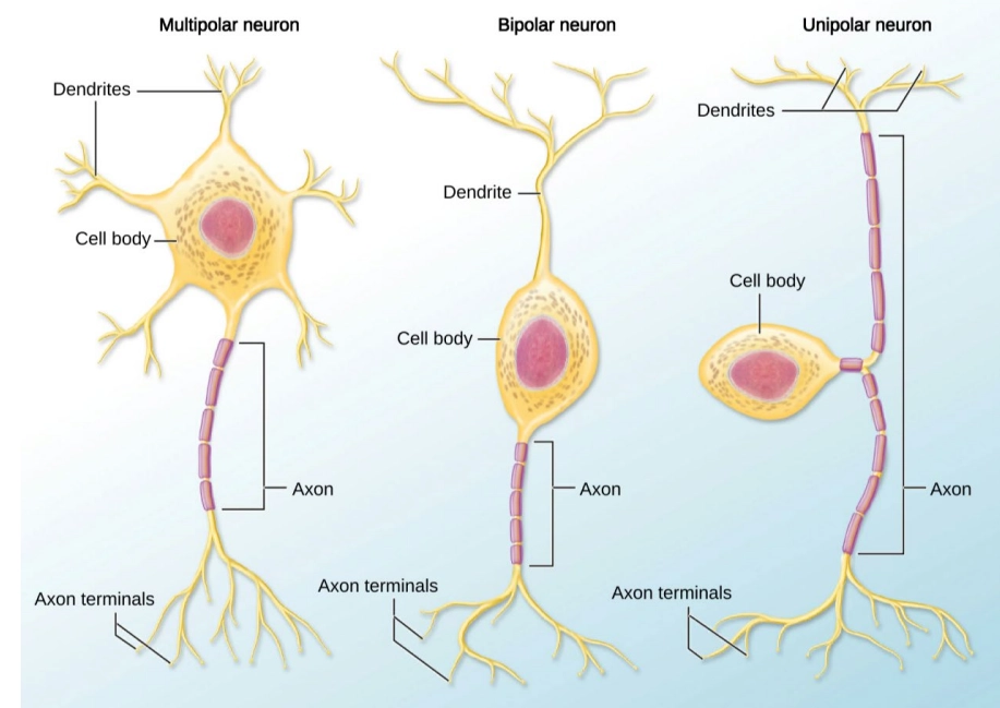
Neuronal shapes: cell body is off to one side of axon
sensory neuron
input & output arm
unipolar or pseudo unipolar
What are the 3 major functional groups of neurons
sensory neurons, motor neurons, interneurons
Which functional group of neurons:
axons are afferent fibers
sends info from PNS to CNS
there are: somatic sensory neurons & visceral sensory neurons
Sensory or afferent neurons
Which function group of neurons:
axons are efferent fibers\
sends info from CNS to PNS
consists of two divisions
somatic motor neurons & visceral motor neurons
motor or efferent neurons
Which functional group of neurons:
located in CNS
situated between motor & sensory neurons
analyze sensory input & coordinate motor outputs
“processes” information
can be excitatory or inhibitory
most common neuron type
interneurons
Which sensory (afferent) neuron:
senses stimuli on the skin & within skeletal muscles
somatic sensory neurons
Which sensory (afferent) neurons):
senses stimuli inside body (organs, tubes, etc)
Visceral sensory neurons
For motor (efferent) neurons:
controls skeletal muscles that move body in voluntary movements
somatic motor neurons
For motor (efferent) neurons:
controls smooth, cardiac, & skeletal muscles for involuntary contractions
visceral motor neurons
For interneurons what does it mean:
messages cause other neurons or cells to be more active
excitatory
For interneurons what does it mean:
messages cause other neurons or cells to be less active
inhibitory
Neuroglia
what cell supports neurons & their activities
insulate myelin on the axon
maintain extracellular environment around neurons → keeps neurons healthy
Glial cells (neuroglia)
Neuroglia
help dilate/constrict blood vessels in response to neural activity
transport blood to neurons
found near neurons & blood vessels (mainly in CNS)
help transfer material from blood to nervous system
blood-drain-barrier
many in the brain
Astrocytes
What is the blood-drain-barrier?
established by astrocytes, protection for neurons from getting exposed to toxins
surround some of PNS neurons
version of astrocytes that are found in the PNS
maintain extracellular environment
act as a bridge between the NS & circulatory system
Satellite cells
found in lining of cavities of brain & spinal cord
AKA ventricles → flow fluid (CSF0 around so neurons have CSF
make cerebrospinal fluid (only in CNS)
Ependymal cells
aren’t actually neural tissue
born in immune system
destroy foreign particles
Microglia

many layers of glial cell plasma membrane wrapped around axon
insulation → makes conduction of impulses faster
Myelin sheaths
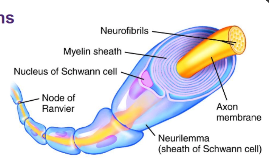
saltatory conduction - action potentials jump from node to node
gaps between myelin
action potentials jump from nodes
nodes of ranvier
Neuronal Communication
uneven distribution of + & - ions across plasma membrane
when there is an action potential there is a rapid change
membrane potential
slower impulse speed
unmyelinated
faster impulse speed
myelinated
faster conduction speed
larger axon diameter
slower conduction speed
smaller axon diameter
Junction between a neuron & another cell
synapse
What are the 2 major types of synapses
chemical synapse and electrical synapse
Type of synapse
communicating cells do not touch
sending cells releases chemical messenger (neurotransmitter) which is received by receiving cell
chemical synapse
Type of synapse
communicating cells physically connected by gap junction
electrical synapse
What is the sending neuron & what is the
Part of the CNS
brings in & processes information
can function independently of the brain
spinal cord
Part of the CNS
integrates & processes info
works with ____
brain
Where is the brain & cavity located (cavities)
posterior cavities
thin specialized membranes that provide protection, physical stability, and shock absorption
covers CNS
made of 3 layers
meninges
Layer of meninges
tough, fibrous outermost layer
stabilizes spinal cord
within the vertebral canal
cranial & sacral attachments stabilize
longitudinal axis of spinal cord
epidural space: space between dura mater & lining of vertebral bones
dura mater
Layer of meninges
middle layer
separated by subarachnoid space
____ trabeculae extend from arachnoid to outer layer of pia mater
spider web-like matrix of trabeculae
cerebrospinal fluid flows within spaces between trabeculae
arachnoid mater
Layer of meninges
innermost layer (adjacent to spinal tissue or brain tissue)
blood vessels are found here
protection in case foreign debris get past
pia mater
consists of neuronal stomata (cell bodies), dendrites, unmyelinated axons
glial cells
found in superficial part of brain
gray matter
neuronal axon (myelinated)
found deep in brain
superficial in spinal cord
where info is sent from one place to another → goes through spaces
white matter
How many pairs of spinal nerves are there?
31 pairs: 8 cervical, 12 thoracic, 5 lumbar, 5 sacral, and 1 coccygeal
What is the difference between the dorsal root and ventral root
dorsal root carries sensory or afferent information to the CNS; ventral root carries motor or efferent commands away from the CNS
What is the Cauda Equina
bundle of spinal nerve roots extending below the end of the spinal cord, look like a horse’s tail
Primary function of the thalamus
acts as a relay station, processing and directing sensory information to the appropriate areas of the cerebral cortex
Whare the 3 regions of the Brainstem
midbrain, pons, and medulla oblongata
function of cerebellum
integrates sensory information to coordinate motor movements, balance and motor learning
Which lobe is responsible for voluntary motor control & personality
frontal lobe
Where is the somatosensory cortex located and what is it responsible for
parietal lobe, responsible for touch, temperature, and pain