Nerve Impulses and Synaptic Transmission
1/72
There's no tags or description
Looks like no tags are added yet.
Name | Mastery | Learn | Test | Matching | Spaced | Call with Kai |
|---|
No analytics yet
Send a link to your students to track their progress
73 Terms
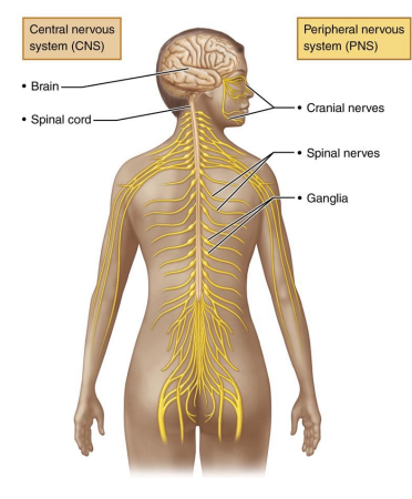
2 principal parts of Nervous system
Central nervous System(CNS)
Peripheral Nervous System (PNS)
Walls of GI tract also contain neurons called the enteric nervous system
CNS
Brain and spinal cord of dorsal cavity
Integration + control center
interprets sensory input and dictates motor output
PNS
The portion of nervous system outside of CNS
Consists mainly of nerves and extend from brain and spinal cord
Spinal nerves to and from spinal cord
Cranial nerves to and from the brain
Nervous System
2 functional divisions of PNS
Sensory
Motor
Sensory(afferent) division
Somatic sensory fibers: convey impulses from skin, skeletal muscles, and joints to CNS
Visceral sensory fibers: convey impulses from visceral organs to CNS
Motor (efferent) division
Transmits impulses from CNS to effectors
Muscles and glands
Two divisions
Somatic nervous system
Autonomic nervous system
2 principal cell types of Nervous tissue
Neuroglia (glial cells): small cells that surround and wrap delicate neurons
Neurons (nerve cells): excitable cells that transmit electrical signals
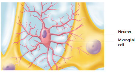
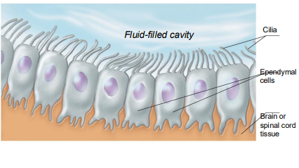
4 main neuroglial cells that support CNS neurons
astrocytes
Microglial cells
Ependymal cells
oligodendrocytes
Astrocytes
Most abundant, versatile, and highly branched of glial cells
Cling to neurons, synaptic endings, and capillaries

6 functions of neuroglia
Support and brace neurons
Play role in exchanges between capillaries and neurons
Guide migration of young neurons
Control chemical environment around neurons
Respond to nerve impulses and neurotransmitters
Participate in information processing in brain
Microglia
mall, ovoid cells with thorny processes that touch and monitor neurons
Migrate toward injured neurons
Can transform to phagocytize microorganisms and neuronal debris
Ependymal cells
Range in shape from squamous to columnar
May be ciliated
Cilia beat to circulate CSF
Line the central cavities of the brain and spinal column
Form permeable barrier between cerebrospinal fluid (CSF) in cavities and tissue fluid bathing CNS cells
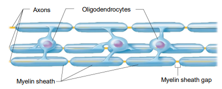
Oligodendrocytes
Branched cells
Processes wrap CNS nerve fibers, forming insulating myelin sheaths in thicker nerve fibers
2 neuroglia of PNS
Satellite cells
Schwann cells
Satellite cells
Surround neuron cell bodies in PNS
Function similar to astrocytes of CNS
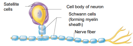
Schwann cells
Surround all peripheral nerve fibers and form myelin sheaths in thicker nerve fibers
Similar function as oligodendrocytes
Vital to regeneration of damaged peripheral nerve fibers

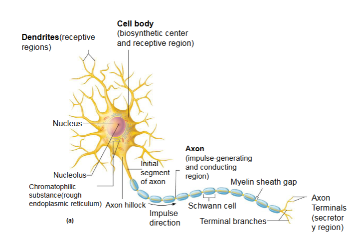
Neurons
(nerve cells) are structural units of nervous system
Large, highly specialized cells that conduct impulses
All have cell body and one or more processes
Special characteristics of neurons
Extreme longevity (lasts a person’s lifetime)
Amitotic, with few exceptions
High metabolic rate: requires continuous supply of oxygen and glucose
Resting membrane potential
~-70 mV for neurons
It is the potential that is generated when neurons are at rest
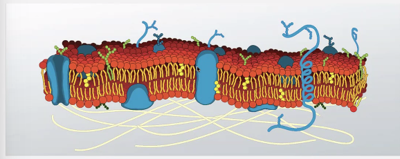
Membranes of neurons
Hydrophilic head+ hydrophobic tails
arranged in a bilayer
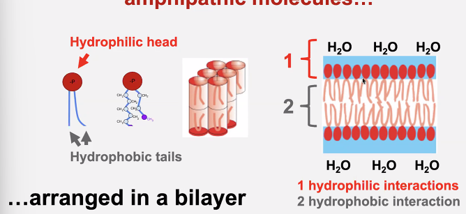
Will either water molecules or ions cross the plasma membrane
No because the hydrophobic area DOES NOT allow charged ions to go freely from one side to the either
Solution: Channels
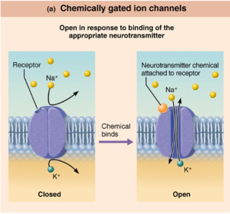
Chemically gated channels
also known as ligand-gated channels,
open when the appropriate chemical (in this case a neurotransmitter) binds
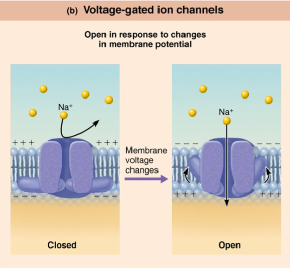
Voltage gated ion channels
open and close in response to changes in the membrane potential
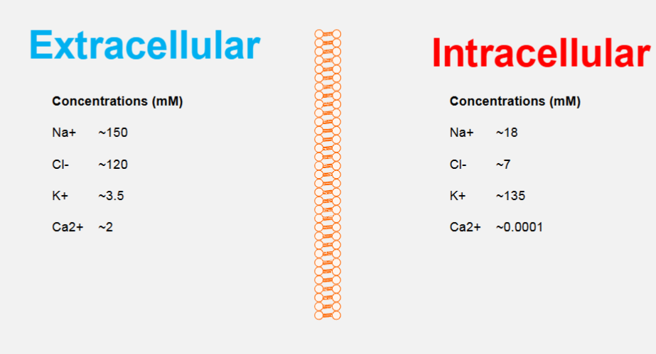
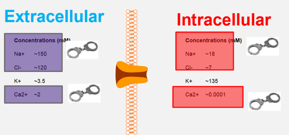
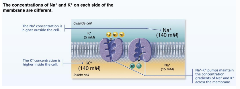
Ions in ECS vs ICS during resting membrane potential
The cell cytosol contains a lower concentration of Na+ and a higher concentration of K+ than the extracellular fluid.
Negatively charged (anionic) proteins balance K+
Na+ and other cations are balanced chiefly by chloride ions .
in both fluids, K+ plays the most important role in generating the membrane potential.
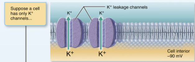
What will K+ do if there were ONLY K+ channels
It will diffuse down its concentration gradient and cross the channel from intracellular to extracellular
results in a negative membrane potential= higher electrical gradient
electrical gradient will pull K+ from extra- to intra
At -90 mV, the concentration+ electrical gradients for K+ are balanced
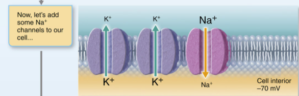
What would happen if N+ channels were added alongside K+ channels
Na+ entry via a FEW leakage channels reduces the negative membrane slightly to -70 mV
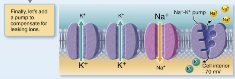
What would happen if Na+/K+ ATPase was channels alongside the K+ and Na+ channels
It will maintain the concentration gradient and maintain the resting membrane potential
It transports any extra K+ in and Na+ out that leak through the channels
prevents the membrane from having equal concentration of both ions
Generating a resting membrane potential depneds on 2 differences
differences in K+ and Na+ concentrations inside and outside cells
differences in permeability of the plasma to these ions
Plasma membrane permeability
At rest= impermeable to anionic proteins
slightly permeable to Na+
25x MORE permeable to K+ than Na+
since K+ diffuses out of cell down its concentration gradient much easier
quite permeable to Cl-
Graded potentials
are short-lived, localized changes in membrane potential
usually in dendrites or the cell body.
They can either be depolarizations or hyperpolarization’s
Purpose: sum together to determine whether or not AP will occur
Why are graded potentials ‘graded’
because their magnitude varies directly with stimulus strength.
The stronger the stimulus, the more the voltage changes and the farther the voltage change extends.
weaker stimulus= decay and less spread of voltage change
due to voltage being lost via leaky channels
3 Types of graded potentials
receptor potential/generator potential
postsynaptic potential
is produced when the stimulus is a neurotransmitter released by another neuron.
The neurotransmitter is released into a fluid-filled gap called a synapse and influences the neuron beyond the synapse.
end-plate potential
Action potentials
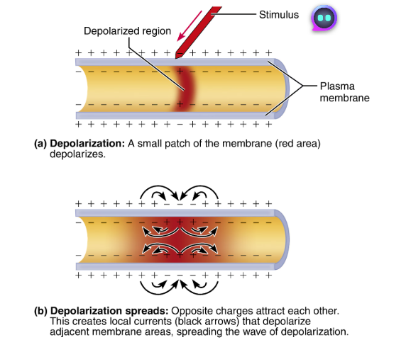
Brief reversal of membrane potential with a change in voltage of ~100 mV (from -70 to +30 mV) in a patch of membrane depolarized by local currents
principal way neurons send signals means of long-distance neural communication
do not decay in amplitude with distance traveled as graded potentials do
occur only in cells with excitable membranes (neurons & muscle cells)
in neurons
only axons can generate action potentials
also referred to as a nerve impulse
Voltage-gated channels on axons open in response to local currents (graded potentials)
K+ channel
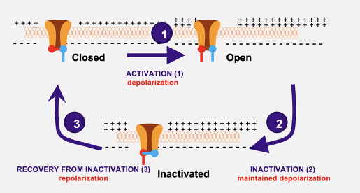
Has an activation gate can either be opened or closed
Na+ channel
Has 2 gates
Both must be open for AP to continue
Gate A opens and Gate B closes during AP
No more flow of ions
Gate and Gate B alternate closing, which takes time
Action Potential steps
Resting states
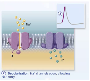
Depolarization
Has a rising phase
Repolarization
Overshoot
Falling phase
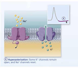
Hyperpolarization
Refractory period
Return to rest
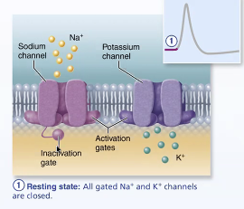
Resting state
ALL voltage-gated Na+ and K+ channels are closed.
Only the leakage channels are open, maintaining resting membrane potential.
Each Na+ channel has two gates:
a voltage-sensitive activation gate that is closed at rest and responds to depolarization by opening,
an inactivation gate that blocks the channel once it is open.
depolarization opens and then inactivates sodium channels.
What must occur for Na+ to enter the cell
Both gates must be open for Na+ to enter,
BUT the closing of either gate effectively closes the channel.
In contrast, each voltage-gated K+ channel has a single voltage-sensitive gate that is closed in the resting state and opens slowly in response to depolarization.
Depolarization
Voltage-gated Na+ channels open.
influx of positive charge(Na+ ions) depolarizes that local patch of membrane further, opening more Na+ channels so the cell interior becomes progressively less negative
When threshold of (-55 and -50 mV), depol. becomes self-generating until ALL Na+ channels are open
permeability of Na+ is 1000x greater than in a resting neuron
eventually membrane potential reaches +30 mV
Rising phase
Rapid opening of all Na+ channels
Occurs only if certain threshold is met or else AP will not be generated
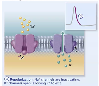
Repolarization
Na+ channels are inactivating, and voltage-gated K+ channels open
Inactivation gates of Na+ begin to close
Na+ permeability declines to resting levels and influx of Na+ eventually stops and AP stops rising
Slow voltage-gated K+ channels open and K+ rushes OUT of cell
restores the negativity of the resting neuron
Both the abrupt decline in Na+ permeability and the increased permeability to K+ contribute to repolarization
Hyperpolarization
Some K+ channels remain open, and Na+ channels reset.
The period of increased K+ permeability typically lasts longer than needed to restore the resting state.
As a result of the excessive K+ efflux before the potassium channels close(hyperpolarization) is
AKA a slight dip following the spike.
At the end of this phase, the Na+ channels have reset to their original position by changing shape to reopen their inactivation gates and close their activation gates.

Falling phase
Na+ channels stay inactivated
Opening of all K+ channels
Vm repolarizes
inactivation gate opens and Na+ closes
2 types of synapses
Chemical synapse
Electrical synapse
Chemical synapses
Most common type
specialized for the release and binding of neurotransmitters
Separated by synaptic cleft (fluid-filled space of 30-50 nm)
2 parts that make up chemical synapses
Axon terminal of presynaptic neuron: contains synaptic vesicles filled with neurotransmitter
Receptor region on postsynaptic neuron’s membrane: receives neurotransmitter
Usually on dendrite or cell body
6 Steps of Information transfer across chemical synapses
AP arrives at axon terminal
Voltage-gated Ca2+ channels OPEN and Ca2+ enters axon terminal
Ca2+ entry causes synaptic vesicles to release neurotransmitters by EXOcytosis
Neurotransmitter diffuses across synaptic cleft and binds to specific receptors on the postsynaptic membrane
Binding of neurotransmitter opens ion channels, resulting in graded potentials
Neurotransmitters effects are terminated
How are neurotransmitter effects terminated
Reuptake by astrocytes or the presynaptic terminal, where the neurotransmitter is stored/destroyed by enzymes(i.e. norepinephrine)
Degradation by enzymes associated with postsynaptic membrane or present in the synaptic cleft (i.e. Ach)
Diffusion away from the synapse
Postsynaptic potentials
Neurotransmitter receptors cause graded potentials that vary in strength
based on:
• Amount of neurotransmitter released
• Time neurotransmitter stays in cleft
• Depending on effect of chemical synapse, there are two types of
postsynaptic potentials
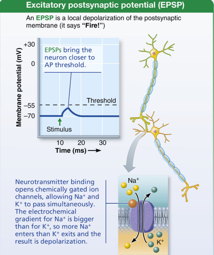
• EPSP: excitatory postsynaptic potentials
• IPSP: inhibitory postsynaptic potentials
What is the effect of neurotransmitters
Excitatory
Inhibitory
To determine if neurotransmitters either of this,
Depends on neurotransmitter and its receptors
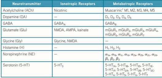
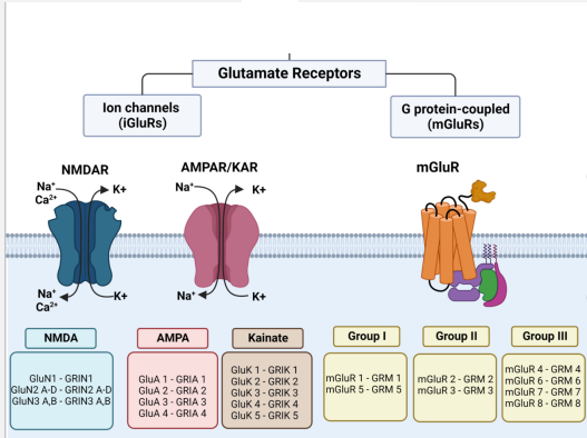
Ionotropic receptors
AKA channel linked receptors are ligand-gated ion channels that mediate DIRECT neurotransmitter action
As ligand binds to 1 or more receptor subunits, proteins change shape
this opens central channel and allows ions to pass
Rapid synaptic transmission
ALWAYS located on opposite of site of neurotransmitter release
At excitatory receptor sites (ie.e receptors for Ach, glutamate, aspartate, ATP), the ionotropic receptors are cation channels(Na+, K+, Ca2+), where Na+ entry contributes the MOST to depolarization
Sensitive to molecules and sometimes, membrane potential
Selective for specific ions
Respond to GABA and glycine
Allows Cl- to pass
mediates hyperpolarization
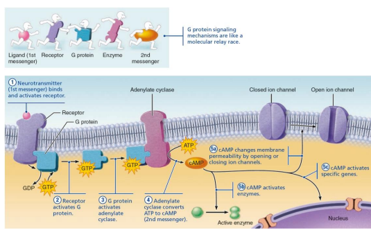
Metabotropic receptors
aka G Protein-coupled receptors
Responses are indirect, complex, slow, and often prolonged
G-protein activates second messenger system
Involves transmembrane protein complexes
Cause widespread metabolic changes, which is why they are termed G protein-coupled receptors
Examples of metabotropic receptors
Biogenic amine receptors
neuropeptide receptors
Muscarinic ACh receptors
WHat happens when neurotransmitters bind to a G-protein-coupled receptor
The G-protein is activated
Activated G proteins typically work by increasing or decreasing the amount of second messengers like cyclic AMP, cyclic GMP, diacylglycerol, or Ca2+ in the cytoplasm
G protein-coupled receptors cause the formation of 2nd messengers
Neurotransmitters
along with electrical signals, are the
language of nervous system
• 50 or more neurotransmitters have been identified
• Classified chemically and functionally
Glutamatergic receptors
Glutamate=Most abundant excitatory neurotransmitter in the brain
Can activate ionotropic and metabotropic receptors
Excitatory synapses and EPSPs
Neurotransmitter binding opens chemically gated channels on POSTsynaptic membranes
Each channel allows simultaneous diffusion of Na+ and K+ into membrane but in opposite directions
Na+ influx greater than K+ efflux, resulting in local net graded potential depolarization called excitatory postsynaptic potential (EPSP) NOT an AP
because APs don’t occur in membrane that have ONLY chemically-gated channels
postsynaptic membranes do not generate APs
EPSPs trigger AP if EPSP is of threshold strength to depolarize axon
Can spread to axon hillock and trigger opening of voltage-gated channels, causing AP to be generated
Excitatory postsynaptic potentials (EPSPs)
Is what is generated by chemically-gated channels of postsynaptic membranes
GABAergic receptors
GABA is responsable for most inhibitory transmission
• GABARs bind ethanol, benzodiazepine, barbiturate
Inhibitory Synapses and IPSPs
Binding of neurotransmitter reduces postsynaptic neuron’s ability to generate an AP
Neurotransmitter binding to receptor opens chemically gated channels that allow entrance/exit of ions that cause hyperpolarization
Makes postsynaptic membrane more permeable to K+ or Cl–
If K+ channels open, it moves out of cell
If Cl– channels open, it moves into cell
Moves neuron farther away from threshold (makes it more negative)
Inhibitory postsynaptic potentials (IPSPs)
are hyperpolarizing changes in potential
ACh receptors
2 subtypes of cholinergic receptors
Nicotinergic
Muscarinic
Each subtype has a different antagonists (curare and atropine)
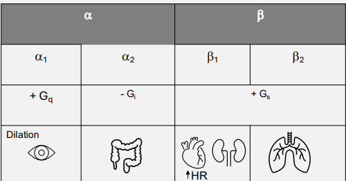
Norpepinephrine receptors
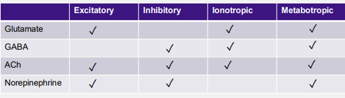
Summary of neurotransmitters
Summation by postsynaptic neuron
A single EPSP cannot induce an AP, but EPSPs can summate (add together) to influence postsynaptic neuron
IPSPs can also summate
Most neurons receive both excitatory and inhibitory inputs from thousands of other neurons
Only if EPSPs predominate and bring to threshold will an AP be generated
Where do excitatory synapses often occur
on dendrites
Where do inhibitory synapses often occur
often on the cell body
2 types of summations
Temporal
Spatial
Temporal summation
One or more presynaptic neurons transmit impulses in rapid-fire order
• First impulse produces EPSP, and before it can dissipate another EPSP is
triggered, adding on top of first impulse
Spatial summation
Postsynaptic neuron is stimulated by large number of terminals simultaneously
• Many receptors are activated, each producing EPSPs, which can then add together