Unit 1 AP Psychology
1/143
Earn XP
Description and Tags
Updated very weekly
Name | Mastery | Learn | Test | Matching | Spaced |
|---|
No study sessions yet.
144 Terms
Evolutionary Psychology
The study of how traits/behaviors evolved to enhance survival & reproductive success
Natural Selection
Process by which organisms with advantageous traits are more likely to survive & reproduce, passing traits to offspring
Nature “Genes”
Biological and genetic factors that influence an individual’s psychological development, traits, & behavior
Example: Traits inherited from parents, chance of inheriting mental health conditions
Nuture “Environment”
The environment influences an individual’s psychological development, traits, & behavior
Example: Social relationships, educational opportunities, socioecnomic status
Twin Studies
Examines similarities & differences between identical (monozygotic) and fraternal (dizygoti) twins to assess the influence of genetics/environment on traits/behaviors
Adoption Studies
Investigates similarities between adopted children & their biological/adoptive families to assess the impact of genetics vs. environment on traits/behaviors
Family Studies
Analyzes the similarities & differences among family members to understand the interplay of genetics & environment in shaping traits & behaviors within a family unit.
Heredity
The transfer of genetic info from parents to offspring
Genetic Predisposition
The inherited likelihood of developing specific traits/conditions due to genetic factors from biological parents
Eugenics
The belief of improving the genetic quality of a population by controlling reproduction to inc desirable traits & dec “undesirable” traits
Cerebral Cortex (Grey Matter)
The outermost layer of the cerebrum, made of grey matter, is the brain processor containing neurons and white matter, the cables that transmit signals between different areas of the brain & the cortex, which is responsible for higher-level cognitive functions like thinking, perceiving, & decision making
Lobes of the Brain
4 main regions into which the cerebral cortex is divided into: frontal, parietal, temporal, and occipital lobe
Association Areas
Parts of the brain that take info from all over the place—like what we see, hear, smell, & touch and put it together to understand the world around us
Frontal Lobes
Located at the front of the brain & involved in higher level cognitive functions like problem solving, planning, & personality expression
Prefrontal Cortex
Region of the brain located in the frontal lobe responsible for higher level cognitive functions & executive functioning
Executive functioning: set of cognitive processes that enable individuals 2 plan/organize
Motor Cortex (front)
Responsible for planning, executing, & controlling voluntary movements of the body by sending signals to the muscles, enabling us to perform actions (walking, talking, holding)
Parietal Lobes
Located at the top of the brain & are primarily responsible for processing sensory information from the body, such as touch, temperature, and spatial awareness
Somatosensory Cortex
Located in the parietal lobe responsible for processing sensations from the skin, muscles, & joints by perceiving sensory stimuli (texture, temp, pressure, pain)
Occipital Lobes
Located at the back of the brain responsible for processing info received from the eyes
The primary visual cortex is situated within the occipital lobes that receive visual info from your eyes
Temporal Lobes
Located on the sides of the brain involved in processing auditory info, language comprehension, & memory
Contains auditory cortex that interprets sound waves from the ears
Corpus Callosum
Thick band of nerve fibers that connects the left & right hemispheres of the brain, facilitating communication between them
Brainstem
Oldest part of the brain responsbile for basic life sustaining functions like breathing, heart rate, & sleep-wake cycles
Pathway for neural signals traveling between the brain & rest of the body by connecting the cerebral cortex to spinal cord
Medulla Oblongata
Located at the base of the brainstem that relays nerve signals between the brain & spinal cord, regulating autonomic functions: heartbeat, breathing, & BP
Example: When working out the medulla oblongate detects the inc in activity & sends signals to adjust your heart rate
Reticular Activating System
Network of neurons in the brainstem that regulates arousal & consciousness
Cerebellum
Located at the back of the brain below the cerebral hemisphere that coordinates movement, balance, & posture to translate into motor movements
Limbic System
Located beneath the cerebral cortex is a set of brain structures involved in emotions, memory, & motivation
Reward Center
Network of brain structures in the limbic system that processes pleasurable experiences & reinforces behaviors associate d with them
Thalamus
Processes & relays sensory information (5 senses info) to the cerebral cortex by directing signals to the appropriate areas of the brain
Hypothalamus
Small structures located before the thalamus responsible for regulating homeostasis in the body
Pituitary Gland
Small pea-sized gland at the base of the brain called the master gland that coordinates hormonal activity for homeostasis bc it regulates hormone production & secretion thru out the body
Hippocampus
Responsible for forming & consolidating new/long-term memories
Amygdala
Almong structure in the temporal lobe that processes emotions by playing a central role in the body’s threat detection system, triggering flight & fight response
The Nervous System
Your body’s communication & control center that sends messages & interprets them to decide how to respond
Central Nervous System
Brain & Spinal cord that is the command center of body processing info, coordinating responses, & regulating bodily functions thru spinal cord transmitting sensory info from the body => brain & motor commands from brain => body
Peripheral Nervous System
The nerves & ganglia outside the brain & spinal cord that transmits sensory info from the body to the CNS
Autonomic Nervous System
Division of PNS that regulates involuntary body functions consisting of 2 main branches: sympathetic & parasympathetic nervous system
Sympathetic Nervous System
Activates the body’s fight or flight response by inc heart rate, dilates airways, & redirects blood 2 essential organs
Parasympathetic Nervous System
Promotes relaxation & restoring the body to a calmer state after experiencing stress by dec heart rate, constrict airways, & enhance digestion
Somatic Nervous System
Division of the PNS responsible for controlling voluntary movements & relaying sensory info from the body => CNS
Neurons
A specialized cell that transmits electrical signals throughout the body
Structure of the Neuron
Cell Body (soma): Main part of the neuron that contains the nucleus
Dendrites: short, branching extensions that receive signals from other neurons & transmit them toward the cell body
Axon: A long, slender projection that carries signals away from the cell body toward other neurons, muscles, or glands
Axon Terminals (Terminal Buttons): At the end of the axon there are small swellings that release neurotransmitters to communicate w/ neighboring neurons
Myelin Sheath: A fatty, insulating layer that surrounds some axons to speed up the transmission of electrical signals
Synapse: Junction between 2 neurons where communication occurs where neurotransmitters are released from the axon terminals of 1 neuron & received by the dendrite/cell body of another neuron
Glial Cells
“Support cells” that provide structural support, insulation, & nourishment to neurons to maintain brain health & functioning
Motor Neurons
Nerve cells that transmit signals from the CNS to the muscles, initiating & controlling voluntary/involuntary movements
Sensory Neurons
Specialized nerve cells that transmit sensory info from sensory receptors that detect physical stimuli & converts into electrical signals processed by the CNS
Interneurons
Nerve cells that are connectors within the CNS that relay signals between sensory neurons & motor neurons
Reflex Arc
Neural pathway that controls reflex actions to sensory stimuli w/o the brain as signals travel from sensory neurons to your spinal cord
Plasticity
When areas of the brain are damaged, the neural connections will re-route so that functions can be regained w/ minimal cognitive damage
Neural Transmission
Process by which neurons communicate w/ each other via electrical & chemical signals
Threshold
The level of stimulation required to produce a response in a neuron
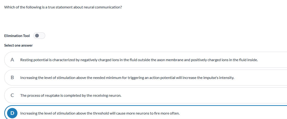
Increasing the lvl of stimulation above the threshold causes more neurons to fire more often

Action Potential
The rapid electrical impulse that travels down the axon with the inside of the cell being more positively charged than the outside
All-or-Nothing Principle
States that neurons fire with a full strength impulse to generate an action potential only if stimulation reaches a minimum threshold
No half firing
Depolarization
When the inside of the neuron becomes less negative due to the influx of positively charged ions (sodium ions), it moves the cell membrane’s potential closer to the threshold = which triggers the neuron to fire an action potential
Refractory Period
A brief period following an action potential during which a neuron is unable to generate another action potential
Resting Potential
The state in which the inside of a neuron has a stable negative charge (more negative) relative to the outside, while the neuron is not actively transmitting signals
Reuptake
The process by which neurotransmitters released during the synapse are reabsorbed by the presynaptic neuron of origin
Multiple Sclerosis “MS”
Chronic autoimmune disease that affects the CNS when the immune system mistakenly attacks the myelin sheath
Myasthenia Gravis
An autoimmune disorder that affects the neuromuscular junction during synapse, where nerve impulses are transmitted to the muscles, when the immune system’s antibodies destroy the neuromuscular connection
Synapse for Neural Transmission
Presynaptic Terminal (Axon terminal): The end of the axon of the presynaptic neuron where NTs are stored in synaptic vesicles
Synaptic Cleft: Tiny gap between the presynaptic terminal & postsynaptic neuron
Postsynaptic Neuron: Neighboring neuron that receives NTs from AT
Postsynaptic Membrane: The membrane of the dendrite/cell body of the postsynaptic neuron, which contains receptors for neurotransmitters
Neurotransmitters: chemical messages released from the axon terminal into the synaptic cleft that bind to receptors on the postsynaptic membrane
Receptors: Protein molecules embedded in the postsynaptic membrane that bind to neurotransmitters, leading to changes in the postsynaptic neuron’s electrical activity
Ion Channels: protein channels in the postsynaptic membrane that allow ions to flow into/out of the neuron in response to neurotransmitter binding, generating postsynaptic potentials
Neurotransmitters
Chemical messengers that transmit signals between neurons for communication are divided into 2 types: Excitatory & Inhibitory
Excitatory Neurotransmitters
Chemicals released by neurons that inc the likelihood of an action potential occurring in the postsynaptic neuron
Glutamate
Primary excitatory neurotransmitter in the CNS involved in learning & memory in the hippocampus
Inhibitory Neurotransmitters
Chemicals released by neurons that decrease the likelihood of an action potential occurring in the postsynaptic neuron
GABA
Primary inhibitory neurotransmitter in the CNS that promotes relaxation (surplus: ED) & reduces anxiety (deficit: insomnia)
Dopamine
Key neurotransmitter in the brain’s reward system that regulates mood causing you to feel good
Surplus: Addiction bc makes u feel good = cocaine
Deficit: Anxiety disorders
Serotonin
Makes you feel happy, which is essential in mental health, and influences emotional well-being
Surplus: Hallucinations
Deficit: Depression
Endorphins
Released in response to intense physical activity, acting as a natural pain reliever & mood enhancer
Surplus: Hurt yourself & not know
Deficit: Body experiences intense pain
Substance P
Contributes to the perception of pain by binding to nerve cells in the spinal cord
Acetylcholine
NT fundamental to muscle movement and function
Surplus: muscle spasms
Deficit: Paralysis
Hormones
Chemical messengers made by endocrine glands that travel through the bloodstream to the pituitary and other target glands, signaling them to release hormones that affect body and brain functions.
Ghrelin
Hormone produced by the stomach that stimulus appetite (in hypothalamus)
Leptin
Hormone produced by fat cells that regulates appetite by acting on the hypothalamus
Melatonin
A hormone that regulates the sleep-wake cycle and circadian rhythm (body’s natural internal 24 hr clock) released from 10pm-2am
Oxytocin
Love hormone in social bonding for trust and intimacy
Adrenaline
Hormone that preps the body for fight or flight response released by adrenal glands by inc HR/breath & dilates pupils
Norepinephrine
A hormone released by the adrenal glands that enhances memory formation during the fight or flight response to sharpen focus
Split Brain Research
A field of neuroscience that studies the functions of the two hemispheres of the brain after they have been surgically separated in a procedure called corpus callosotomy to treat epilepsy
Contralateral Hemispheric Organization
The left side of the brain primarily controls and receives sensory information from the right side of the body, and the right side of the brain controls the left side of the body
Hemispheric Specialization
Explored thru split brain research describing how left hemisphere of brain handles language and logical processing while right hemisphere focuses on creativity and emotion
Linguistic Processing
The complex cognitive processes involved in understanding & producing language
Broca’s Area
Located in frontal lobe responsible for producing speech
Broca’s Aphasia
Language disorder caused by stroke, leading to difficulty in producing speech & forming sentences
Wernicke’s Area
In temporal lobe responsible for understanding speech
Wernicke’s Aphasia
Language disorder caused by stroke leading to diffulcyt understanding spoken & written language
Electroencephalogram “EEG”
Non-invasive neuroimaging technique used to record the electrical activity of the brain to diagnose epilepsy & sleep disorders
Functional Magnetic Resonance Imaging “fMRI”
Neuroimaging technique used to measure brain activity via changes in blood flow & oxygen lvls to observe the areas of the brain active during stimuli
Show brain activity by showing different regions in brighter or darker colors indicating diff amounts of blood flow
Lesioning
Research technique used to study brain function by intentionally damaging areas of the brain in test animals
Consciousness
The state of being aware to perceive one’s thoughts and surroundings
Circadian Rhythm
The 24-hour internal biological clock that regulates the sleep-wake cycle that responds to light (awake) and darkness (night) in an organism's environment.
Jet Lag
Temporary disruption of the body’s circadian rhythm due to travel across multiple time zones, resulting in sleep disturbances as the body adjusts to a new time zone
Shift Work
Employment schedules outside of typical daytime hours that disrupt the body’s circadian rhythm
Inc risks of health problems b/c mismatch between work hrs & body’s internal clock
NREM Stage 1
The first stage of non-rapid eye movement sleep by beginning to fall asleep that lasts 5-10 minutes characterized by falling in/out of sleep:
Brain waves slow down, muscles relax, & hypnic jerks (sudden muscle contractions)
Theta waves present
During this time, you are just falling asleep and can be easily awakened. Unless you are awakened, you will only be in Stage 1 once during the night
NREM Stage 2
Light stage of sleep lasting around 20 minutes characterized by autonomic functions decreasing:
Brain waves further slow down, sleep spindles (brief bursts of high frequency periodically appear that show a person is asleep) appear as well as k complexes (bursts of low frequency and slightly higher amplitude waves)
Theta Waves
Note: this is the stage that the cycle repeats back to (not Stage 1)
NREM Stage 3
Deepest stage of sleep characterized by the presence of delta waves, (lower in frequency & higher in amplitude) becoming least responsive to external stimuli where restoration of resources occur: process during sleep where the body/brain replenishes energy, repair tissues, & remove waste products
Critical for declarative/explicit memories (facts, vocabulary, studying for a test).
REM Sleep
Paradoxical stage of sleep characterized by rapid eye movements, vivid dreams, & muscle paralysis of voluntary muscles associated w/ increased brain activity
Important in processing EMOTIONAL memories & connections
After Stages 3-4, you move into a lighter sleep known as REM sleep. This very important stage of sleep replaces Stage 1. Each cycle (Stages 1-3) lasts approximately 90 minutes, and as the night progresses, your time in REM sleep increases. Each cycle has periods of both light and deep sleep.
REM Rebound
The phenomenon where the body increases the time spent in REM sleep after a period of REM deprivation = more intense REM sleep episodes
Activation-Synthesis (Dreams) Theory 1
First theory proposing that dreams are a result of random neural activity in the brainstem during REM sleep which is interpreted & synthesized by the cerebral cortex into a story
Consolidation-Theory (Dreams) Theory 2
Second theory suggesting that dreams play a role in memory consolidation & processing
During sleep, the brain organizes info acquired throughout the day, contributing to memory storage & learning
Insomnia
Sleep disorder characterized by difficulty in faling/staying/restorative asleep leading to daytime impairment
Must occur for at least a couple of weeks to properly diagnose
Narcolepsy
Sleep disorder characterized by excessive daytime sleepiness
Sleep Apnea
Sleep disorder in which breathing repeatedly stops and starts