methods of studying cells?
1/13
There's no tags or description
Looks like no tags are added yet.
Name | Mastery | Learn | Test | Matching | Spaced | Call with Kai |
|---|
No analytics yet
Send a link to your students to track their progress
14 Terms
what is the magnification of an object?
Making the image appear larger or bigger to see more clearly
What is resolution?
The minimum distance apart that 2 objects can be distinguished as separate objects in an image.
the greater the resolution, the more clear the image will be
give 4 ways a student could ensure they produce a correct biological drawing?
no sketching
show labels
no shading
show magnification/scale bar
How to calculate magnification?
Magnification = image size / actual size
What are the 2 main types of electron microscopes?
Scanning electron and transmission electron
How do electron microscopes work?
they use electrons to form an image
they have a higher resolution than optical microscopes and give a more detailed image
they have a maximum resolution of 0.0002 micrometres
the maximum useful magnification is about x1 500 000
They send a beam of electrons that are focused by electromagnets inside a vacuum environment.
the vacuum environment is needed so that particles in the air do not deflect the electrons out of the beam alignment.
How does the transmission electron microscope work?
beam of electrons is transmitted through specimen
denser parts of the specimen absorb more electrons, making them appear darker
they give high resolution images, enabling you to see internal structures like organelles
can only be used on thin specimens
How does the scanning electron microscope work?
scan a beam of electrons across the specimen
this knocks off electrons from the specimen, which are gathered in a cathode ray tube to form an image
the images show the surface of the specimen
SEMs can be used on thick specimens
they give lower resolution images than TEMs
difference between TEM and SEM?
TEM:
has a higher resolution
has a resolution in the range 0.05nm to 2nm
shows the cell interior
shows the ultrastructure of the cell
SEM:
has a resolution in the range 5nm to 50nm
shows a 3D image
shows the cell surface
Limitations of the electron microscopes?
whole system must be in a vacuum so living specimens cannot be observed
A complex staining process is required which may introduce artefacts into the image
Specimens have to be very thin, particulary for TEM so that electrons can pass through
SEM has a lower resolving power than TEM, but both have greater resolving power than a light microscope because electrons have a short wavelength
image produced is not in 3D/ only 2D images produced
How does the light microscope work?
they use light to form an image
light has a longer wavelenght
have a maximum resolution of 0.2 micrometres
therefore, you cannot see ribosomes, SER and RER, lysosomes (because they are smaller than 0.2 micrometres)
you may be able to see mitochondria
you can see the nucleus
the maximum useful magnification is about x1500
What is cell fractionation?
The process in which different parts and organelles of a cell are separated so that they can be studied in detail
the most common method of cell fractionation is differential centrifugation
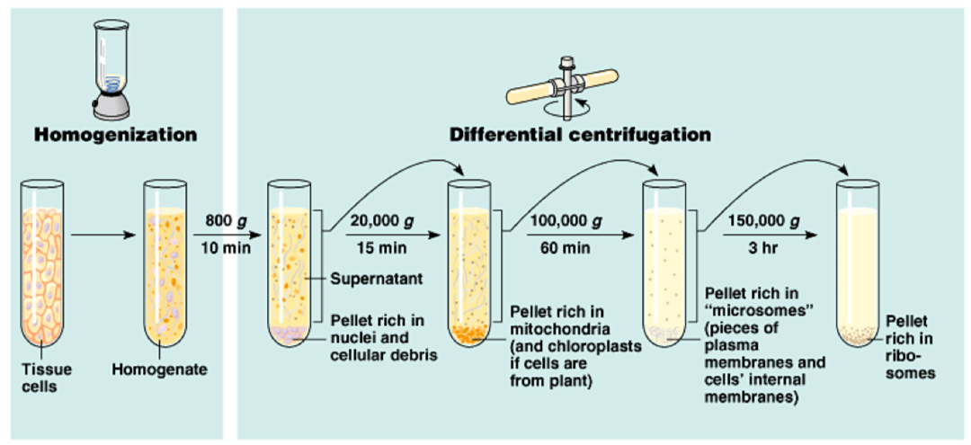
Process of homogenation?
cells are blended first in a homogeniser to break open the cells
then filtered to remove whole cells/ large debris forming the resultant fluid called the homogenate. This tube of homogenate is then placed in a centrifuge and spun at low speed
The heaviest organelles, the nuclei, are forced to the bottom of the tube, where a thin sediment or pellet forms
The fluid at the top, called the supernatant, is removed, which leaves just the sediment of the nuclei.
The supernatant is then transferred to another tube and spun at a slightly faster speed. This time, the pellet that forms contains the next heaviest organelle, the mitochondria
This process continues so that each time the speed is increased, the next heaviest organelle is sedimented and separated out.
Properties of the homogenate at the beginning?
placed in a cold, buffered solution, of the same water potential as the cells
This is to prevent the organelles from bursting under osmotic pressure
, to inactivate any enzymes from breaking down organelles/ inhibit enzyme action
and so that the pH does not fluctuate, so stopping enzymes from denaturing