Unit 12 - Adrenal Medulla Tumors
1/23
There's no tags or description
Looks like no tags are added yet.
Name | Mastery | Learn | Test | Matching | Spaced | Call with Kai |
|---|
No analytics yet
Send a link to your students to track their progress
24 Terms
What is Pheochromocytoma?
Rare, Adrenal medulla tumor
Is Pheochromocytoma benign or malignant?
Usually benign in adrenal gland (10% malignant)
Which mass has an excessive secretion of epinephrine & norepinephrine?
Pheochromocytoma
What does a Pheochromocytoma mass produce?
Intermittent hypertension
What is the US appearance of Pheochromocytoma?
Unilateral or bilateral
Highly vascular
Poor through-transmission
May be large & bulky
Varied appearance
Cystic, solid, calcified components
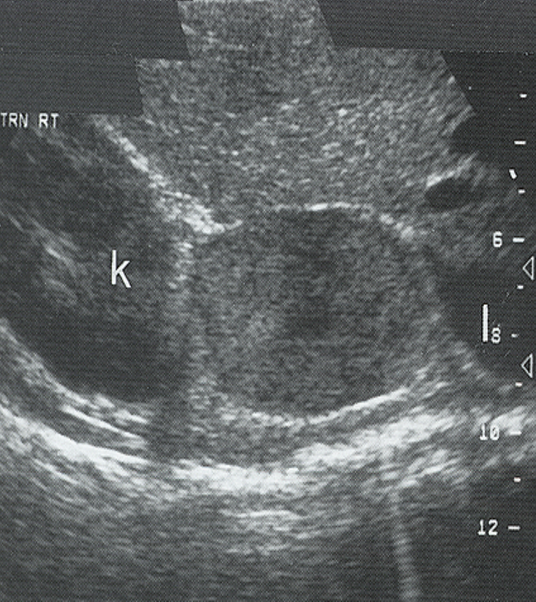
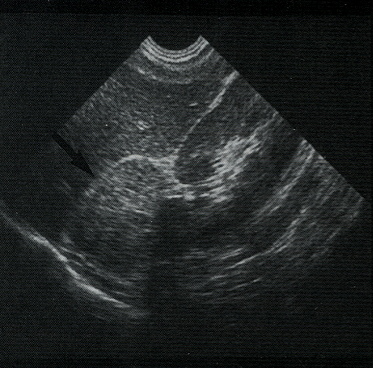
What is this image showing?
Pheochromocytoma
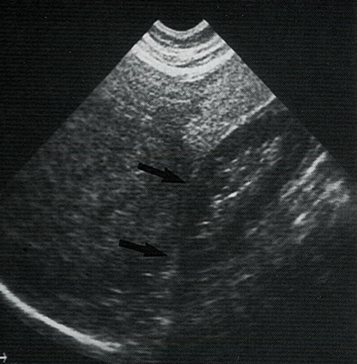
What is this image showing?
Pheochromocytoma Calcified Adrenal Gland
Most common adrenal gland malignancy in childhood?
Adrenal Neuroblastoma
Most common tumor of infancy?
Adrenal Neuroblastoma
Where would an Adrenal Neuroblastoma be located in an adrenal gland?
Medulla
Adrenal Neuroblastoma’s are usually __________ in children.
Asymptomatic
What other symptom may be seen in children with an Adrenal Neuroblastoma?
Palpable abdominal mass
What is the US appearance of an Adrenal Neuroblastoma?
Well-encapsulated tumor
Usually displaces kidney inferiorly & laterally
Large
Heterogeneous
Echogenic mass
Poorly defined margins
May have cystic or calcified components
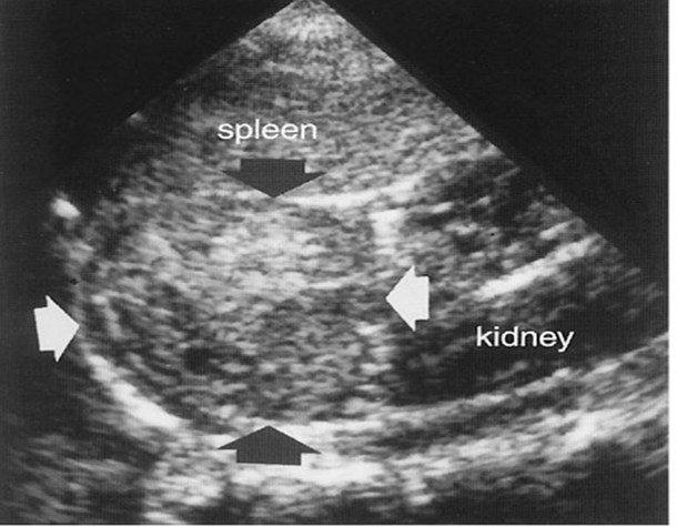
What is this image showing?
Adrenal Neuroblastoma
A Myelolipoma is ______ & _______.
Rare & Benign
A Myelolipoma is composed of:
Adipose tissue
Lymphocytes
Primitive myeloid cells
Primitive myeloid cells are derived from the _____ ______.
Bone marrow
What is this image showing?
Myelolipoma
Lesions in the liver or Morison’s pouch displace fat echoes ________ & _________.
Posteriorly & inferiorly
Extrahepatic masses shift the IVC ____________.
Anteriormedially
Extrahepatic masses displace the right kidney ___________.
Anteriorly
Retroperitoneal lesions in the adrenal gland & the kidney displace fat echoes _________ & __________.
Anteriorly & Superiorly
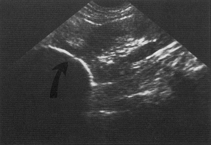
What is this image showing?
Shift in “fat line” —> Adrenal mass
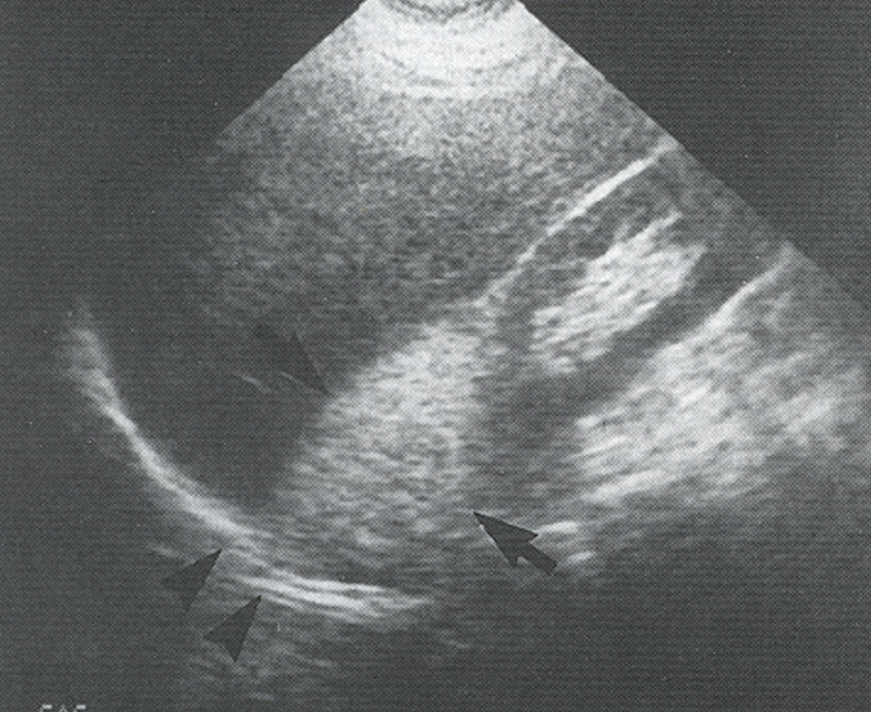
What is this image showing?
Shift in “fat line” —> Liver mass distinction