Diagnostic Imaging
1/57
There's no tags or description
Looks like no tags are added yet.
Name | Mastery | Learn | Test | Matching | Spaced |
|---|
No study sessions yet.
58 Terms
RPA
radiation protection advisor
RPS
radiation protection supervisor
mSv
milisievert
maximum permissable dose
20msv annually
classified person- 30msv
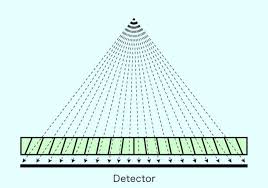
grids
fits over cassette to absorb scatter radiation
kV
kilovoltage
PPE
Gown, thyroid guard, gloves(0.35mm), glasses(0.75mm)
dosimeter
contains lithium fluoride and is sent to HPA for readings every 1-3months
fluorence
crystals that emit visible light which is utilised in the composition of intensifying screens
types of xray machine
portable, mobile, fixed tube heads
xray tube head
where protons are generated
contains anode and cathode
anode
small metal disc that is afixed to a large copper rotor with an electric motor
xray photon production
mAs applied, filament heated, electrons produced, kV applied, electrons stopped/slowed down
fine focus
small filament produces small effective focal spot
cathode
expels electrons from the circuit and focus them in a beam on a focal spot on the anode
Understanding kV
photons have higher energy/frequency when kV is increased
kV increased to increase the amount of radiation coming out of xray tube
photographic density
measurement of how much a substance, such as film, transmits light or electromagnetic radiation
inverse square law
scattering exposure levels decrease proportionally with the inverse of the distance squared fro. the xray source
mA
milliamp
half press button
cathode mateiral
tungsten filament
kV
fully pressed button
anode material
tungsten and copper stem
S
seconds
half pressed + full pressed button
FFD
film focal distance
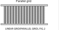
parrellel linear grid
has lead strips which are vertical and parralel to one another
cheap
can be used at any FFD

grid cut off
uneven density or loss of density on the xray due to undesirable absorption of the primary beam by the radiographic grid
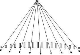
focused linear grid
lead strips are tilted with an increasing angle from the center of the grid
must be specific FFD
prevents grid cut off

pseudofocused grid
lead strips are parallel by height but decreases toward the edge
correct FFD
reduced grid cut off

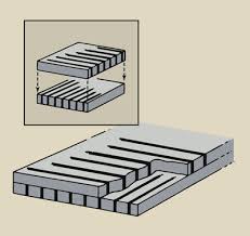
crossed grid
two parallel linear grids stacked on top of each other
expensive

grid factor
usually 2.5-3
Contrast study
medium that is in contrast to the tissues being imaged, in order to visualise the tissue
Positive
shows up white on xrays (barium sulphate and iodinated contrast media)
negative
shows up black on xray
double
first use a positive medium then negative
negative contrast media
commonly used agent is air, mainily in bladder
negative pressure complications
bladder rupture, intramural air, fatal air embolism, introgenic bladder
barium sulphate properties
all poms
used for GI studies
not absorbed through GI tract
insoluble in water
Barium sulphate comes in
paste
mixed with food
liquid
granules
powder
barium impregnated polyethelene spheres (BIPS)
Barium Sulphate contrainidications
not allowed to enter bloodstream
leakage in body can cause adhesions
risk of aspiration pneumonia
can cause GI irritation
iodinated contrast media properties
all poms
higher atomic number than the tissues so is radiopaques
water soluble
can be ionic/nonionic
Ionic
dissociates in solutions
very high osmotic pressure
should not be used in myelography
cheaper than non ionic
Non Ionic
remains a single molecule
lower osmotic pressure
less effect on nervous tissue
expensive
can be used for everything
iodinated contrast media contraindications
only non ionic can be used for myelography
uncontrolled hyperthyroidism in humans
renal disease due to fluid being passed through kidneys to be excreted
Negative Contrast media
radiolucent
used for bladder, intestinal tract and joints
Air
shows up black
Positive Contrast Media
radiopaque
used for GI tract
barium sulphate
shows up as white
Ionic Iodinated CM
Radiopaque
used for GI, blood vessels, joints, salivary glands
high osmotic pressure
non ionic iodinated cm
used for myelography
low osmotic pressure
rules for contrast studies
no sedation for GI studies
plain xrays first
starve for 24hours
enema 2-3 hours before
catheterise
retrograde large intestinal study
diluted barium given slowly into ascending colon with enema pump or funnel
purse string suture to tighten the anus and keep tube in place
timings for GI studies
Oesphagus- immediatly after
stomach - immediatly after
small intestine - taken in intervals
Intersuseption
block necking of intestine
Angiography
iodinated contrast media
into peripheral vein
60-90mins before it reaches aortic arch
IV Urography
heavily sedated
bladder must be empty
never use on a patient in renal failure
retrograde urethrography
catheterise the urethra with widest catheter possible
tip of catheter is postioned distal to area of interest
retrograde vaginourethrography
pass foley catheter, prefilled with saline
take xray immediatly after admin
myelography
patient must be under GA
Plain xray taken of the area of suspicion
cisternal puncture is required if lesion is from the neck down
lumbar puncture is required if lesion is from the lumbar down
scintigraphy
radioactive isotopes given to the patient
they are then detected by the gamma camera
HYPERTHROIDISM CATS