Week 12 (Chapter 28): Normal anatomy: Intraoral Images
1/203
There's no tags or description
Looks like no tags are added yet.
Name | Mastery | Learn | Test | Matching | Spaced | Call with Kai |
|---|
No study sessions yet.
204 Terms
Which type of bone is also known as compact bone and forms the dense outer layer?
Cortical bone
How does cortical bone typically appear on a processed dental radiograph?
Radiopaque
In cancellous bone, the lattice-like network of bone pieces is called:
Trabeculae
Why do marrow spaces appear radiolucent on a dental image?
Because they contain soft tissue and lack dense bone structure
Which of the following best describes the structural arrangement of cancellous bone?
Soft, spongy bone located between two layers of dense cortical bone
Which term is synonymous with cortical bone?
Compact bone
All bone prominences appear ___________ on a dental image because they are composed of dense ___________ bone.
Radiopaque; cortical
Which term describes a sharp, thornlike projection of bone?
Spine
A 'Ridge' is defined as a ___________ prominence or projection of bone.
Linear
How does a Tuberosity differ from a Tubercle in terms of description?
A tuberosity is rounded, while a tubercle is a small bump or nodule
Which of the following describes a 'Process' in anatomical terms?
A marked prominence or projection
How would a Tubercle appear on a dental radiograph?
As a radiopaque small bump
Which of the following describes the Coronoid process in a dental radiograph?
A marked radiopaque projection of the mandible
The coronoid process is part of which anatomical structure?
The mandible
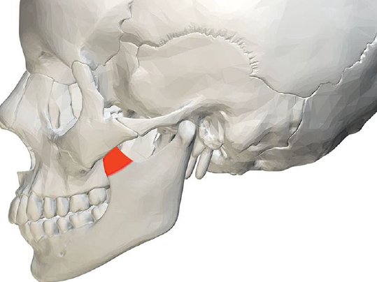
What is this part?
Process
The external oblique ridge is a ___________ prominence that appears ___________ on a mandibular molar image.
Linear; radiopaque
Which anatomical structure is described as a sharp, thornlike projection located at the midline of the maxilla?
Anterior nasal spine
On a mandibular molar radiograph, you see a dense radiopaque line extending downward and forward from the ramus. This is likely the:
External oblique ridge
Which visual characteristic distinguishes a 'Spine' from a 'Ridge' on a dental radiograph?
A spine is a sharp, thornlike point, while a ridge is a long, linear projection
The Anterior Nasal Spine is located at the midline of the __________.
Maxilla
The term 'Thornlike' is most closely associated with which anatomical feature?
Spine
Which of the following describes the genial tubercles?
Small, radiopaque bumps located on the lingual aspect of the mandibular midline
How does the maxillary tuberosity typically appear on a radiograph?
As a radiopaque, rounded prominence posterior to the third molar region
Which term describes a tube-like passageway through bone that houses nerves and blood vessels?
Canal
A 'Fossa' is best described as a ____________ area of bone.
Broad, scooped-out
On a radiograph, why do spaces like the maxillary sinus appear radiolucent?
Because they are hollow cavities filled with air
Which of the following describes the difference between a foramen and a canal?
A foramen is a simple opening, while a canal is a long, tube-like passageway
The Maxillary Sinus is an example of a:
Hollow space or cavity within bone
What structure is described as a 'scooped-out' area of bone?
Fossa
Which structure is a radiolucent, tube-like passageway that travels through the mandible?
Mandibular canal
The Submandibular fossa is a scooped-out area where the __________ rests.
Submandibular salivary gland
Where is the Incisive Foramen typically located on a dental image?
At the midline of the anterior maxilla
Radiographically, a Fossa appears radiolucent because:
The bone is thinner than in the surrounding areas
A 'Septum' is described as a bony wall that divides two spaces. How does it typically appear on a radiograph?
Radiopaque
Which structure represents an immovable joint between bones and appears as a thin radiolucent line?
Suture
The two paired bones of the maxilla meet at the ___________ of the face.
Midline
Which of the following is NOT a structure formed by the maxilla?
Mandibular ramus
The maxilla forms the _________ and _________ of the nasal cavities.
Sides; floor
Where is the incisive foramen located in the oral cavity?
At the midline of the anterior hard palate
What is the typical radiographic appearance of the nasopalatine foramen?
A small ovoid or round radiolucent area
Which specific nerve exits the maxilla through the incisive foramen?
Nasopalatine nerve
Which of these best describes the radiographic appearance of the incisive foramen?
A radiolucent area between the roots of the central incisors
What is another name for the incisive foramen?
Nasopalatine foramen
Which term describes the 'sculpted' part of the maxilla that forms the side of the nose?
Lateral wall of the nasal cavity
The superior foramina of the incisive canal are located on the floor of the:
Nasal cavity
Radiographically, how many superior foramina are typically seen, and where are they located relative to the teeth?
Two small round RLs; superior to the apices of central incisors
The superior foramina join together to form which anatomical structure?
Incisive canal
The median palatine suture represents the line of union between which two parts of the maxilla?
Palatine processes
How does the median palatine suture typically appear on a maxillary central incisor radiograph?
As a thin radiolucent line between the central incisors
The median palatine suture extends from the alveolar bone to the:
Posterior hard palate
Between which two teeth is the lateral fossa typically located on a radiograph?
Maxillary lateral incisor and canine
Where is the lateral fossa located relative to the infraorbital foramen?
Inferior and medial
What is the characteristic shape of the nasal cavity as described in bony anatomy?
Pear-shaped
The floor of the nasal cavity is primarily formed by which structures?
Palatal processes of the maxilla and horizontal portions of palatine bones
On a dental radiograph, where is the radiolucent area of the nasal cavity typically seen?
Above the maxillary incisors
Which two bones primarily form the bony portion of the nasal septum?
Vomer and ethmoid bone
How does the nasal septum appear on a maxillary anterior radiograph?
As a vertical radiopaque partition
The nasal septum divides the nasal cavity into the right and left:
Nasal fossae
The nasal cavity is divided into two separate chambers known as:
Nasal fossae
The floor of the nasal cavity is described as a dense band of bone. How does this appear radiographically?
Radiopaque
Which of the following defines the inferior border of the nasal cavity?
Floor of the nasal cavity
The floor of the nasal cavity is composed of the horizontal portions of the palatine bones and the __________.
Palatal processes of the maxilla
The anterior nasal spine is described as a sharp projection of which bone?
Maxilla
What is the characteristic radiographic shape of the anterior nasal spine?
V-shaped
Where is the anterior nasal spine located on a radiograph relative to other landmarks?
At the intersection of the nasal floor and the nasal septum
Which directional terms best describe the location of the anterior nasal spine within the nasal cavity?
Anterior and inferior
The term 'conchae' refers to which specific shape characteristic of these bones?
Shell-shaped or scroll-shaped
Where do the inferior nasal conchae originate from within the nasal cavity?
The lateral walls of the nasal cavity
Why do the inferior nasal conchae appear as a 'diffuse' radiopaque mass rather than a sharp line?
Because they are wafer-thin and curved plates
Which of the following describes the nature of the maxillary sinus?
Paired cavities located within the maxilla
The maxillary sinus is typically located above the apices of which teeth?
Maxillary premolars and molars
How does the maxillary sinus appear on a dental radiograph?
As a large radiolucent area with a thin radiopaque border
What is the primary function or nature of the septa found within the maxillary sinus?
They are bony partitions that divide the sinus into compartments
How do sinus septa typically appear on a periapical or panoramic image?
As radiopaque lines crossing the radiolucent sinus space
Which statement is true regarding the presence of septa in the maxillary sinus?
The number and location vary depending on the individual's anatomy
What is the primary content found within the nutrient canals of the maxillary sinus?
Blood vessels and nerves
How do nutrient canals appear radiographically, and what defines their borders?
A narrow radiolucent band bounded by two thin radiopaque lines
Which areas receive their blood supply and innervation from the vessels within these canals?
Maxillary teeth and interdental areas
The 'Inverted Y' is formed by the intersection of which two anatomical structures?
The lateral wall of the nasal fossa and the anterior border of the maxillary sinus
How does the Inverted Y typically appear on an intraoral radiograph?
Radiopaque
Where is the Inverted Y most commonly located in a periapical radiograph?
Above the maxillary canine
Which specific type of bone tissue forms the radiopaque lines of the Inverted Y?
Dense cortical bone
If a clinician observes an Inverted Y on a radiograph, which tooth is most likely centered in that image?
Maxillary canine
Where is the maxillary tuberosity located in relation to the third molar?
Posterior (distal) to the third molar region
How is the appearance of the maxillary tuberosity described on a radiograph?
A radiopaque (RO) bulge
Which of the following describes the shape and composition of the maxillary tuberosity?
A rounded prominence of bone
The hamulus is a projection of bone that extends from which larger bone of the skull?
Sphenoid bone
How is the hamulus most commonly identified on a dental radiograph?
As a radiopaque hooklike projection
Which specific portion of the sphenoid bone gives rise to the hamulus?
Medial pterygoid plate
If you are looking at a periapical radiograph of the maxillary third molar, where exactly should you look for the hamulus?
Posterior to the maxillary tuberosity
The radiographic density of the hamulus is identical to other bony processes. This means it appears:
Radiopaque
What is the characteristic shape of the zygomatic process of the maxilla on a periapical radiograph?
A J- or U-shaped radiopacity
The zygomatic process of the maxilla appears radiopaque because it is composed of which type of tissue?
Dense cortical bone
In which radiographic area would you most likely find the zygomatic process of the maxilla?
Maxillary molar region
What is the primary anatomical function of the zygomatic process of the maxilla?
To articulate with the zygoma or malar
Which term is used interchangeably with the 'Zygoma' to describe the cheekbone?
Malar bone
How does the Zygoma appear on a periapical radiograph compared to the zygomatic process of the maxilla?
As a diffuse, radiopaque band extending posteriorly
The Zygoma articulates with which specific part of the maxillary bone?
Zygomatic process of the maxilla
Which portion of the mandible is described as the vertical part located posterior to the third molar region?
Ramus
The 'Angle' of the mandible is formed by the junction of which two borders of the ramus?
Posterior and lower borders