Cell Biology flashcards
1/97
There's no tags or description
Looks like no tags are added yet.
Name | Mastery | Learn | Test | Matching | Spaced |
|---|
No study sessions yet.
98 Terms
What are the three parts of the cell theory?
1) All living things are made of cells. 2) Cells are the basic unit of structure and function in living things. 3) All cells come from pre-existing cells.
How does deductive reasoning apply to cell theory?
It allows scientists to make predictions about cells based on the theory. Any newly discovered organism will be made up of one or more cells, if something is not made of cells it isn’t alive, and new cells observed in growth or healing must have originated from existing cells.
Define micrograph.
An image taken through a microscope showing a magnified specimen.
What are the main parts of a light microscope?
Eyepiece (ocular lens), objective lenses, stage, light source, base, coarse adjustment knob, fine focus knob and diaphragm, etc.
How do you focus a microscope on a sample?
Start with the lowest power objective and slowly adjust the coarse focus knob until the sample is in view. Then, switch to a higher power objective and refine focus with the fine focus knob.
Define magnification.
The ratio of an object's image size to its real size. It indicates how much larger an object appears under a microscope compared to its actual size.
How do you calculate total microscope magnification?
Total magnification = ocular lens magnification × objective lens magnification.
How do you measure field of view (FOV) diameter under low power?
Place a ruler under the microscope and measure the visible diameter in millimeters.
How do you calculate FOV diameter under higher power?
FOV high = diameter (LP) × magnification (LP) / high magnification.
How can you estimate the size of a sample under the microscope?
Estimate how much of the field of view the object occupies and multiply by the field diameter.
What formula is used to calculate magnification of a micrograph?
Magnification = image size ÷ actual size of specimen. (AIM TRIANGLE)
Define resolution.
smallest interval distinguishable by the microscope, which then corresponds to the degree of detail visible in an image created by the instrument.
Compare light and electron microscopes.
Light microscopes use light and lenses for live or stained cells (lower resolution); electron microscopes use electron beams (higher resolution)
What is freeze-fracture electron microscopy?
A technique where frozen cells are split along membranes and coated with metal to reveal internal surfaces. With the cell membrane's bilayer in half, electron microscopes can look at the inside surface of the membrane*. This technique allowed us to better understand the structure and function of cell membranes.
Define fluorescence.
The emission of light by a substance that has absorbed light or other radiation.
How is fluorescence used in microscopy?
Fluorescent dyes or proteins bind to specific cell structures and glow under certain wavelengths of light.
What is immunofluorescence?
A method using fluorescently labeled antibodies to locate specific proteins in cells.
What is cryogenic electron microscopy (cryo-EM)?
A technique where samples are rapidly frozen to preserve structures for high-resolution imaging.
Give an example of technology leading to discovery in microscopy.
The invention of the electron microscope led to the discovery of organelles like mitochondria and ER in detail.
What structures are common to all cells?
Plasma membrane
What are the two main types of cells?
Prokaryotic and eukaryotic.
What are the main differences between prokaryotic and eukaryotic cells?
Prokaryotes lack a nucleus and membrane-bound organelles; eukaryotes have both.
Functions of prokaryotic cell wall
provides structure, prevents lysis (bursting), offer protection, anchors pili and flagella
Define extracellular.
Located or occurring outside a cell.
Contrast eukaryotic and prokaryotic ribosomes.
Eukaryotic: 80S; Prokaryotic: 70S.
Describe nucleoid DNA structure and function.
The nucleoid contains the prokaryote’s circular, naked DNA that controls cell activities, stores hereditary information, and replicates before division.
Compare genetic material of prokaryotes vs eukaryotes.
Prokaryotes: circular naked DNA; Eukaryotes: linear DNA wrapped around histones in a nucleus.
Outline structural diversity among prokaryotes.
Prokaryotes vary in shape, cell wall composition, external structure, internal organization, and environmental adaptations— giving them immense structural and functional diversity.
What does it mean for eukaryotic cells to be compartmentalized?
Organelles are membrane-bound to separate incompatible processes.
List functions of eukaryotic organelles.
Plasma membrane: barrier; Cytoplasm: reactions; Ribosomes (80S): protein synthesis; Nucleus: controls genes; Mitochondria: ATP; Chloroplasts: photosynthesis; ER: synthesis/transport; Golgi: packaging; Vesicles: transport; Vacuoles: storage; Lysosomes: digestion; Cytoskeleton: structure/movement.
What are the advantages of compartmentalization?
Increases efficiency by isolating incompatible reactions and maintaining optimal conditions.
What distinguishes living from nonliving things?
Living things carry out 7 processes of life, non living things don’t
What are the 7 functions of life?
Metabolism, growth, reproduction, response, homeostasis, organization, and adaptation.
Name 3 atypical eukaryotic cells.
Skeletal muscle fibers, Aseptate Fungal Hyphae, Red Blood cells
Why are skeletal muscle fibers atypical?
They are multinucleated and very long.
Why are aseptate fungal hyphae atypical?
They have many nuclei in a continuous cytoplasm without dividing walls.
Why are red blood cells atypical?
They lack a nucleus and most organelles.
Why are phloem sieve tube elements atypical?
They lack nuclei and depend on companion cells.
What is a micrograph?
A photograph taken through a microscope.
What structures can you identify in prokaryotic micrographs?
Cell wall, plasma membrane, nucleoid region, 70S ribosomes, Flagella, pili,
What structures can you identify in eukaryotic micrographs?
Nucleus, plasma membrane, cytoplasm, nucleolus, mitochondria, chloroplasts, Smooth/Rough ER, Golgi, 80S ribosomes, vesicles/vacuoles, lysosomes, cilia, flagellum, cell wall
What are proper rules for biological drawings?
Draw clear
What molecule makes up most of the cell membrane?
Phospholipids.
Define amphipathic.
Molecules with both hydrophilic and hydrophobic regions.
Why do phospholipids form bilayers in water?
Hydrophilic heads face water; hydrophobic tails avoid it
Why are phospholipids good barriers?
They prevent free passage of polar molecules while allowing selective transport.
Define diffusion.
Passive movement of particles from high to low concentration.
Give one example of simple diffusion.
Oxygen diffusing into cells or carbon dioxide diffusing out.
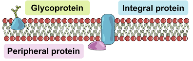
Define membrane protein.
A protein embedded in or attached to the cell membrane.
Contrast integral and peripheral proteins.
Integral: span the bilayer; Peripheral: attached to membrane surface.
Define solute
substance dissolving into another body. Salt dissolving into water. Salt is solute, water is solvent
Define osmosis.
Passive movement of water across a selectively permeable membrane from low to high solute concentration.
Why does osmosis not require energy?
It occurs due to natural kinetic movement of water molecules.
Explain the movement of water in osmosis.
Water moves toward higher solute concentration until equilibrium.
Define aquaporin.
A channel protein that specifically facilitates water movement across membranes.
Define facilitated diffusion.
Passive transport of molecules through membrane proteins.
Define channel protein and carrier protein.
Channel: forms pore for ions/water; Carrier: changes shape to move specific molecules.
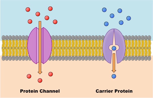
How do channel proteins make membranes selectively permeable?
They only allow specific ions/molecules to pass.
Give an example of a selective channel protein.
Sodium or potassium ion channels.
Define active transport.
Movement of molecules against concentration gradient using ATP energy.
Give one example of active transport.
Sodium-potassium pump moving Na⁺ out and K⁺ into cells.
Define selective permeability.
The ability of a membrane to allow some substances to pass more easily than others.
Define glycosylation.
Addition of carbohydrate groups to proteins or lipids.
What are the functions of glycolipids and glycoproteins?
Cell recognition
Explain the fluid mosaic model.
Membrane is a dynamic bilayer of phospholipids with embedded proteins moving freely.
Give an example of technology aiding organelle discover.
Electron microscopy revealed details of mitochondria and ER. These are both examples of organelles.
Explain cell fractionation.
Cells are broken up and organelles separated by centrifugation to study their functions.
Define organelle.
A specialized structure within a cell performing a specific function.
What is the advantage of separating nucleus and cytoplasm?
Allows control of gene expression and protects DNA from cytoplasmic enzymes.
What is the advantage of compartmentalization using lysosomes and vacuoles?
Lysosomes isolate digestive enzymes; phagocytic vacuoles confine harmful substances.
Define solvation.
The process where solvent molecules surround and interact with solute particles.
Give an example of solvation.
Na⁺ and Cl⁻ ions surrounded by water molecules when salt dissolves.
Define osmolarity
The total concentration of solute particles in a solution. Specifically, it is defined as the number of osmoles of solute per liter of solution
What are the units of osmolarity?
Osmoles per liter (Osm/L).
What happens to cells in an isotonic solution
The concentration of solutes outside the cell is equal to the concentration inside the cell.
How do hypotonic (causes cells to absorb water) or hypertonic solutions (causes cells to release water) affect plant tissues?
Hypotonic: cells become turgid (swollen); Hypertonic: plasmolysis (shrinkage).
Define osmoregulation.
Maintenance of water balance in cells/organisms.
What is the role of contractile vacuole in freshwater unicellular organisms?
It expels excess water to prevent bursting.
How is normal saline used medically?
Isotonic to body fluids (doesn’t cause shrinkage or swelling); used for IV fluids which help maintain fluid and electrolyte balance.
Define cellular differentiation.
Process by which unspecialized cells develop into specialized cells.
Explain differential gene expression.
Only certain genes are activated in specific cell types.
Define morphogen gradient.
A concentration gradient of signaling molecules guiding cell differentiation.
What is the role of morphogen gradients?
They determine cell fate in developing embryos.
Define stem cell.
An undifferentiated cell capable of self-renewal and differentiation.
Define stem cell niche.
Microenvironment that supports and regulates stem cell behavior.
Give two examples of stem cell niches.
Bone marrow (hematopoietic stem cells) and intestinal crypts.
Define totipotent
Type of cell that has the ability to develop into any cell type in an organism, including both the embryonic (body) cells and extaembryonic tissues like the placenta.
Why are stem cells most common in early embryos?
They give rise to all specialized cell types.
What is the range of cell sizes in humans?
From ~5 µm (RBCs) to 100 µm (oocytes).
Why are cells limited in size by surface area-to-volume ratio?
Smaller cells exchange materials more efficiently.
Give examples of scientific models.
Molecular models
What are limitations of models in science?
They simplify complex systems and may not represent all factors.
Why are cubes used to model SA:V ratio?
They’re simple for measuring surface area and volume changes.
Why are repeated measurements important?
They increase reliability and accuracy of results.
Why use standard deviation?
To show how spread out data values are from the mean.
What do error bars represent?
Variability or uncertainty in data (often ±1 SD or SE).
Why are controlled variables necessary?
To ensure only the independent variable affects results.
List 3 cell adaptations that increase SA:V ratio.
Microvilli,Flattening or thin cell shape (ex. red blood cells, long or elongated projections (ex. nerve cells)