The heart
1/72
There's no tags or description
Looks like no tags are added yet.
Name | Mastery | Learn | Test | Matching | Spaced | Call with Kai |
|---|
No analytics yet
Send a link to your students to track their progress
73 Terms
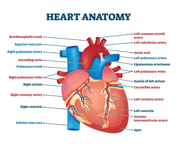
The heart is surrounded by a
pericardium, consisting of two parts:
– An outer _______ pericardium
– An inner ________ pericardium, which
consists of two layers
fibrous and serous
attached to
the surface of the heart
inner visceral layer(epicardium)
adjacent to the fibrous
pericardium
outer parietal layer
The space between the two
serous layers is called the
pericardial cavity, it contains
pericardial fluid
Epicardium
External surface, consists of visceral pericardium
Myocardium
Consists of cardiac tissue, including cardiac muscle cells, connective
tissue, blood vessels, and nerves
Endocardium
Internal, endothelial surface
Cardiac muscle cells contract
without information coming
from the
CNS (involuntary)
move directly from one cell to
another creating a direct, electrical
connection
Ions
The Cardiac Skeleton is where
each cardiac cell is wrapped in an elastic sheath and muscle layers is wrapped in a fibrous sheet
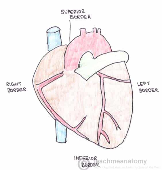
The base is
the superior border of the heart
The apex is
the inferior portion of the heart

The right border is
formed by only the right atrium

The inferior border is
formed by the right ventricle
Interatrial groove
separates the left and right atria
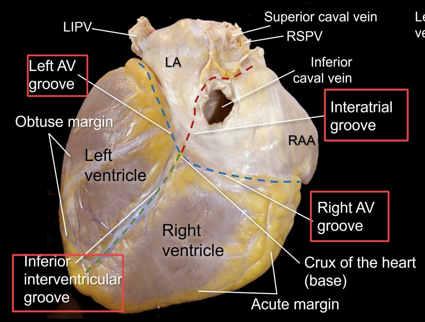
Coronary sulcus
separates the atria and the ventricles
Anterior interventricular sulcus
separates the left and right ventricles
Posterior interventricular sulcus
also separates the left and right
ventricles
The left and right atria
– Positioned superior to the coronary sulcus
– Both have thin walls
– Both contain an expandable anterior portion called an auricle
The left and right ventricles
– Positioned inferior to the coronary sulcus
– Both have thicker walls than the atria
▪ Left wall is thicker than the right wall
The Right Atrium Receives oxygen-poor venous
blood via
superior vena
cava, inferior vena cava, and
coronary sinus
which contains Contains pectinate muscles right or left atrium?
Right atrium
Blood enters the right ventricle
by passing through the
atrioventricular valve(tricuspid valve)
Blood leaves the right ventricle
by passing through the
pulmonary valve(pulmonary
semilunar valve)
The right AV valve is connected
to
papillary muscles via
chordae tendineae
Trabeculae carneae
Muscular ridges
Moderator band
Muscular band that
extends from the
interventricular septum
to the ventricular wall only found in the right ventricle
The Left Atrium Receives oxygenated blood
from the lungs via
the right and
left pulmonary veins
in the left atrium Blood passes through the left
atrioventricular valve(mitral
valve)
which has the thickest wall right or left ventricle?
left ventricle
Blood leaves the left
ventricle by passing through
the
aortic valve(aortic
semilunar valve)
Blood enters the
ascending aorta
Blood then travels to the
_______ and then
down the _________ and to all body parts
(systemic)
aortic arch and descending aorta
AV valve function during the
cardiac cycle
Papillary muscles relax, Due to pressure in the atria,
the valves open, and Blood flows from atria to
ventricle
When the ventricles
contract
pressure causes
the AV valves to close and
semilunar valves to open
– Atrial branches
– Right marginal branch
– Posterior interventricular
branch
Right coronary artery (RCA)
– Circumflex branch
– Left marginal branch
– Anterior interventricular
branch
Left coronary artery (LCA)
Great cardiac vein
Delivers blood to the
coronary sinus
Middle cardiac vein
Delivers blood to the
coronary sinus
Coronary sinus
Drains directly into the
posterior aspect of the right
atrium
Which layer of the heart wall contains the cardiac muscle cells, connective tissues, and blood vessels?
Myocardium
The intercalated discs of cardiac muscle tissue contain gap junctions. What is the primary function of these gap junctions?
to allow ions to move between cells for a direct eletrical connection
What are the two circuits the heart pumps blood into?
1. Pulmonary circuit 2. Systemic circuit
Arteries
Transport blood away from the heart.
Veins
Transport blood toward the heart
Capillaries
Vessels that interconnect arteries and veins.
What are the four parts of every AV valve?
1. Ring of connective tissue. 2. Cusps. 3. Chordae tendineae. 4. Papillary muscles (contract to prevent valve inversion).
Describe the Left Ventricle and how it compares to the Right.
Thickest wall for strong contractions (systemic circuit).
Does not have a moderator band.
Left AV valve (bicuspid/mitral) has 2 cusps and 2 papillary muscles.
Contraction is 6 to 7 times more powerful than the right ventricle.
Explain the function of AV valves during the Cardiac Cycle.
1. Papillar muscles relax. 2. Pressure in atria opens the valves; blood flows to ventricle. 3. Ventricles contract; pressure closes AV valves and opens semilunar valves. 4. Closure of AV valves prevents regurgitation (backflow) into atria.
Contraction is
systole
Atrial systole
Blood flows into the
ventricles
Ventricular systole
Blood is ejected into
the pulmonary trunk
and the ascending
aorta
Relaxation is
diastole(Chambers are filling with
blood)
Cardiac contractions of the cardiac
cycle are coordinated by
conducting
cells
Nodal cells(conducting cell)
Establish the rate of
contractions, SA and AV nodes
Conducting cells
Distribute the contractile
stimulus to the myocardium
Sinoatrial node (SA node)location and common name?
Located in the posterior wall of the right atrium near the entrance of
the superior vena cava, Also called the cardiac pacemaker
Bradycardia
slower-than-normal heart rate
Tachycardia
faster-than-normal heart rate
Atrioventricular node (AV node) location
Sits within the floor of the right atrium
Cardiac conducting system
1. Impulse travels from the SA node to the AV node
▪ Impulse conducted by internodal pathways
▪ Atrial contraction occurs
2. AV node slows impulse
3. Impulse travels from the AV node to the AV bundle
4. The AV bundle conducts impulse along the interventricular septum and
then divides to form the right and left bundle branches
▪ Serve right and left ventricle
5. The bundle branches conduct impulses to the Purkinje fibers
▪ Purkinje fibers connect to cardiac muscle cells
▪ Ventricular contraction occurs
Norepinephrine from the
sympathetic division of the
ANS causes:
An increase in the heart rate
An increase in the force of
contractions
Acetylcholine from the
parasympathetic division of the
ANS causes:
A decrease in the heart rate
decrease in the force of
contractions
Stimulation of
cardioacceleratory center:
activates sympathetic
neurons
Heart rate increases
Stimulation of cardioinhibitory
center
activates parasympathetic
neurons
Vagus (N X) is involved
Heart rate decreases
Which of the following statements
regarding the cardiovascular system
is TRUE?
When the heart beats, the atria contract first, followed by the
ventricles.
Which of the following vessels sends
blood to the lungs from the right
ventricle?
pulmonary artery
The myocardium _____.
consists of multiple, interlocking layers of cardiac muscle tissue
Which of the following contains the
fetal remnant of the foramen ovale?
right atrium
Which layer of the heart consists of
simple squamous epithelium tissue
that is continuous with the
endothelium of the attached greater
vessels?
endocardium
Which describes the inferior, pointed
tip of the heart that is formed mainly
by the left ventricle?
apex
Which of the following is the
function of the papillary muscles?
They prevent valve inversion into the atria.
Which of the following receives
deoxygenated blood from the
systemic circuit via the superior and
inferior vena cava?
right atrium