Unit 3: Anatomy Lecture Slides (work in progress)
1/53
Earn XP
Description and Tags
cranial nerves and hormones on matching questions, which are half of test put hormones on card primary sensory areas
Name | Mastery | Learn | Test | Matching | Spaced | Call with Kai |
|---|
No analytics yet
Send a link to your students to track their progress
54 Terms
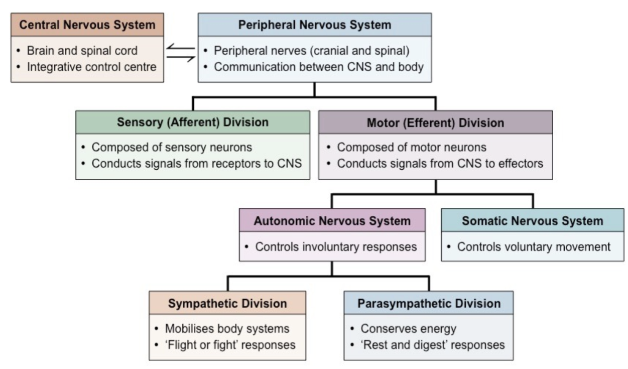
Introduction to the Nervous System [ch 13: Nervous System: Nervous Tissue]
• The nervous system controls & adjusts the body’s activities
• provides swift but brief responses
• Functions
• sensory: detects changes within & outside body
• integrative: analyzes & stores information; determines appropriate
responses
• motor: responses
• Divisions
• Central Nervous System (CNS)
• brain & spinal cord
• integrates, processes & coordinates sensory input w/ motor output
• seat of intelligence, memory, learning & emotion
• Peripheral Nervous System (PNS)
• consists of structures outside of CNS → nerves (inc. roots & branches),
neuromuscular junctions
• provides sensory information to CNS + carries motor commands away from
CNS

Peripheral Nervous System Divisions
• Peripheral nervous system (PNS) divided into:
• Sensory (afferent) division: brings sensory info to CNS
• begins at receptors → categorized as somatic, visceral, special sensory
• Motor (efferent) division: carries out motor commands from CNS
• ends at effectors`
• subdivided into autonomic & somatic nervous systems
• Somatic NS: voluntary; skeletal muscle effectors
• Autonomic: involuntary; glands, smooth & cardiac muscle effectors
• divided into sympathetic (“fight-or-flight”) & parasympathetic (“rest-or-digest”) NS
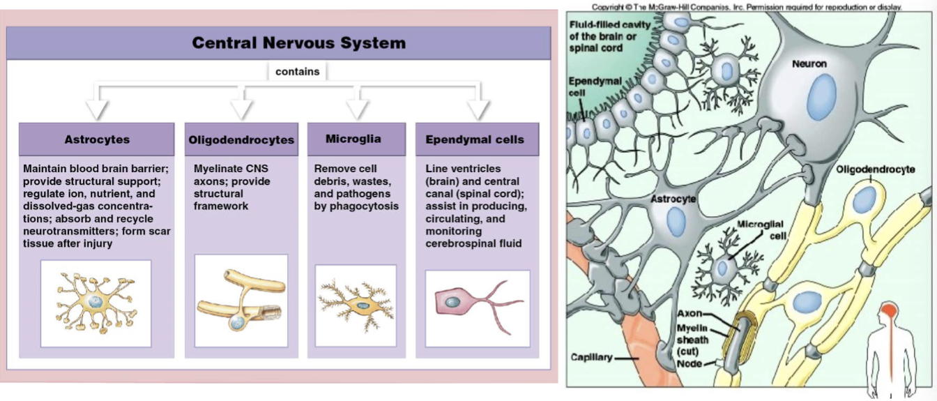
Nervous Tissue: Cells
• Nervous tissue consists of two cell types:
• neurons
• responsible for transfer & processing of information
• consists of a soma (cell body), axon, dendrites, and axon terminals
• neuroglia (brain tumors)
• supporting cells; maintain homeostasis
• mitotically active → can lead to gliomas
• protect the neuron
CNS Neuroglia ***
• Astrocytes
• largest & most numerous
• has multiple processes
• involved in neurotransmitter metabolism
• comprises the blood-brain barrier
• imp’t for brain dev’t
• link bet. neuron & blood vessels → nutrition &
metabolism
• Microglia
• small phagocytic cells
• derived from
monocytes
• Ependymal cells
• make up the ependyma: lines brain
ventricles & spinal cord central canal
• produces
cerebrospinal fluid
(CSF)
• squamous to columnar epithelia w/ cilia
• Oligodendrocytes
• forms a support network for neurons (has branches)
• produce myelin sheaths for
portions of up to 15 neurons
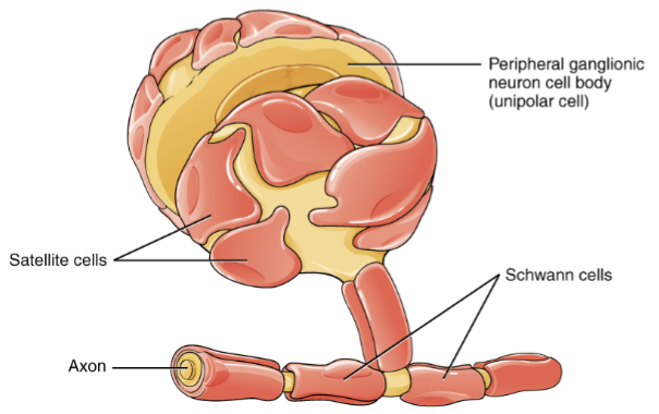
PNS Neuroglia: Satellite Cells & Schwann Cells
• Satellite cells
• support neurons in PNS
ganglia
• wrapped around neuronal
cell bodies
• Schwann cells: aka neurolemmocytes (pancakes that wrap themselves around ONE axon)
• myelinate PNS axons
• 1 myelin sheath around a
portion of 1 PNS neuron
Neurons: Structure
• cell body (soma): processes information; contains...
• perikaryon: cytoplasm
• neurofilaments/neurotubules: cytoskeleton
• neurofibrils: bundles of neurofilaments
• Nissl bodies: clusters of rough ER → chromatophilic substance
• dendrites: highly branched processes; receiving (input) portion
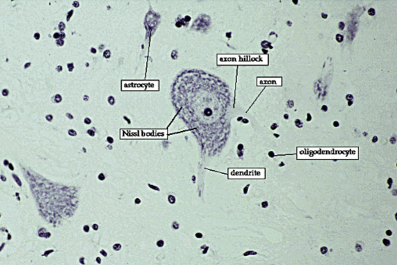
Neurons: Structure
• axon: long, cylindrical projection; transmitting portion → transmits
nerve impulses to target cell; contains...
• axoplasm & axolemma: cytoplasm & cell membrane
• axon hillock: cell body-axonal junction → site of action potential generation
• axoplasmic transport: movement of material bet. cell body & axon
• collaterals: branches off the axon
• telodendria: ends of axon & collaterals
• axon terminals: ends of telodendrias
• synaptic end bulbs: contain synaptic vesicles w/ neurotransmitters
• Axons typically myelinated
• myelin: insulative covering
• increases speed of action potential conduction
• node of Ranvier: gaps in myelin sheath
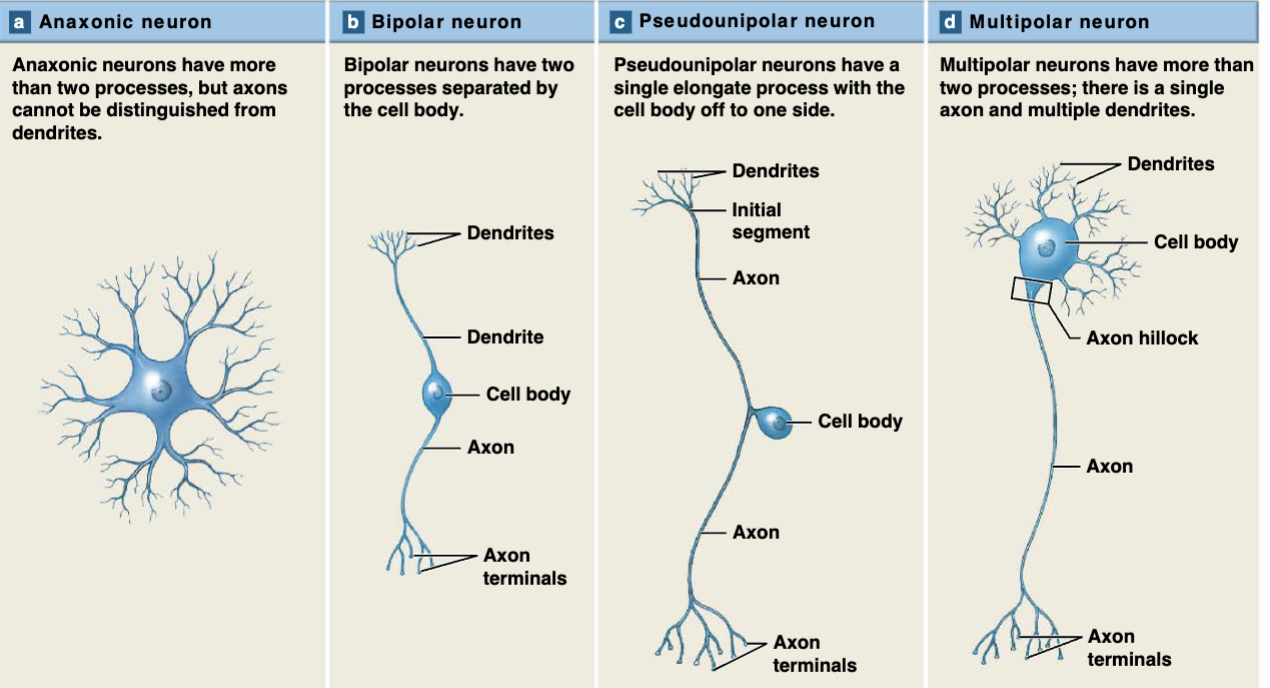
Neurons: Classification****
Neurons can be classified based on:
• structure: # of processes extending from cell body
• function: sensory, motor, or interneuron
• Structural Classifications
• anaxonic: many processes, but axons & dendrites indistinguishable
• only in CNS
• multipolar: multiple dendrites, 1 axon
• most brain & spinal cord neurons
• bipolar: 1 dendrite, 1 unmyelinated axon
• retina, inner ear, olfactory areas of brain
• pseudounipolar: cell body off to side; 1 process, but distinctly separates into dendrite & axon
WE WILL GET THIS PICTURE AND WILL HAVE TO IDENTIFY WHICH NEURON IS WHICH**********
Neurons Classification
• Functional Classifications
• sensory (afferent): transmit from receptors (PNS) → CNS
• motor (efferent): transmit from CNS → effectors (PNS)
• interneurons (association neurons): analyze sensory input & coordinate
motor outputs; located entirely in CNS, bet. motor & sensory neurons
• most neurons in body (~90%)
• can be excitatory or inhibitory
Nervous System Terminology***
• nerve fiber: any neuronal process → primarily the axon
• nerve: bundle of many fascicles → comprised of
many nerve fibers that course along the same path in PNS
• includes both sensory & motor fibers surrounded by CT = endo-, peri- & epi- neurium
• ganglion (pl. ganglia): cluster of nerve cell bodies in PNS
• tract: bundle of nerve fibers in CNS
• nucleus: cluster of nerve cell bodies in CNS
• white matter: myelinated axons
• grey matter: unmyelinated fibers (dendrites & axons) and cell bodies
Neuron Organization and Processing***
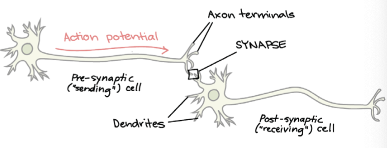
• Recall: Neurons connect w/ other neurons or effectors via synapses
• presynaptic neuron: conducts impulses toward a synapse
• postsynaptic neuron: receives impulses & conducts them away from a synapse
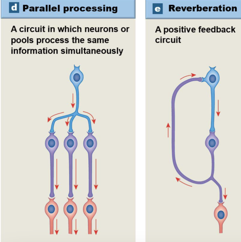
• Neurons can be organized into smaller organized groups called neuronal pools → identified by their neural circuits
• Divergence
• Convergence
• Serial processing
• Parallel processing
• Reverberation
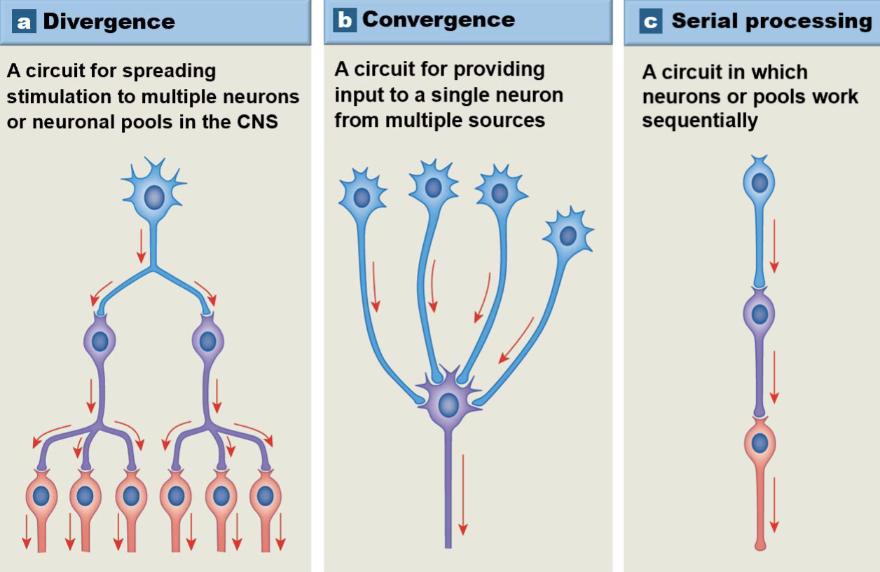
Neural Circuits: Types****
• Divergence: 1 presynaptic neuron → many postsynaptic neurons
• permits broad distribution of a specific input
• info enters CNS → spreads to brain & spinal cord simultaneously
• Convergence: many presynaptic neurons → 1 postsynaptic neuron
• Serial processing: 1 presynaptic → 1 postsynaptic in a sequence
• info from 1 part of the brain goes to the next part then another part, etc.
• Parallel processing: 1 neuron → many neurons → 1 neurons
• several neurons process the info at the same time
• e.g. If you step on a nail, you typically move your foot, shout “ouch,”
and dance a bit, all at the same time
• Reverberation: positive feedback arrangement
• collateral axons extend back toward origin of impulse → impulse
continued and/or enhanced
ON TEST THIS IS A FILL IN THE BLANK USING PICTURES************
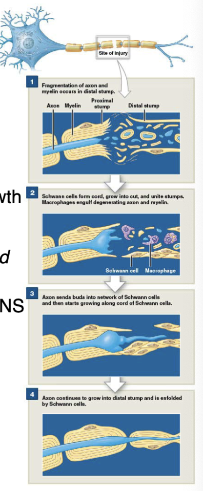
Regeneration of Nervous Tissue***
• Neurons have limited ability for regeneration
• CNS
• little to no repair
• Neuroglia inhibit axonal regrowth
• Astrocytes proliferate rapidly following injury →
scar tissue develops = physical barrier to regrowth
• PNS****
• Intact cell body & active Schwann cells required
for repair/regrowth to occur
• Wallerian degeneration: repair mechanism (nerves) in PNS
• S1: axon distal to injury site deteriorates
• S2: macrophages phagocytize debris
• S3: Schwann cells divide → form solid cord that
follows original axon path
• S4: Schwann cells release axonal growth factors
• cut axons: regrowth in hours
• crushed/torn axons: 1 or more weeks
Motorcyclist injures nerve in leg, why doesn’t it repair itself? Cell body not in tact & no active Shwann cells. PNS is for legs and has nerves
Introduction to the Central Nervous System
• Central Nervous System (CNS) consists of:
• brain & spinal cord
• integrates & processes information
• can function w/ one another and independently of each other
• Spinal cord anatomy
• vertebral column: forms the canal
• meninges: connective tissue coverings that encircle spinal cord & brain
• cerebrospinal fluid
Gross External Anatomy of the Spinal Cord
• Spinal cord has 31 spinal segments; each segment has:
• dorsal roots
• dorsal root ganglia
• ventral root
• spinal nerve: mixed nerves consisting of afferent & efferent fibers
• posterior median sulcus: shallow,
longitudinal groove on posterior surface
• anterior median fissure: deep crease on anterior surface
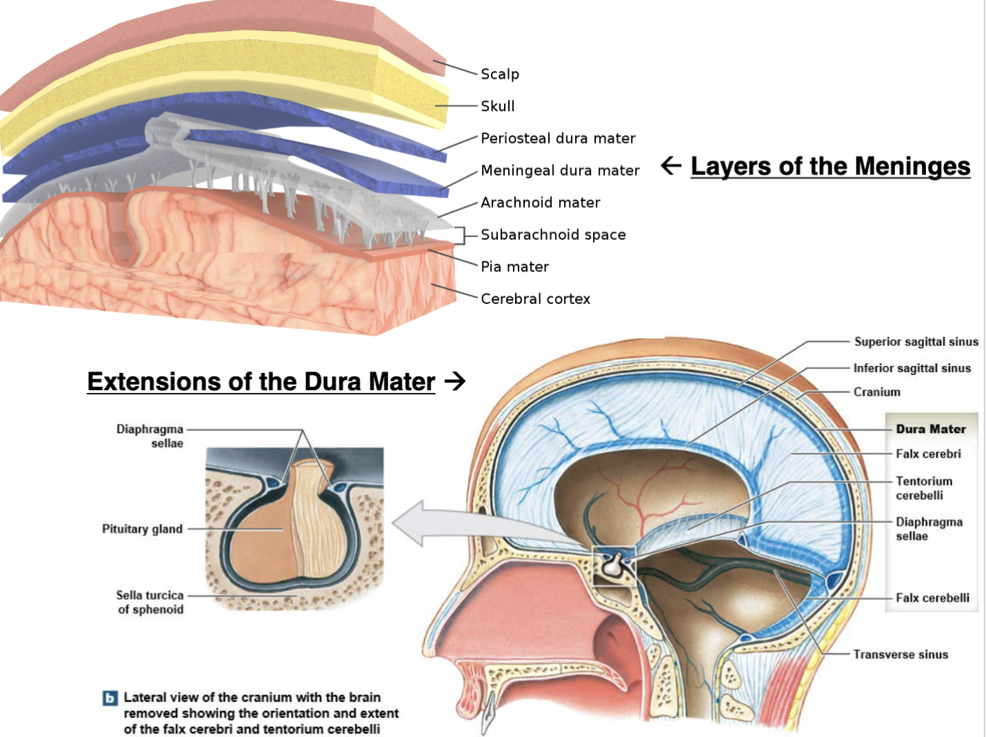
Spinal Meninges***
• Spinal meninges: specialized membranes for protection, physical stability & shock absorption
• continuous w/ cranial meninges
• 3 layers
• dura mater: tough, fibrous outermost layer
• arachnoid mater: middle layer
• pia mater: innermost layer (adjacent to spinal or brain tissue)
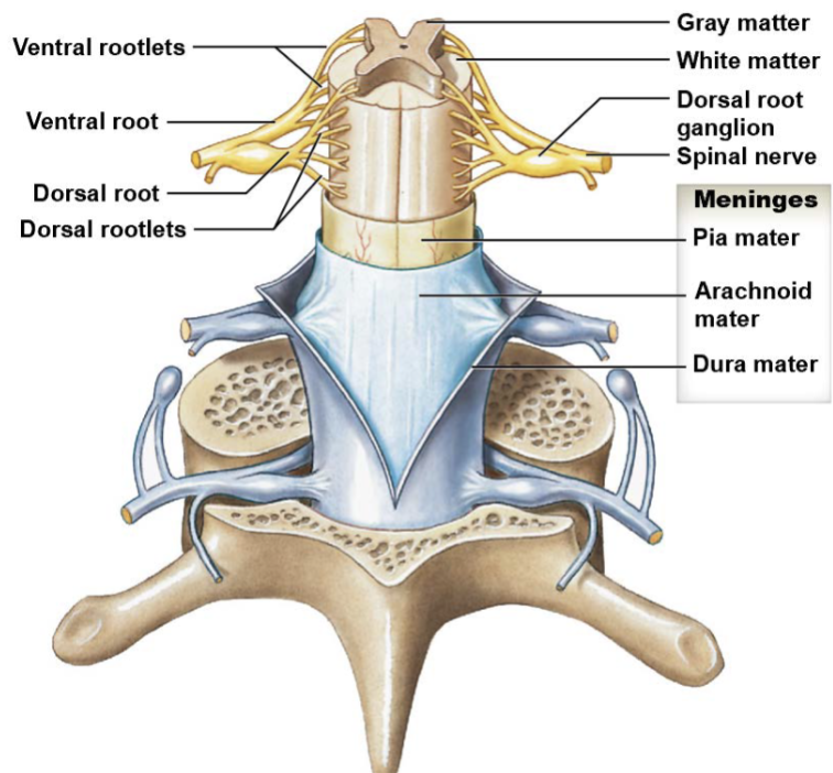
Dura Mater & Arachnoid Mater: Spinal Meninges***
• Dura mater: superficial layer of dense irregular CT
• stabilizes spinal cord w/in vertebral canal
• cranial & sacral attachments stabilize longitudinal axis of spinal cord
• forms a sac from level of foramen magnum to 2 nd sacral vertebra
• fat & CT lie in epidural space bet. dura mater & vertebral canal
• blends w/ filum terminale to form coccygeal ligament
• Arachnoid Mater: avascular middle layer of collagen & elastic fibers
• continuous w/ brain’s arachnoid mater
• separated from pia mater by subarachnoid space
• contains cerebrospinal fluid
• subdural space: bet. dura mater & deeper meninges
• contains interstitial fluid
• only in CADAVERS; not in living people
Pia Mater: Spinal Meninges***
• Pia mater: vascular deepest layer of collagen & elastic fibers
• firmly attached to brain & spinal cord tissue
• denticulate ligaments: membranous extensions of pia mater that attach
pia mater & arachnoid mater to dura mater
Sectional Anatomy of the Spinal Cord
• Gray matter: H-shaped region of neuronal cell
bodies, neuroglia, unmyelinated axons & dendrites
• surrounds a central canal → extends entire length of spinal cord
• central canal continuous w/ 4 th ventricle of brain
• called horns due to their shape
• White matter: consists primarily of myelinated axons
(some unmyelinated)
• axons organized into tracts or columns
• located outside gray matter area
Sectional Anatomy: Organization of Gray Matter
• Cell bodies organized into nuclei (sensory & motor)
• Gray commissures***: connect right & left sides
• consists of axons crossing from 1 side to the other
• Horns
• posterior (dorsal) horns: sensory somatic & visceral nuclei
• lateral (intermediate) horns: visceral motor nuclei
• anterior (ventral) horns: somatic motor nuclei
Spinal Cord Tracts (c15)***
• Gray matter receives & integrates information
• White matter tracts conduct the nerve impulses
• Communication involves sensory & motor tracts
• ascending tract: sensory (delivers info to brain)
• descending tract: motor (delivers info to periphery)
• All tracts involve both brain & spinal cord
Somatosensory Tracts: Spinal Cord Tracts (c15)***
3 major (somato) sensory tracts: ASCENDING tracts
SPINOTHALAMIC (STT): pain, temp, crude touch, deep pressure
POSTERIOR COLUMN: proprioception (body position), 2-point discrimination (fine touch), vibration
• gracile fasciculus (fasciculus gracilis): transmits info to cerebrum from
areas inferior to T6
• cuneate fasciculus (fasciculus cuneatus): transmits info to cerebrum from
areas superior to T6
SPINOCEREBELLAR (SCT): proprioception
Motor Tracts: Spinal Cord Tracts (c15)
2 major motor tracts: DESCENDING tracts
• Pyramidal: originate in cerebral cortex, transmits to spinal cord
• precise voluntary movements
• Extrapyramidal (subconscious motor): originate in brain stem, transmits to spinal cord
• involuntary & automatic control of musculature = skeletal muscle tone,
balance, posture, locomotion, equilibrium
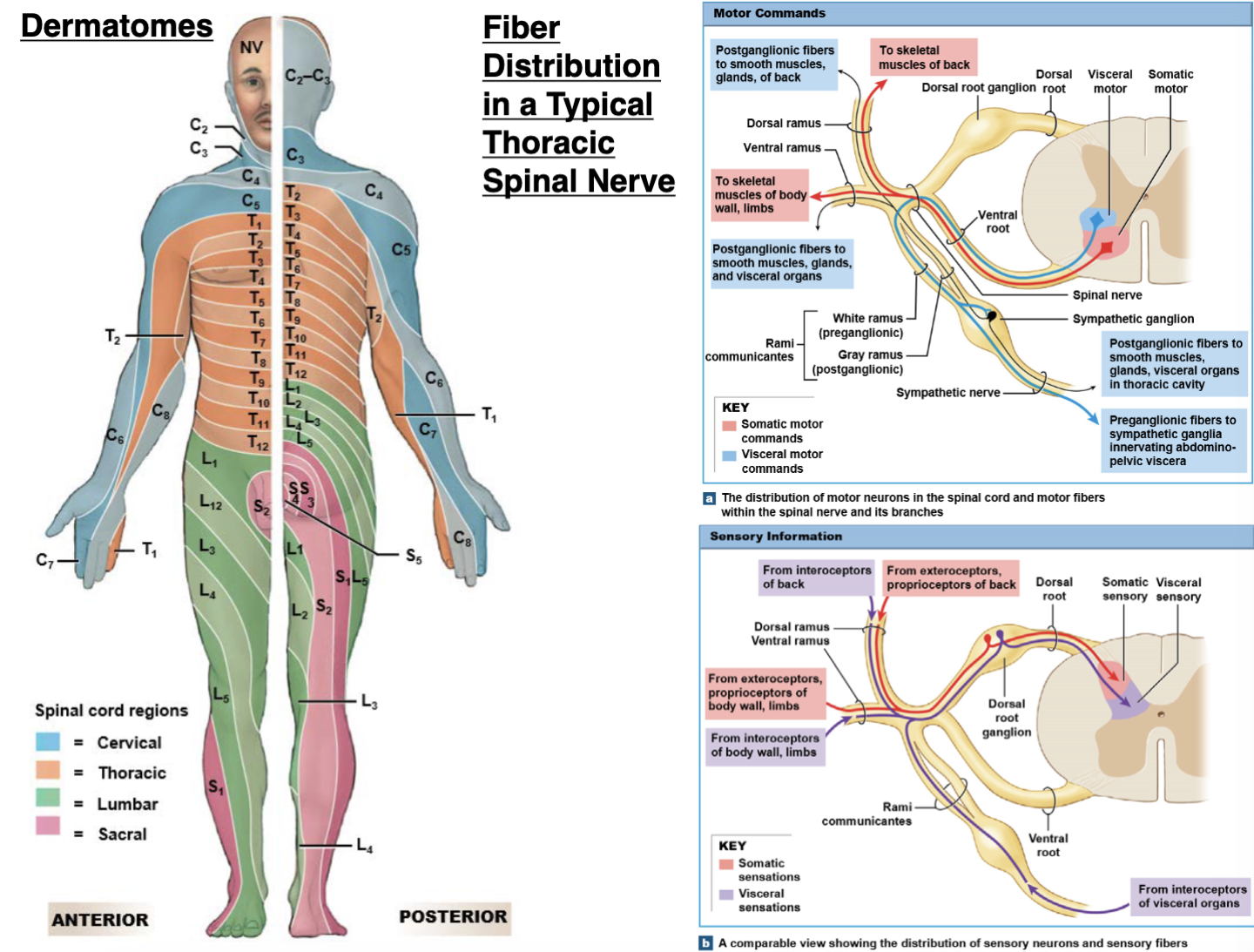
Spinal Nerves: Organization***
• Each peripheral nerve consists of:
• epineurium: outer layer; fuses w/ dura mater as nerve exits through
intervertebral foramen
• perineurium: surrounds each fascicle (bundle of axons)
• endoneurium: layer surrounding each axon
Peripheral Distribution of Spinal Nerves***
• Rami communicantes: 2 additional branches on spinal nerves T 1 → L2 that
carry visceral motor fibers to & from nearby autonomic ganglia
• white ramus communicans
• gray ramus communicans
• associated w/ sympathetic division of autonomic nervous system
• Dermatomes: area of skin that provides sensory input to posterior roots of 1
pair of spinal nerves or to 1 spinal cord segment
• adjacent dermatomes may overlap
Nerve Plexuses
• Nerve plexus: interwoven network of nerves formed from
all adjacent ventral rami (except T2 → T12 )
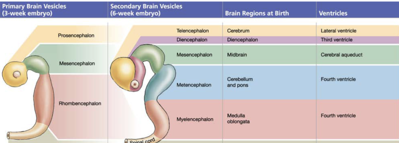
Embryology of the Brain [ch16: The Brain]
• The CNS begins as a hollow neural tube w/ a fluid-filled lumen
(neurocoel)
• Neurocoel expands & forms the brain’s various ventricles
• 4 th wk of dev’t: 3 primary brain vesicles form in the neural tube:
• prosencephalon: forebrain
• mesencephalon: midbrain
• rhombencephalon: hindbrain
• 6 th wk of dev’t: prosencephalon & rhombencephalon subdivide
• prosencephalon
• diencephalon: thalamus & hypothalamus
• telencephalon: cerebrum
• mesencephalon: does not divide
• rhombencephalon
• myelencephalon: medulla oblongata
• metencephalon: pons & cerebellum
Know how to diagram this out***
Cerebrum
conscious
thought & intellectual
functions; memory;
conscious regulation of
skeletal muscle contractions
• paired cerebral
hemispheres separated by
longitudinal fissure
• Contains sulci (grooves) &
gyri (ridges)
Cerebellum
coordinates
somatic motor function;
adjusts output of somatic
motor centers smooth
operation
Midbrain
(midbrain): processes visual & auditory data;
maintains consciousness & alertness; reflexive somatic motor
responses to stimuli
Medulla Oblongata
relays info to thalamus & brainstem;
regulates heart rate, blood pressure & digestion
Pons
relays info to cerebellum & thalamus; regulates somatic
& visceral motor centers
Protection and Support of the Brain: Cranial Meninges***
• Meninges surround brain & spinal cord; 3 layers
• dura mater: most superficial; 2 layers
• thicker, outer periosteal layer –
attached to cranial bones
• thinner, inner meningeal layer
• extensions of dura mater
• falx cerebri: separates cerebral
hemispheres
• falx cerebelli: separates
cerebellar hemispheres
• tentorium cerebelli: separates
cerebellum from cerebrum’s
occipital lobes
• diaphragma sellae: lines sphenoid’s sella turcica
• arachnoid mater: middle layer
• pia mater: deepest layer
Protection and Support of the Brain: Cerebrospinal Fluid***
• Cerebrospinal fluid (CSF): provides support for the brain
• located in subarachnoid space & brain ventricles
• 2 lateral ventricles (1 st & 2nd): cerebrum
• 3rd ventricle: vertical midline slit inferior to thalamus
• 4th ventricle: bet. brainstem & cerebellum
• Functions of CSF
• mechanical protection: floats the brain
• chemical protection: optimal for neuronal signaling
• circulation: medium for exchange
Formation & Circulation: Cerebrospinal Fluid***
• Formation of CSF
• produced by ependymal cells covering the choroid plexus
(capillary networks in ventricle walls)
• Circulation of CSF
• S1: CSF made in choroid plexus of lateral ventricles flows into 3 rd
ventricle via interventricular foramen
• S2: 3 rd ventricle adds CSF → flows through cerebral aqueduct,
passing through midbrain & into 4th ventricle
• S3: 4 th ventricle adds more CSF → flows into subarachnoid space
via 1 median & 2 lateral apertures of 4 th ventricle
• S4: CSF reabsorbed through arachnoid granulations &
superior sagittal sinus into cerebral veins
• Most CSF flows into subarachnoid space w/ some flowing into
spinal cord’s central canal
Rhombencephalon » Myelencephalon = Medulla Oblongata
• Medulla oblongata: continuous w/ spinal cord
• bet. foramen magnum & pons
• pathway for communication: contains all ascending & descending
tracts that relay info bet. brain & spinal cord
• Pyramids in the medulla oblongata
• found on ventral side → contains largest motor tracts from cerebral
cortex → spinal cord
• cross at decussation of pyramids*** (responsible for contralateral control of body)
• fibers end in anterior gray horns & synapse w/ skeletal muscle motor
neurons
• Olives: lateral bulges → control precise voluntary movements
& pressure
• Autonomic nuclei = reflex centers: receive input from cranial
nerves, cerebral cortex, diencephalon & brain stem
• cardiovascular centers (cardiac & vasomotor)
• respiratory rhythmicity centers (rhythmic breathing)
Rhombencephalon » Metencephalon = Pons***
• Pons: prominent bulge superior to
medulla oblongata & anterior to
cerebellum
• consists of nuclei & tracts
• acts as bridge bet. spinal cord &
brain + diff. parts of brain
• nuclei involved w/ involuntary control
of breathing
• pneumotaxic (inhibition) &
apneustic (prolonged inspiration)
centers
Reticular Formation: Brain Stem
• Reticular formation: interconnected nuclei w/in brainstem; extends into spinal cord & diencephalon
• small areas of gray matter separated by white fibers
• SENSORY function
• reticular activating system (RAS)***: relays info to cortex
• responsible for consciousness & awakening
• MOTOR function
• major relay center for muscle tone
Mesencephalon » Midbrain: Brain Stem
• Mesencephalon: bet. pons & diencephalon (hypothalamus)
• Tectum: aka roof; surface posterior to cerebral aqueduct
• contains corpora quadrigemina***: 2 pairs of sensory nuclei responsible for
processing auditory & visual stimuli
• substantia nigra***: regulates motor output of basal nuclei → subconscious
muscle activities → releases dopamine (inhibitory neural transmitter that stops muscle contraction) [dopamine counteracts serotonin (excitatory: contracts muscles)]
Parkinson’s disease: there is no dopamine b/c substantia nigra is destroyed
Prosencephalon » Diencephalon: Introduction***
• The diencephalon consists of:
• epithalamus: superior to thalamus
• contains pineal gland*** → makes melatonin → regulates day/night cycles (and stops gametes from maturing)
• forms roof of diencephalon
• thalamus: paired structures above midbrain; 80% of diencephalon
• hypothalamus: inferior to thalamus
• infundibulum***: connects hypothalamus to pituitary gland
• forms floor & parts of lateral walls of 3rd ventricle
Prosencephalon » Diencephalon
• epithalamus: superior to thalamus
• contains pineal gland → makes melatonin → regulates day/night cycles
• forms roof of diencephalon
• hypothalamus: inferior to thalamus
• infundibulum: connects hypothalamus to pituitary gland
• forms floor & parts of lateral walls of 3rd ventricle
Prosencephalon » Diencephalon: Hypothalamus************************
• Functions of the hypothalamus
• controls & integrates voluntary & autonomic NS activities
• regulates:
• body temperature (pre-optic area)
• food intake/digestive functions,
• waking state & sleep patterns (aka circadian rhythms - suprachiasmatic
nucleus)
• heart rate
• blood pressure
• respiration
• acts as a thirst center
• part of the limbic system: emotions (rage & aggression)
• secretes hormones (ADH - supraoptic nucleus & oxytocin –
paraventricular nucleus)
Rhombencephalon » Metencephalon: Cerebellum
• Cerebellum
• posterior to medulla & pons; inferior to occipital lobe of cerebrum
• separated from cerebrum by tentorium cerebelli
• 2 cerebellar hemispheres
• cerebellar nuclei & cortex: subconscious coordination of movements
• arbor vitae: “tree of life;” branching array of white matter in internal
portion; connects cerebellar cortex & nuclei w/ cerebellar peduncles
Rhombencephalon » Metencephalon: Cerebellum***
• Cerebellum
• coordinates your semantic motor function based on learned patterns of movement
• Functions*
• regulating posture & balance
• programming + fine-tuning of voluntary & involuntary movements
• Information Relay*
• Cerebellum receives sensory input from proprioceptors in muscles,
joints, etc.
• Cerebellum detects variations & sends feedback to motor areas
• has no direct connections w/ skeletal muscles
Prosencephalon » Telencephalon: Cerebrum - Structures & Lobes***
• Cerebrum
• 2 hemispheres separated by longitudinal fissure
• several lobes
• frontal: conscious control of skeletal muscles
• central sulcus: separates frontal from parietal
• precentral gyrus: anterior to central sulcus = primary motor area
• parietal: conscious perception of touch, pressure, vibration, pain, temperature & taste
• postcentral gyrus: primary somatosensory area
• parietooccipital sulcus: separates parietal from occipital
• temporal: conscious perception of auditory & olfactory stimuli
• lateral cerebral sulcus: separates temporal from frontal
• insula: deep to temporal lobe; “island” of cortex
• occipital: perception of visual stimuli
• transverse fissure: separates cerebrum from cerebellum
• gyri: elevated ridges
• sulci: depressed grooves
• corpus callosum: allows left & right hemispheres to communicate w/
each other
Telencephalon: Cerebrum - Gray & White Matter***
• cerebral cortex: outer gray
matter
• cerebral white matter:
myelinated axons extending
in 3 directions
• association fibers: connect
cortical areas w/in same
hemisphere
• commissural fibers:
connect corresponding
lobes of diff. hemispheres
(crosses over)
• corpus callosum
• anterior commissure
• posterior commissure
• projection fibers: form
descending & ascending
tracts (internal capsule)
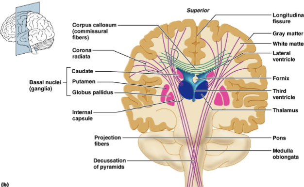
Telencephalon: Cerebrum - Basal Nuclei ***
• Basal nuclei: groups of nuclei in each hemisphere
• corpus striatum: major input site for basal ganglia
• caudate nucleus: controls large automatic movements of skeletal muscles
• lentiform nuclei
• putamen: lateral; same function as caudate nucleus
• globus pallidus: medial; regulation of muscle tone
• claustrum: subconscious processing of visual info
• amygdaloid body: limbic system
• Functions
• subconscious control & integration of skeletal muscle tone
• coordination of learned movement patterns
• processing, integration & relay of info from cerebral cortex
Telencephalon: Cerebrum - Limbic System***
• Limbic system: encircles brain stem on inner border of cerebrum &
floor of diencephalon, superior to corpus callosum
• Functions
• establishes emotional states
• memory storage & retrieval
• links conscious functions w/ unconscious autonomic functions
Telencephalon: Cerebral Cortex Sensory Areas: ONE OF MATCHING QUESTIONS************ worth 10% of test
• Sensory areas: primary vs. secondary & association
• primary areas: have most direct connections w/ peripheral sensory
receptors
• secondary & association areas: receive input from primary areas &
participate in interpretation of sensory info
• Primary somatosensory: postcentral gyrus (parietal lobe)
• nerve impulses for touch, pain, temperature & proprioception
• localize exact points of body where sensations originate
• Primary visual: occipital lobe
• impulses from eye receptors sent to thalamus and then to visual cortex
• Primary auditory: temporal lobe
• basic characteristics of sound (e.g. pitch & rhythm)
• Primary olfactory: temporal lobe
• interprets smell
• Primary gustatory: parietal lobe
• interprets taste
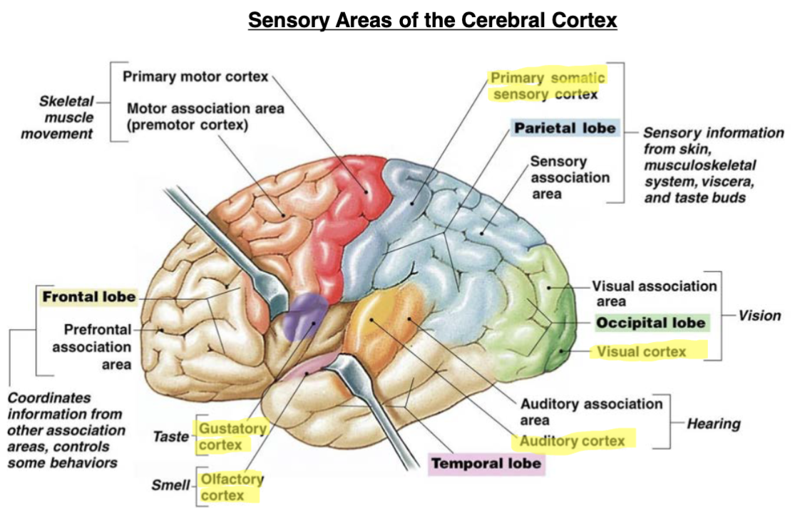
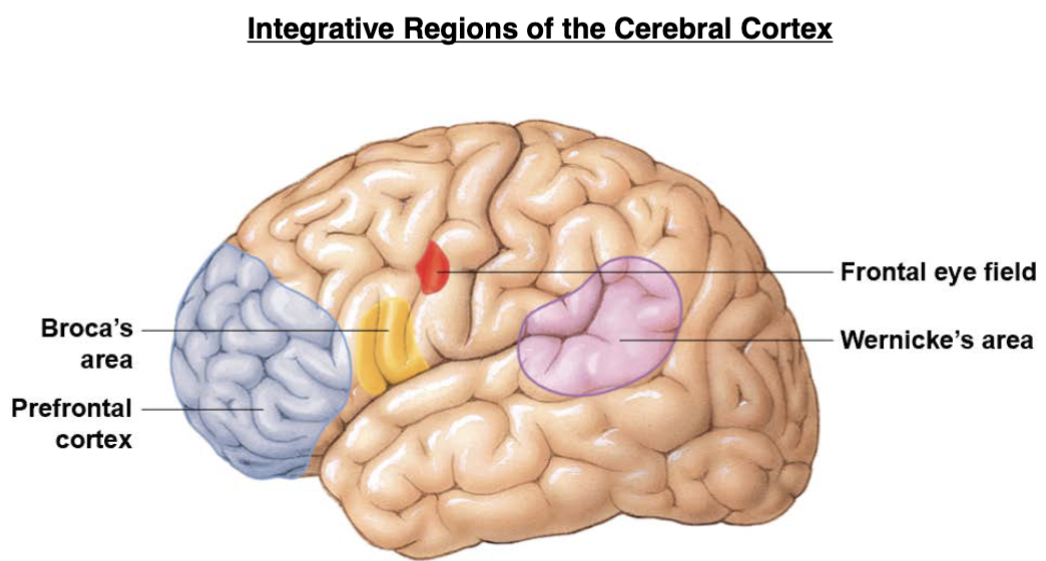
Telencephalon: Cerebral Cortex Motor & Language Areas****************
• Primary motor cortex: precentral gyrus (frontal lobe)
• voluntary contractions of skeletal muscles
• Language areas: associated w/ left cerebral hemisphere
• Broca’s area: aka speech center = motor speech area
• regulates patterns of breathing & vocalization for normal speech
• nerve impulses from Broca’s area sent to premotor regions → control
muscles of larynx, pharynx & mouth
• impulses to primary motor area regulate breathing
• Wernicke’s area: analytical center = language comprehension
• inferior to auditory cortex in temporal lobe
• interprets sound as speech, music, or tone
• translates words/sounds into thoughts
• plays role in personality → integrates sensory info & coordinates access to
visual & auditory memories
auditory association area***
Telencephalon: Association Areas***
• Association areas: associated w/ integrating & understanding sensory or motor info (cause and effect, decision making)
• Prefrontal association area (frontal lobe)
• integrates info from sensory association areas & predicts consequences
of possible responses
• Somatosensory association area (posterior to primary
somatosensory area)
• processes input from thalamus, inferior portions of brain & primary
somatosensory area
• integrates & interprets sensations (inc. storage of memories for
comparison w/ new ones, tactile interpretation of objects → size, form &
texture)
• Visual association area (occipital lobe)
• receives input from primary visual cortex & thalamus
• relates past visual experiences w/ present for evaluation
Telencephalon: Association Areas***
• Premotor cortex (immediately anterior to primary motor cortex)
• uses memories of learned movement patterns to coordinate complex &
sequential motor activities
• complicated learning & reasoning functions
• Auditory association area (within Wernicke’s area)
• discerns auditory cues as sounds, speech, or music
• Gnostic area
• common integrative area → among somatosensory, visual & auditory
association areas
• integrates many sensory inputs into a common thought
• transmits results to appropriate effectors
• Frontal eye field (frontal cortex)
• controls voluntary scanning eye movements
Telencephalon: Cerebral Higher-Order Functions***
• Higher-order functions
• performed by cerebral cortex
• involves communication bet. cerebral cortex & other areas of brain
• requires conscious & unconscious info processing
• subject to modifications & adjustments
• Integrative centers of cerebral cortex
• integrate complex sensory stimuli & motor responses
• prefrontal cortex
• general interpretive area
• Broca’s area
• Wernicke’s area
• Hemispheric lateralization: each hemisphere has different functions
• left: speech center, writing, language, mathematics
• right: analysis by touch, spatial visualization
Integrative Regions of the Cerebral Cortex***
The Cranial Nerves and Functions MATCHING QUESTION 12 POINTS*************
don’t need to know the sensory/andor motor
know the red box
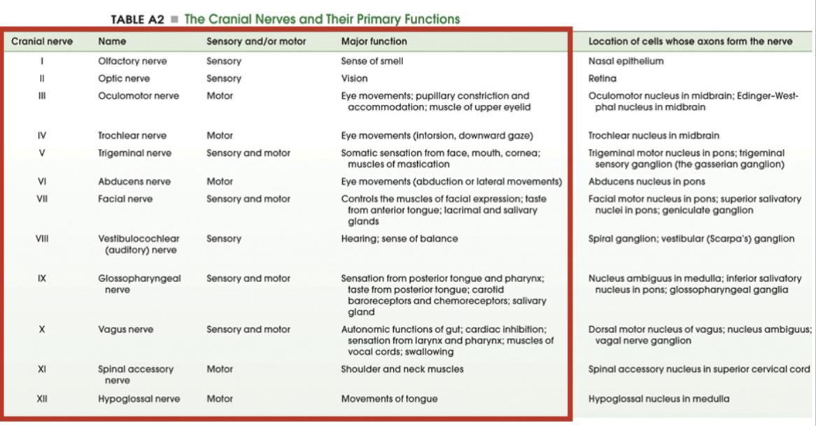
Cranial Nerves
• 12 pairs (I-XII) of cranial nerves
• innervate the periphery emerging from the brain (not the spinal cord)
• located on brain’s ventrolateral surface
• numbered beginning at anterior aspect of brain
put on card*