lecture 56: urinary incontinence and pelvic relaxation
1/65
There's no tags or description
Looks like no tags are added yet.
Name | Mastery | Learn | Test | Matching | Spaced | Call with Kai |
|---|
No study sessions yet.
66 Terms
95%
___% of children achieve urinary continence by 5
•achieved some degree of continence prior to incontinence
•consider stress of psychological or physical abuse
define secondary urinary incontinence
nocturnal enuresis
the most common cause of functional urinary incontinence in children
-genetic risk (chromosome 12 and 13q)
-attention deficit disorders
-small bladder, nocturnal polyuria, nocturnal detrusor over-reactivity, disorder of sleep or arousal states
risk factors of nocturnal enuresis
•Comorbid constipation is common and effective treatment is integral to management of incontinence
comorbid condition of daytime incontinence
reassurance and fluid restriction before consideration of treatment
evaluation of urinary incontinence in children under 5
urine dipstick
screening tool for infection, kidney disease ,and glucosuria
serum electrolytes, calcium, glucose and creatinine
screenings for polyuria
stress incontinence
•involuntary leakage of urine that occurs when intra-abdominal/intra-vesical pressure exceeds urethral pressure
urge incontinence
•involuntary leakage that occurs due to involuntary contractions of the bladder, uninhibited detrusor contractions
•occurs when both stress incontinence and urge incontinence are present
define mixed incontinence
overflow incontinence
occurs when the bladder is unable to contract thus overfills and overspills past urethra, ineffective detrusor muscle
vaginitis, UTI
infectious ddx of urinary incontinence
•Stress incontinence
•Detrusor overactivity
•Mixed types
•Overflow incontinence
ddx of filling and storage disorders
urethral diverticulum
-diverticulum collects small volumes of urine
-distal to sphincter--> intermittent leaking without trigger of small volumes
•Typically, post obstetric trauma, surgery or radiation
a fistula causing incontinence is usually secondary to
fistula
•Urine not leaking out of urethra meatus, leaking into alternate space and making its way to outside
urethral diverticulum
intermittent leaking without triggering of small volumes
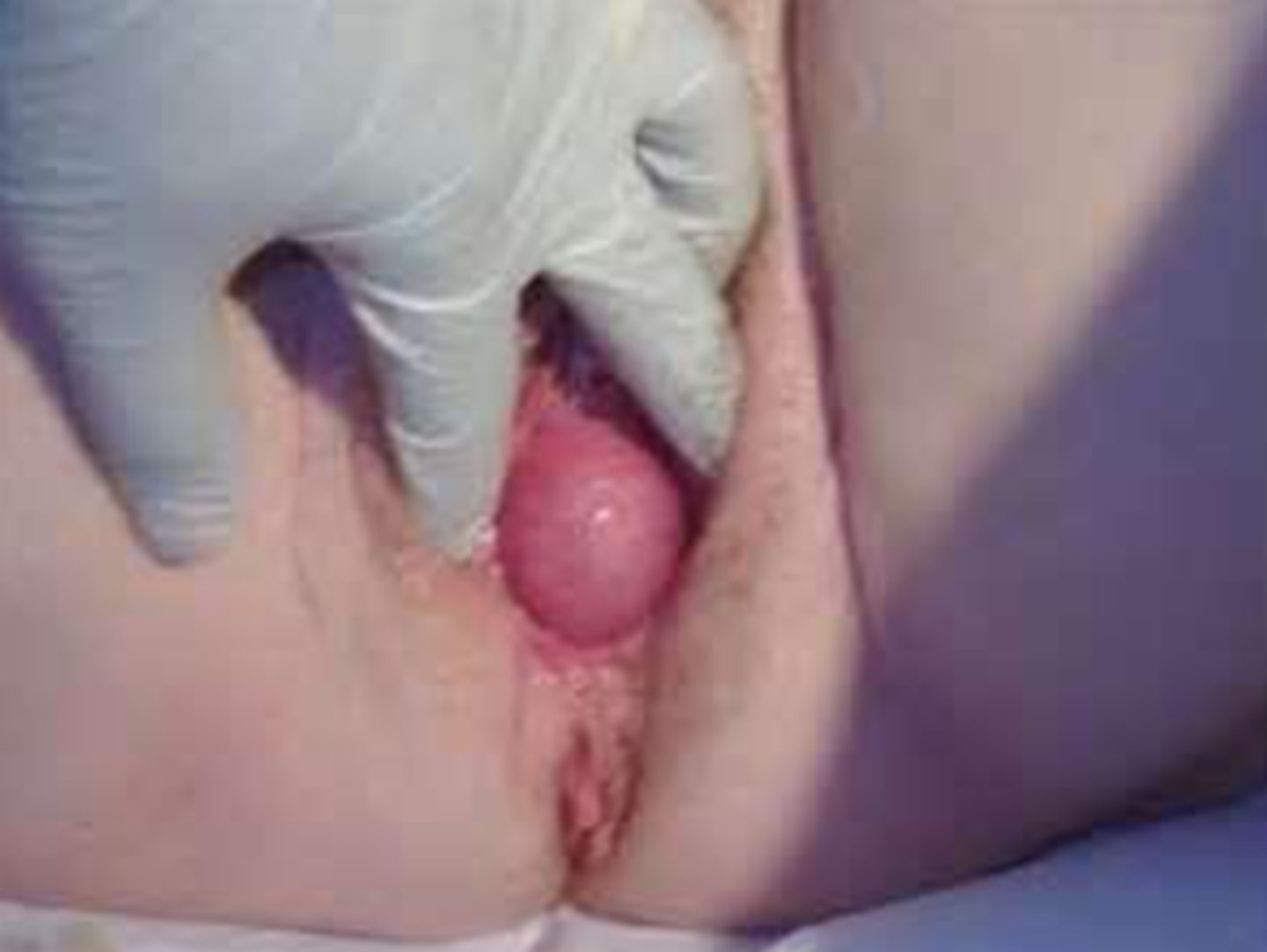
cystocele
vagina weakness of anterior fascia--> bulging of anterior vaginal wall
•As enlarges cystocele may kink bladder neck and result in obstruction of urine outflow and present as urinary retention
later complication of cystocele
anal wink and bulbocavernosus reflexes to assess sacral reflex pathway
how do you assess motor and sensory function of pelvis/urethra
urinalysis-- all should be screened for UTI
screening test for ALL PTS with complaints of incontinence
stress test
ask patient to Valsalva and observe for urine leakage, preferably standing
after patient empties bladder, measure remaining volume by catheter drainage or ultrasound calculation
how do you do a post-void residual to evaluate for urinary incontinence in-office
post void residual
in office procedure that only documents partial emptying, not incontinence
normal is less than 35 ml
->100 ml should be investigated
normal value of post void residual
urinary diary
1.documents triggers for incontinence, bladder irritants
Lubricated Q-tip is placed in the urethral meatus. With Valsalva, in healthy woman little movement occurs and no leakage of urine. In woman with stress incontinence, with Valsalva, the visible Q-tip end will move up toward the ceiling and a positive test is defined by movement of at least 30 degrees. Often urine loss is observed, but not required to have a positive test.
how do you perform the Q tip test
visible Q-tip end will move up toward the ceiling and a positive test is defined by movement of at least 30 degrees.
define a positive Q tip test
activieis (runninng, jumpping laughing)
-obesity--> atrophy of bladder tissues, intrinsic weakness of pelvic support
most common etiologies of stress incontinence
urinary stress incontinence
incontinence that is primarily an anatomic problem--> loss of support of the urethral vesical junctionn
-associated with anterior vaginal wall prolapse
•With valsalva the urethra is displaced downward thus decreasing intraurethral pressure
•When pressure in bladder exceeds that in urethra, leakage occurs because sphincter can not hold urine back
anatomy of stress incontinence
•Age
•Multiparity- previous vaginal deliveries or vaginal trauma
•Body weight
•Previous pelvic surgery ex. Hysterectomy
•Smokers
•Constant straining
risk factors for stress incontinence
urge incontinence
•Occurs when bladder becomes unstable and has contractions that are not controlled, small continual contractions of detrusor.
urge incontinence
-no bladder inhibition, there is a large volume of leaking, complete emptying
stress incontinence
•Results in small spurts of urine loss and incomplete emptying
urge incontinence
pt usually has intense urge to void then leaks prior to making it to the restroom
-usually associated with frequency, worse at night
-voiding diary very helpful in diagnosis
urge incontinence
most common incontinence in men
mixed incontinence
urodynamic testing is very helpful to assess for
urodynamic testing
•Evaluation of urine storage, bladder emptying and sphincter control mechanisms
•Multichannel urethral pressure profiles demonstrate urethral leak point
•Measure bladder capacity, volume at first urge, volume at incontinence
•Demonstrates repetitive detrusor muscle contraction in urge incontinence
overflow incontinence
-incomplete bladder emptying due to detrusor weakness or obstruction
-usually neuro issue with no bladder contraction
-no perception of bladder fullness
overflow incontinence
•Leaks when bladder pressure is higher than urethral pressure
•Continuous small amount of leaking – few, or no triggers to incontinence
-weight loss minimum 8%
-pelvic floor training/kegel exercises
-fluid management: max 2 L per day
-vaginal pessarrries
-bulking agents
main treatments for stress incontinence
vaginal pessaries
-provides mechanical support to weakened tissues in tx of stress incontinence
-fitted to individual pt
•vaginal trauma, vaginitis, urinary retention, retention
of foreign body, abrasions, adhesions
risks of vaginal pessaries
bulking agents
pyrolytic carbon-coated beads and calcium hydroxylapatite
• injected periurethrally or transurethrally
•less invasive but less effective than surgery
-tx of stress incontinence
-anticholinergics
-antimuscarinnics: act on bladder M2 and M3 receptors to inhibit involuntary detrusor contractions
-onabotulinumtoxin A: inhibit Ach
Rx tx for urge incontinence
bladder traininng
neurostimulation using tibial nnerve
non pharm tx for urge incontinence
•Bladder retraining
•Self cath
•Will not respond to incontinence surgery, prolapse surgery may be indicated
mannagement of overflow incontinence
•Primarily a break or tear in connective tissue and endopelvic fascia which results in loss of support of vaginal wall and organs
most common cause of pelvic organ prolapse
•Age
•Multiparity- previous vaginal deliveries or vaginal trauma
•Large infant birth weights
•Body weight
•Previous pelvic surgery ex. Hysterectomy
•Smokers
•Constant straining
•Connective tissue disease
•Family history
risk factors for pelvic prolapse
cystocele (bladder)
anterior vaginal wall prolapse
enterocele (small bowel)
apical vaginal wall prolapse
rectocele
posterior wall prolapse
Vaginal bulge, pressure, incontinence, prolonged urination, partial emptying, frequency, nocturia
symptoms of cystocele
•Some women report “splinting” – compressing vaginal wall to accomplish rectal emptying
•Constipation, rectal fullness, increases straining and promotes circular pattern
• Also produces pressure on the bladder
symptoms of rectocele
•support of cardinal and uterosacral ligaments, lengthening these ligaments
uterine prolapse is due to a defect in
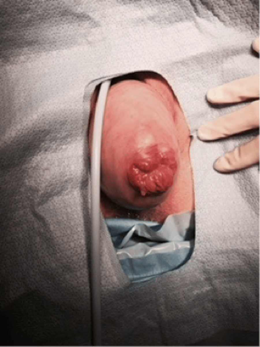
procedentia
complete prolapse of cervix or uterus below the vestibule
1st degree- noticeable defect of support
2nd degree- half of vaginal wall involved
3rd degree- prolapse to vaginal opening
4th degree- prolapse beyond vestibule
what are the degrees of pelvic organ prolapse?
•Untreated risk includes urinary retention, UTI, hydroureter, obstruction.
risks of not treating pelvic organ prolapse
1.Pelvic floor exercises for the mild cases
2.Changes in activities (i.e. lifting and straining). Treat constipation.
3.Pessaries- requires adequate levator muscle tone
non surgical treatments for pelvic organ prolapse
colporraphy
plication of connective tissue – tying together colpometrium, vaginal incision, recurrence rates high, augmentation with mesh or animal graft
-surgical treatment of pelvic organ prolapse
At time of hysterectomy, augment support to vaginal apex
how do you prevent pelvic organ prolapse in hysterectomy?
NE through B receptors
NT that inhibits detrusor muscle contraction
alpha receptors
receptor that stimulates internal sphincter contraction
spinobulbospinal reflex
Micturition is fundamentally a ___________ facilitated and inhibited by higher brain centers such as the pontine micturition center and, like defecation, subject to voluntary facilitation and inhibition.