2. Heart + Vessel Anatomy
5.0(1)
Card Sorting
1/40
Earn XP
Description and Tags
Study Analytics
Name | Mastery | Learn | Test | Matching | Spaced |
|---|
No study sessions yet.
41 Terms
1
New cards
Heart location + bordering structures
* Location: thorax (middle mediastinum)
* Bordered by:
* Lungs (laterally)
* Diaphragm (inferiorly)
* Sternum (anteriorly)
* Great vessels (superiorly)
* Esophagus (posteriorly)
* Bordered by:
* Lungs (laterally)
* Diaphragm (inferiorly)
* Sternum (anteriorly)
* Great vessels (superiorly)
* Esophagus (posteriorly)
2
New cards
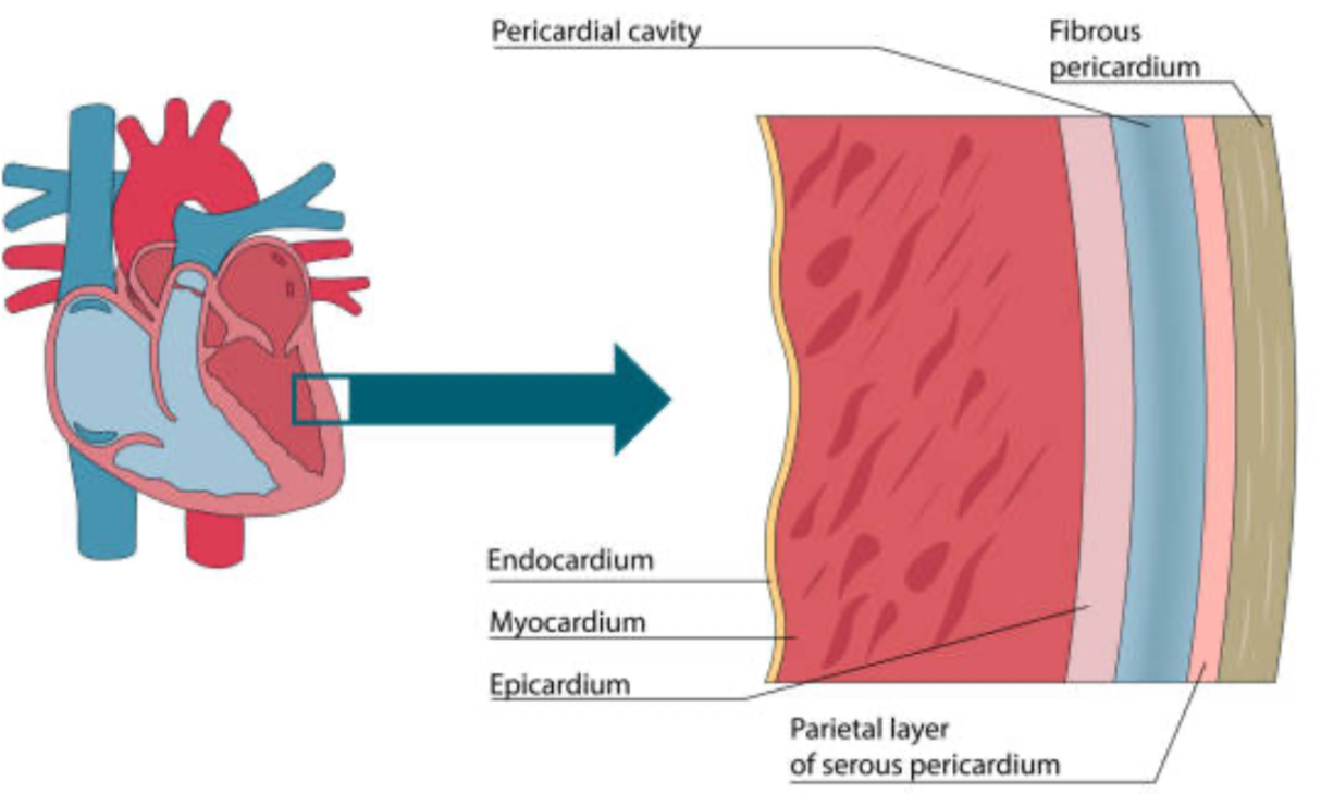
Layers of the heart
1. Pericardium (outer membrane enclosing heart)
2. Myocardium (heart muscle)
3. Endocardium (inner layer, wall of heart chambers)
3
New cards
Layers of the pericardium
1. Fibrous layer
2. Parietal layer
3. Space/cavity
4. Visceral layer (epicardium)
4
New cards
Chambers of the heart
1. Right atrium
2. Right ventricle
3. Left atrium
4. Left ventricle
5
New cards
Flow of blood through heart
1. Superior + inferior vena cava
2. Right atrium
3. Tricuspid valve
4. Right ventricle
5. Pulmonary valve
6. Pulmonary arteries
7. **Lungs**
8. Pulmonary veins
9. Left atrium
10. Mitral/bicuspid valve
11. Left ventricle
12. Aortic valve
13. Aorta
14. **Body**
6
New cards
Function of valves in heart
Prevent back flow
7
New cards
Heart valves
AV valves:
1. Tricuspid valve
2. Mitral/bicuspid valve
Semilunar valves:
1. Pulmonary valve
2. Aortic valve
1. Tricuspid valve
2. Mitral/bicuspid valve
Semilunar valves:
1. Pulmonary valve
2. Aortic valve
8
New cards
Auscultating the four valves (remember the pneumonic)
@@A@@ll Pigs **E**at %%T%%oo ^^M^^uch
* @@Aortic@@: __2nd__ intercostal space; right sternal border
* Pulmonic: __2nd__ intercostal space; left sternal border
* **Erb’s point**: __3rd__ intercostal space; left sternal border
* %%Tricuspid%%: __4th__ intercostal space; left sternal border
* ^^Mitral^^: __5th__ intercostal space; left midclavical line
9
New cards
What is Erb’s point good for?
Listening to the entire heart
10
New cards
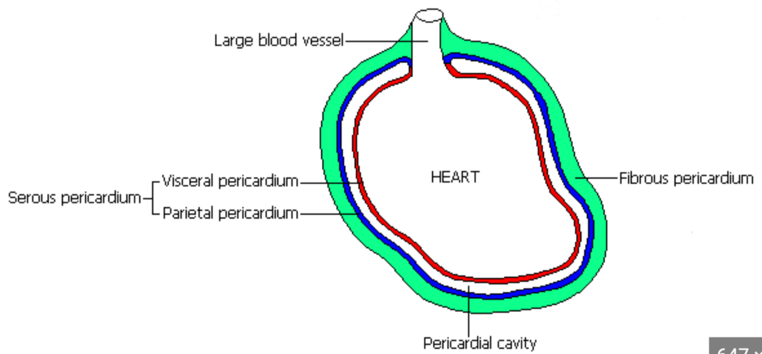
Heart wall
* Heart is surrounded by tough membrane: __fibrous pericardium__
* Inside fibrous pericardium is __serous (fluid) pericardium__ (protection and insulation)
* __Parietal layer__ of serous pericardium lines fibrous pericardium
* __Visceral layer__ is fused to heart surface
* Potential cavity between layers called __pericardial cavity__
* Inside fibrous pericardium is __serous (fluid) pericardium__ (protection and insulation)
* __Parietal layer__ of serous pericardium lines fibrous pericardium
* __Visceral layer__ is fused to heart surface
* Potential cavity between layers called __pericardial cavity__
11
New cards
Epicardium
* Outer layer of heart wall (innermost layer of pericardium)
* Same as visceral pericardium
* Same as visceral pericardium

12
New cards
Myocardium
* Middle layer
* Made of cardiac muscle
* Made of cardiac muscle

13
New cards
Endocardium
**Epithelium** that lines the heart
14
New cards
Are atria or ventricles larger?
Ventricles
* Require more muscular force
* Require more muscular force
15
New cards
Why are left and righthand chambers of the heart separated?
So there is no mixing of blood from one side to the other
16
New cards
Interatrial septum
Wall that separates the two atria
17
New cards
Interventricular septum
Wall between the ventricles
18
New cards
Are the atrial or ventricular walls thinner?
Atrial walls are thinner
* Need to create less force than ventricular walls do
* Need to create less force than ventricular walls do
19
New cards
Do atria or ventricles generate higher pressure?
Ventricles
20
New cards
Are the walls of the left ventricle or right ventricle thicker? Why?
* Left ventricle walls = thicker
* Right ventricle only pumps blood to lungs
* Left ventricle pumps blood throughout entire body
* Right ventricle only pumps blood to lungs
* Left ventricle pumps blood throughout entire body
21
New cards
How does blood get to the right atrium?
2 veins:
1. Superior vena cava
1. Blood from head, neck, chest, upper extremities
2. Inferior vena cava
1. Blood from trunk, organs, abdomen, pelvic region, and lower extremities
1. Superior vena cava
1. Blood from head, neck, chest, upper extremities
2. Inferior vena cava
1. Blood from trunk, organs, abdomen, pelvic region, and lower extremities
22
New cards
Pulmonary veins
Bring blood back to left atrium (from lungs)
23
New cards
Pulmonary trunk
Carries blood from right ventricle to the lungs
24
New cards
Aorta
Carries blood from left ventricle to the body
25
New cards
Atrioventricular (AV) valves (location, names)
* Between each atrium and ventricle
* Right side: tricuspid valve
* Left side: mitral/bicuspid valve
* Right side: tricuspid valve
* Left side: mitral/bicuspid valve
26
New cards
Semilunar valves (location, names)
* Between the ventricles and large arteries that carry blood away from heart
* Right: pulmonary valve
* Left: aortic valve
* Right: pulmonary valve
* Left: aortic valve
27
New cards
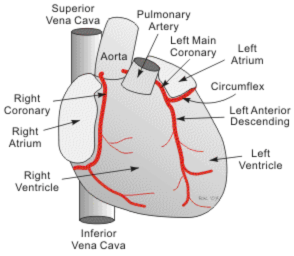
Coronary vessels (location, function, main arteries)
* Location: arise just above aortic valve
* Function: provide oxygenated blood to the heart muscle
* Two main arteries:
* Right coronary artery (RCA)
* Left coronary artery (LCA)
* Left anterior descending (LAD)
* Left circumflex (LCX)
* Function: provide oxygenated blood to the heart muscle
* Two main arteries:
* Right coronary artery (RCA)
* Left coronary artery (LCA)
* Left anterior descending (LAD)
* Left circumflex (LCX)
28
New cards
Right Coronary Artery (RCA)
Supplies:
1. Right atrium
2. Right ventricle
3. Inferior wall of left ventricle
1. Right atrium
2. Right ventricle
3. Inferior wall of left ventricle

29
New cards
Left Anterior Descending (LAD)
Supplies:
1. Anterior wall of left ventricle
1. Anterior wall of left ventricle
30
New cards
Left Circumflex (LCX)
Supplies
1. Left atrium
2. Lateral and posterior walls of left ventricle
1. Left atrium
2. Lateral and posterior walls of left ventricle

31
New cards
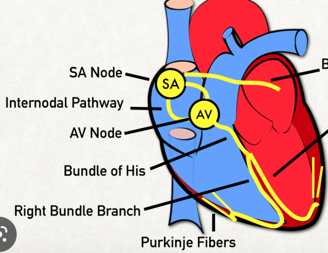
Coronary circulation for SA node
* RCA (55% of population)
* LCX (45% of population)
* LCX (45% of population)

32
New cards
Coronary circulation for AV node
* RCA (90% of population)
* LCX (10% of population)
* LCX (10% of population)
33
New cards
Coronary veins
Parallel arterial supply
34
New cards
Describe how the tricuspid valve works
* When the right ventricle is full of blood, the ventricle contracts
* The one-way tricuspid valve shuts as right ventricular pressure increases so blood doesn’t squirt back into right atrium
* The one-way tricuspid valve shuts as right ventricular pressure increases so blood doesn’t squirt back into right atrium
35
New cards
Describe how the mitral and aortic valves work to pump blood into aorta
* Ventricular pressure increases
* Mitral valve is forced shut
* Blood ejected out of left ventricle through aortic valve to ascending aorta to body
* Mitral valve is forced shut
* Blood ejected out of left ventricle through aortic valve to ascending aorta to body
36
New cards
Heart innervation
* Supplied by **autonomic nerve fibers from cardiac plexus**
* Sympathetic and parasympathetic fibers
* Visceral fibers
* Sympathetic and parasympathetic fibers
* Visceral fibers
37
New cards
Visceral fibers
For pain and reflexive information
38
New cards
Heart innervation: sympathetic supply
* From __superior 5 or 6 thoracic spinal nerves__
* Increases rate, conduction, force of contraction, and blood flow through coronary arteries
* Vasodilation
* Increases rate, conduction, force of contraction, and blood flow through coronary arteries
* Vasodilation
39
New cards
Heart innervation: parasympathetic supply
* From __vagus nerve (CN 10)__
* Vasoconstriction
* Vasoconstriction
40
New cards
Sympathetic effects on heart (innervation, hormone, effects: HR, AP conduction, contraction force)
Innervation:
* __Sympathetic neurons__ innervate entire heart
* Release __norepinephrine__
Effects:
* Inc. HR (positive chronaotropic effect)
* Inc. contraction force (positive inotropic effect)
* Inc. rate of AP (action potential) conduction (positive dromotropic effect)
* __Sympathetic neurons__ innervate entire heart
* Release __norepinephrine__
Effects:
* Inc. HR (positive chronaotropic effect)
* Inc. contraction force (positive inotropic effect)
* Inc. rate of AP (action potential) conduction (positive dromotropic effect)
41
New cards
Parasympathetic effects on heart (innervation, hormone, effects: HR, AP conduction, contraction force)
Innervation:
* __Vagus nerve (CN 10)__ innervates atria (does not really effect ventricles)
* Releases __acetylcholine__
Effects:
* Dec. HR (negative chronaotropic effect)
* Dec. rate of AP (action potential) conduction (negative dromotropic effect)
* No real effect on contraction force because ventricles are not innervated