MICROBIO: EUKARYOTES
1/22
Earn XP
Description and Tags
Identify the specimen
Name | Mastery | Learn | Test | Matching | Spaced |
|---|
No study sessions yet.
23 Terms
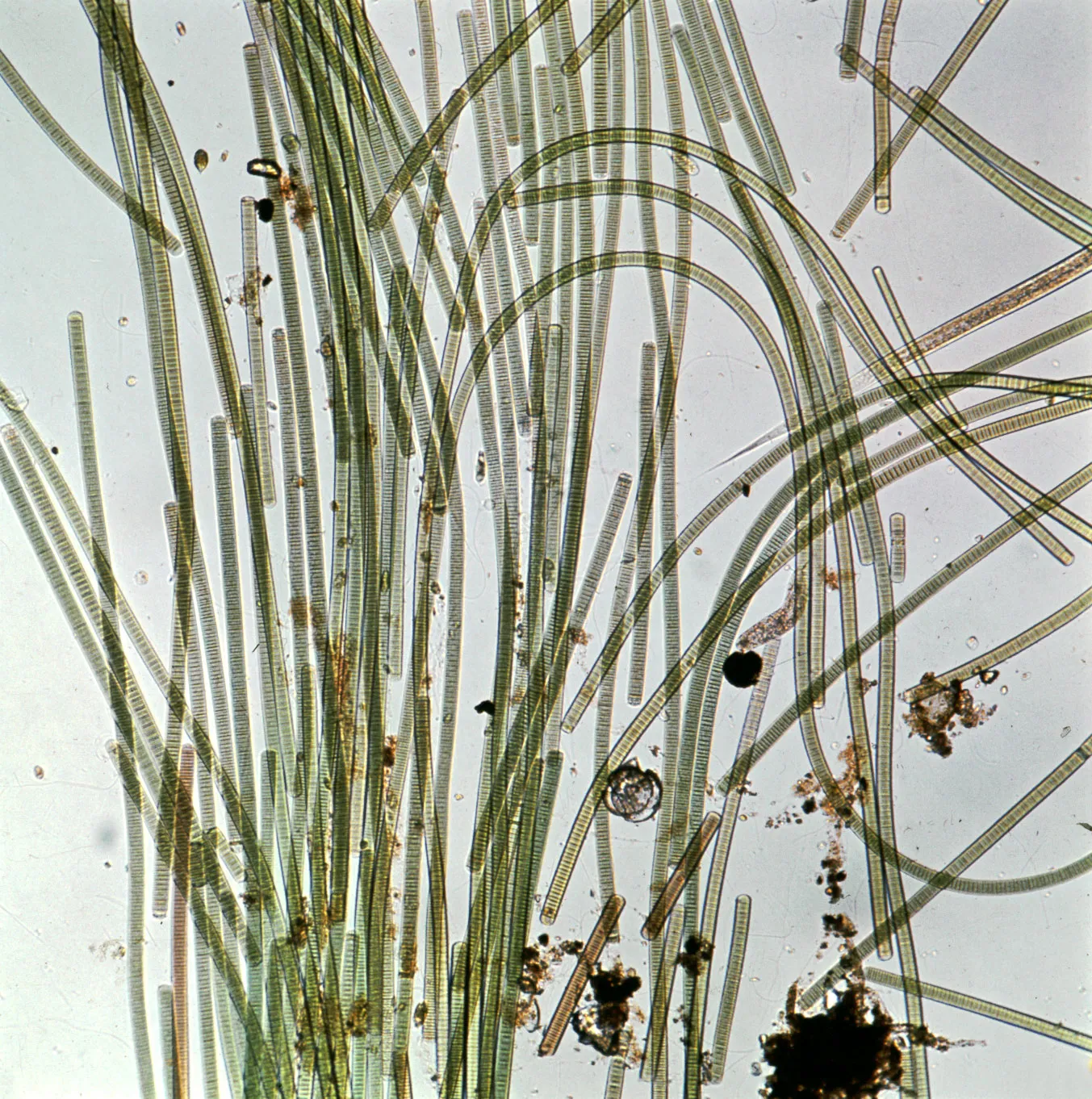
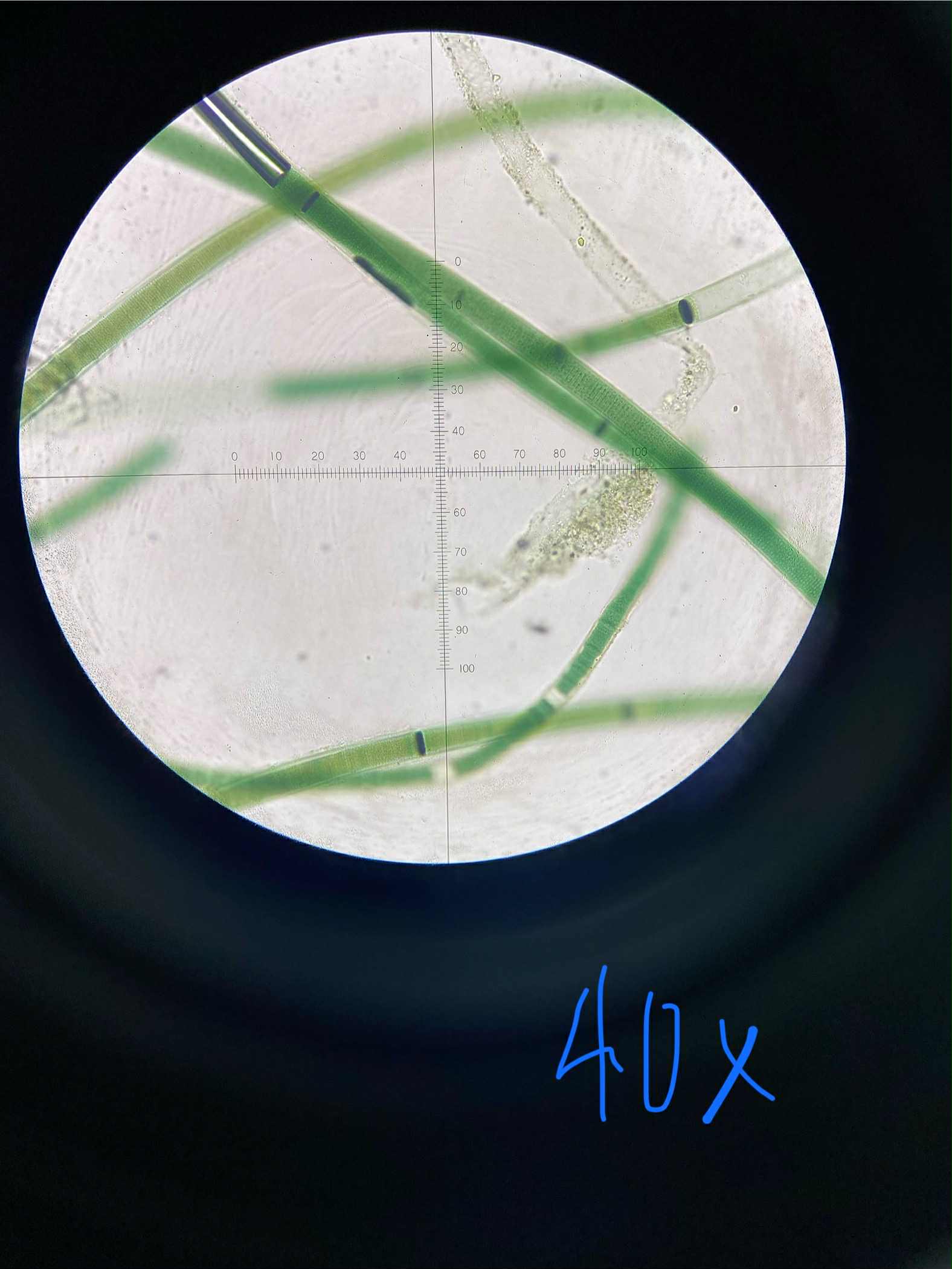
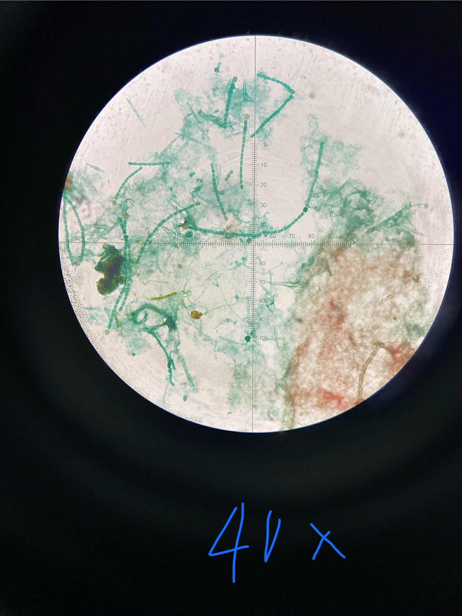
Oscillatoria
cyanobacteria
blue-green
filamentous; unbranched
cylindrical cells arranged in a single row
no flagella; but gliding motility
distinctive oscillating/waving movement; nitrogen fixation
specimen
phylum
color
shape
cell arrangement
motility
characteristic
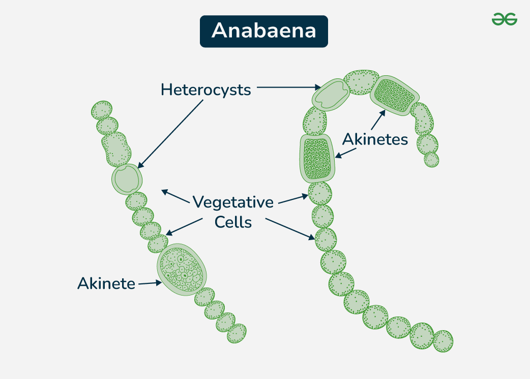
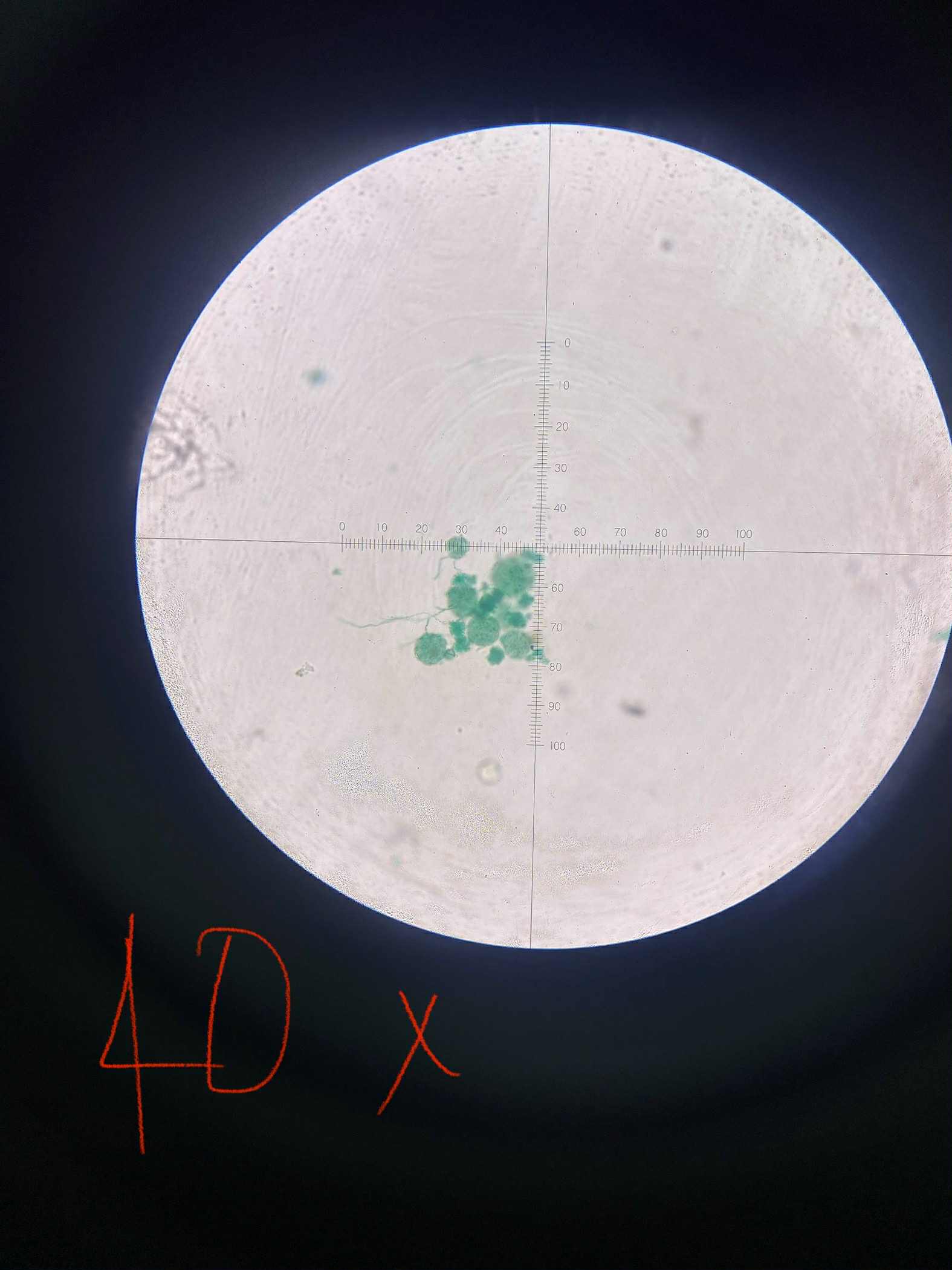
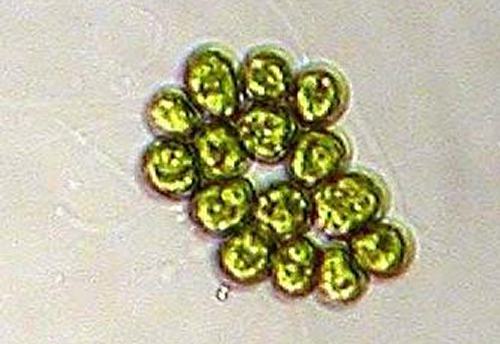
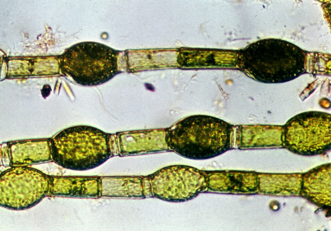
Anabaena
cyanobacteria
blue-green
filamentous
cells in a single row resembling a string of beads
no flagella; gliding movement from slime secretion
presence of heterocysts for nitrogen fixation and akinetes
specimen
phylum
color
shape
cell arrangement
motility
characteristic
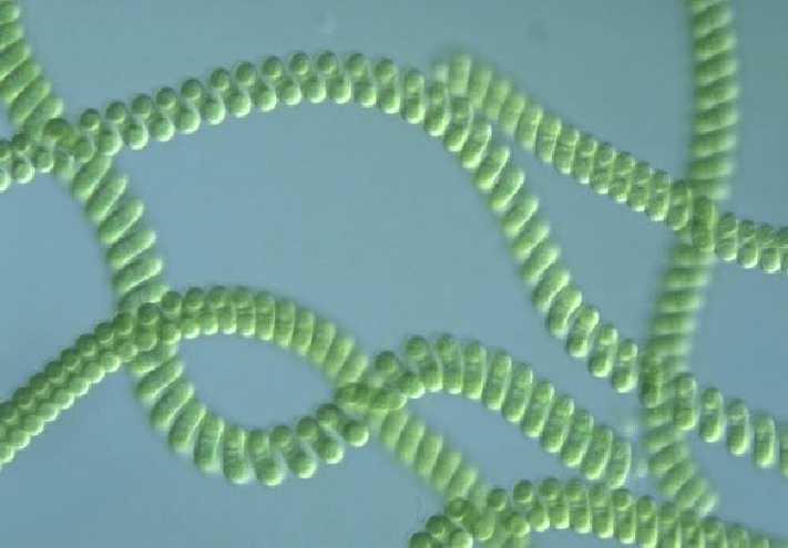
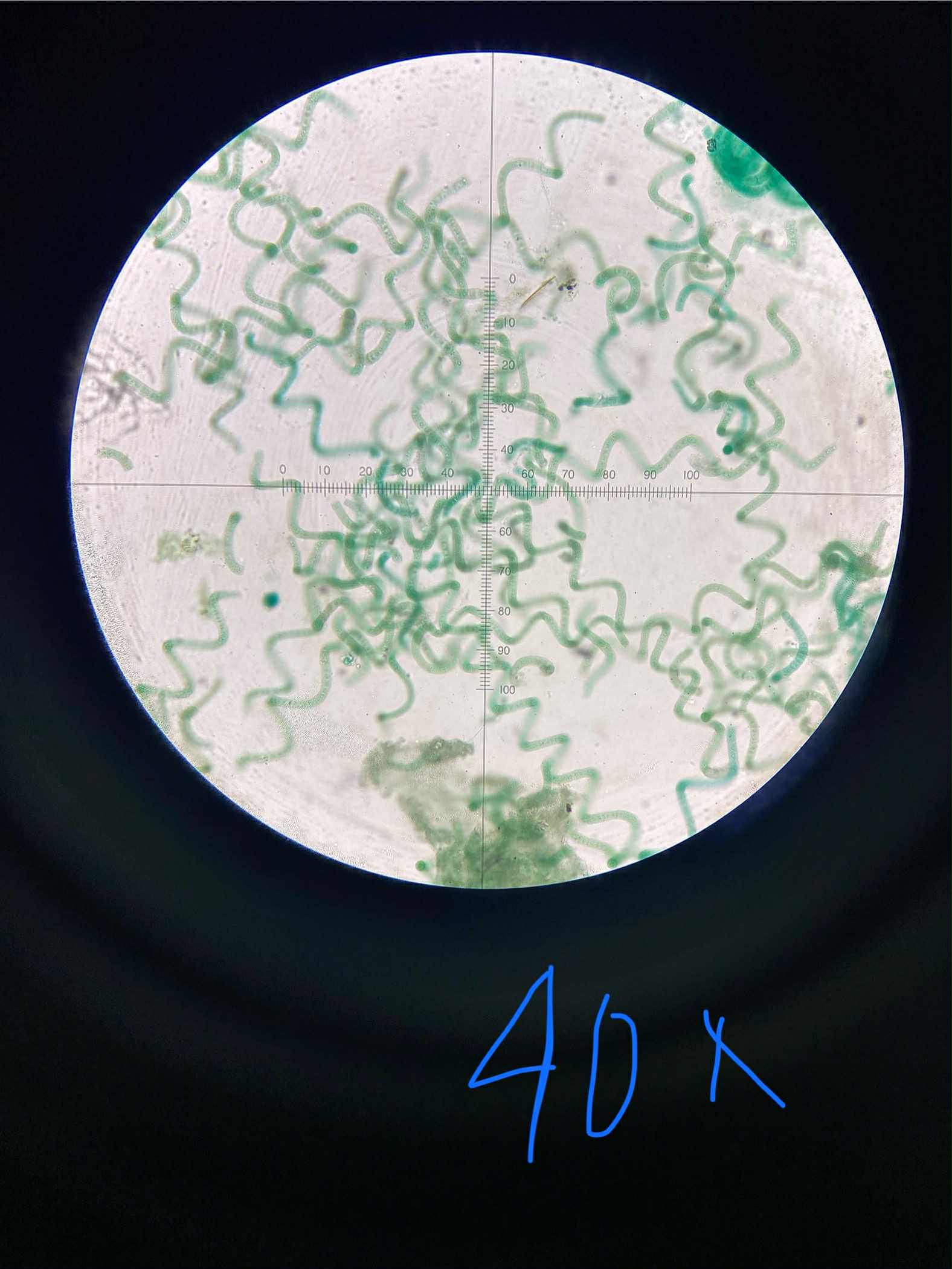
Spirulina
cyanobacteria
blue-green
filamentous; spirally coiled
cells in a single row along the helix held together by a thin sheath
no flagella; gliding movement dependent on Ca2+ and Na+ ions
distinct pigment comes from phycocyanin
specimen
phylum
color
shape
cell arrangement
motility
characteristic
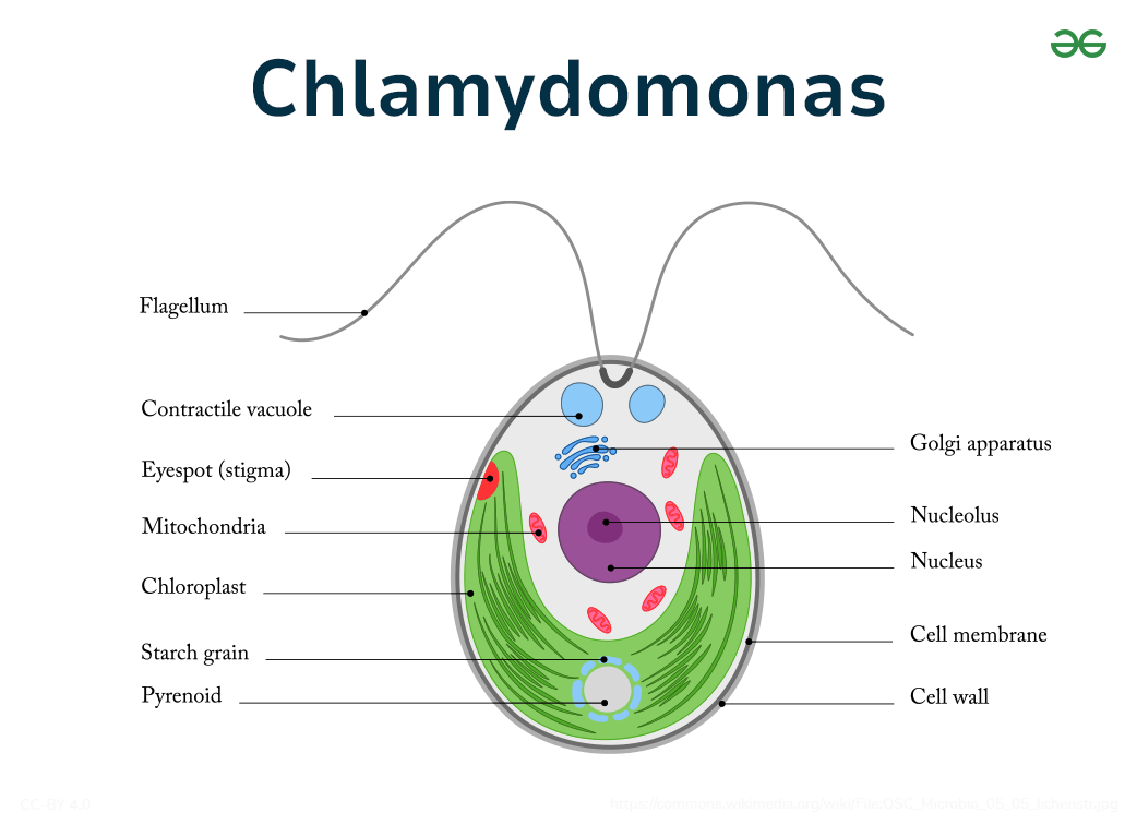
Chlamydomonas
chlorophyta
green
spherical/ovoid
single
motile by means off two anterior flagella
has an eyespot (stigma) with photoreceptors for photosynthesis
specimen
phylum
color
shape
cell arrangement
motility
characteristic
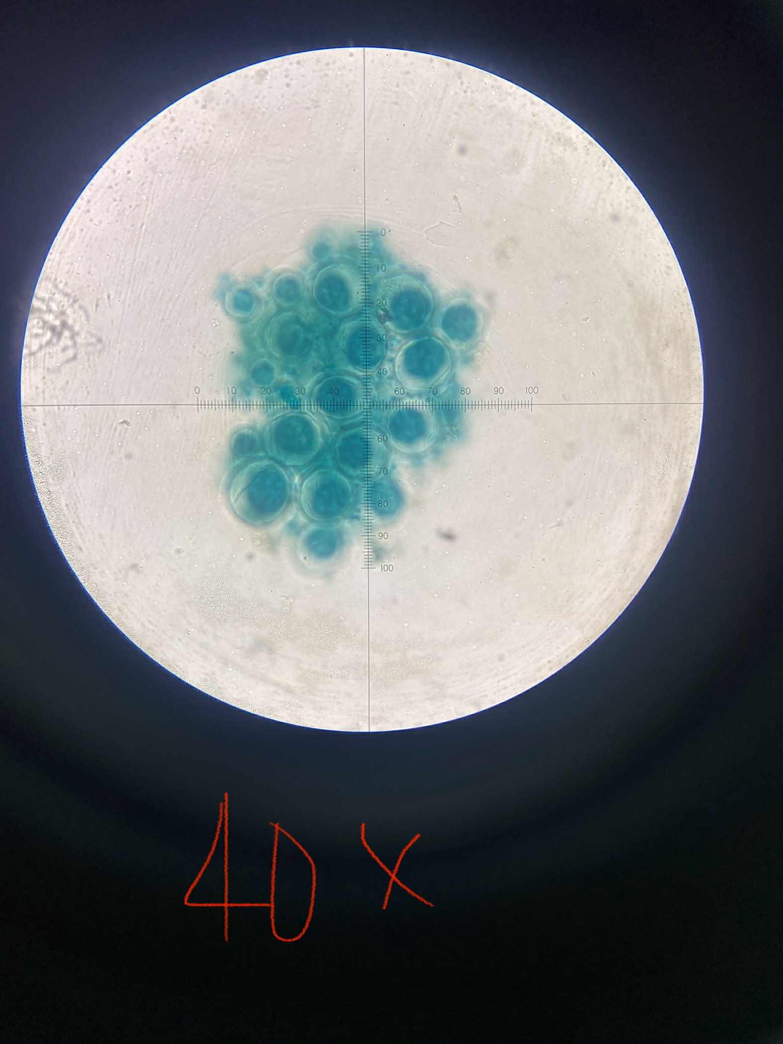
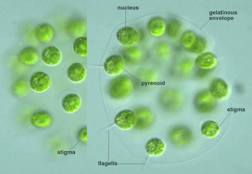
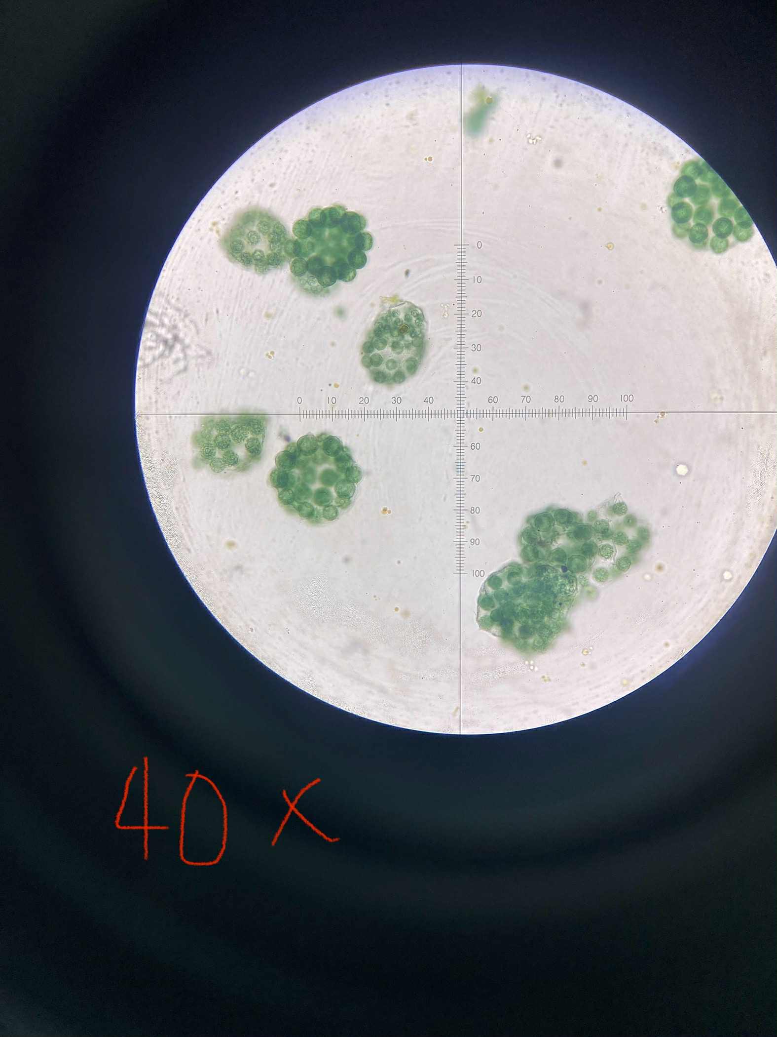
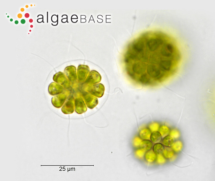
Eudorina
cholrophyta
green
spherical/ovoid colonies
colonies of 16 / 32 cells radially embedded within a matrix
highly motile; coordinated beating of the two flagella on each cell
each cell has a stigma; hollow colonies surrounded by a gelatinous matrix
specimen
phylum
color
shape
cell arrangement
motility
characteristic
Pandorina
chlorophyta
green
globular/spherical colony
radial compacted mass; held together at their bases in a gelatinous mix
2 flagella on each cell to create the rolling motion of colony
each cell has an eyespot
specimen
phylum
color
shape
cell arrangement
motility
characteristic
Gonium
chlorophyta
green
flat and spherical/curved colony
4/8/16 cells arranged in flat colonies (spherical/curved) held together by extracellular matrix
2 flagella per cell; pinwheel motion
exhibits phototaxis
specimen
phylum
color
shape
cell arrangement
motility
characteristic
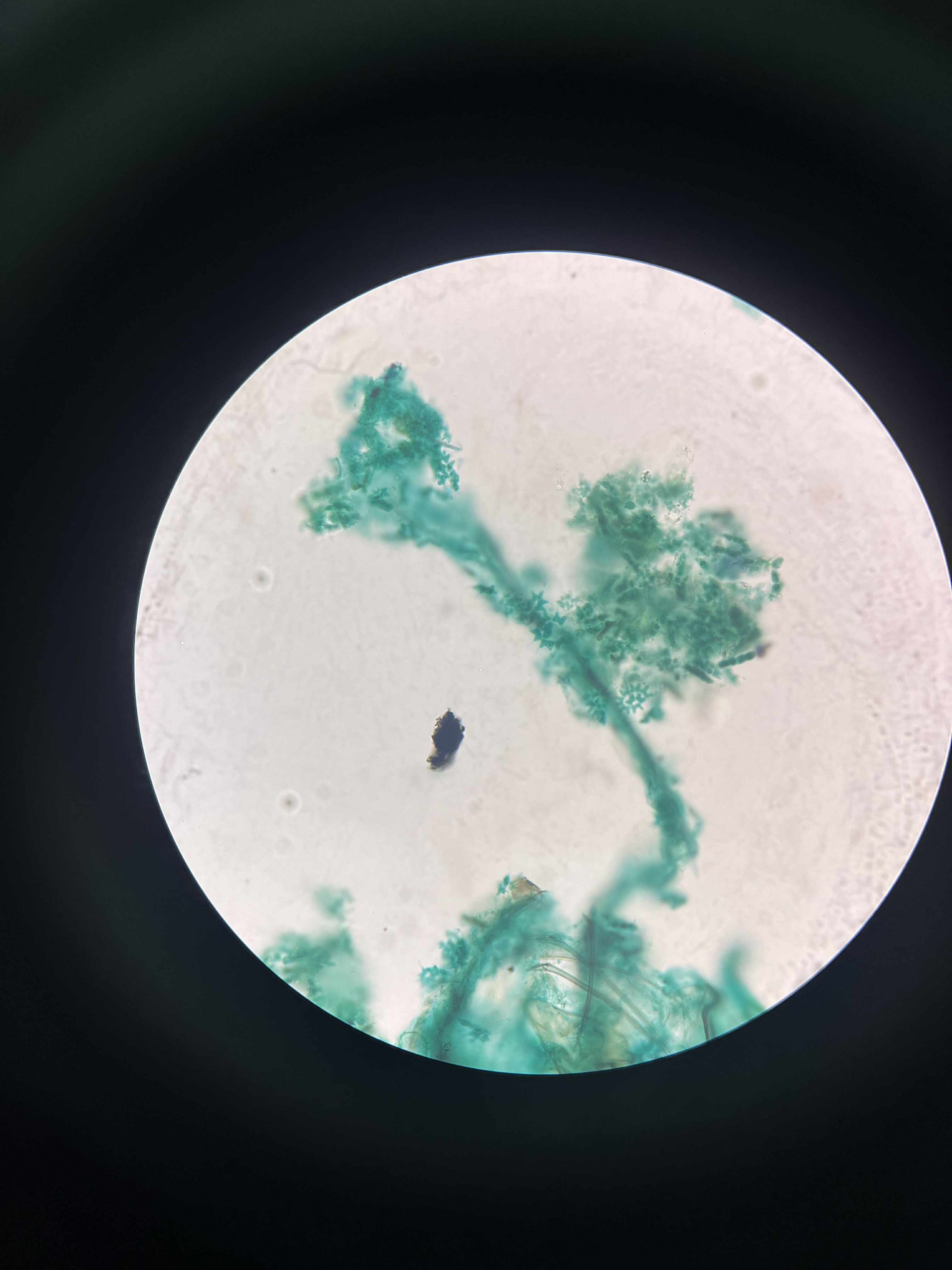
Cladophora
chlorophyta
green
branched filamentous colonies thallus
multinucleate cells arranged into uniseriate filaments
vegetative form non-motile; asexual zoospores have 4 flagella; sexual gametes have 2
forms cladophora balls; ability to grow attached or unattached freshwater and marine envi
specimen
phylum
color
shape
cell arrangement
motility
characteristic

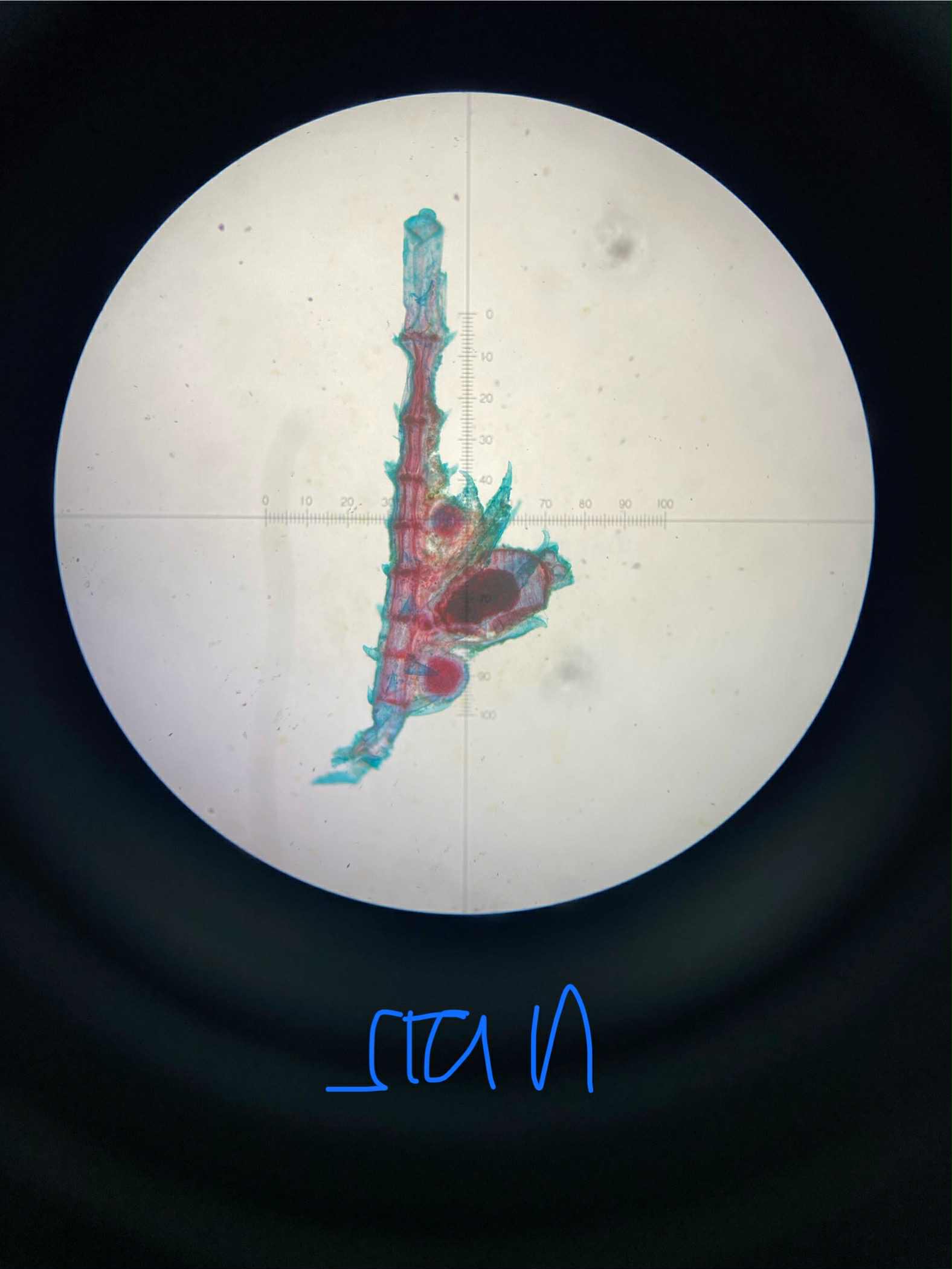
Chara
charophyta
green
thallus with main axis (w distinct nodes and internodes) and lateral branches
multicellular thallus
sessile organism; motile sperm
a stonewort; globules and nucules at nodes; calcification on their surfaces
specimen
phylum
color
shape
cell arrangement
motility
characteristic
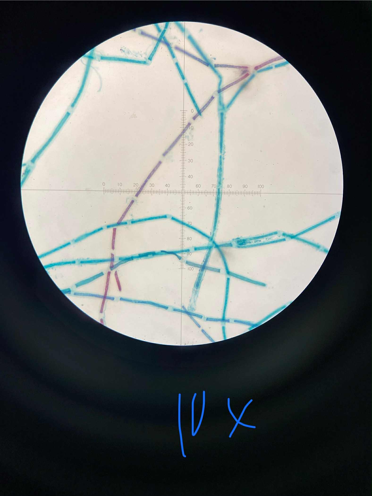
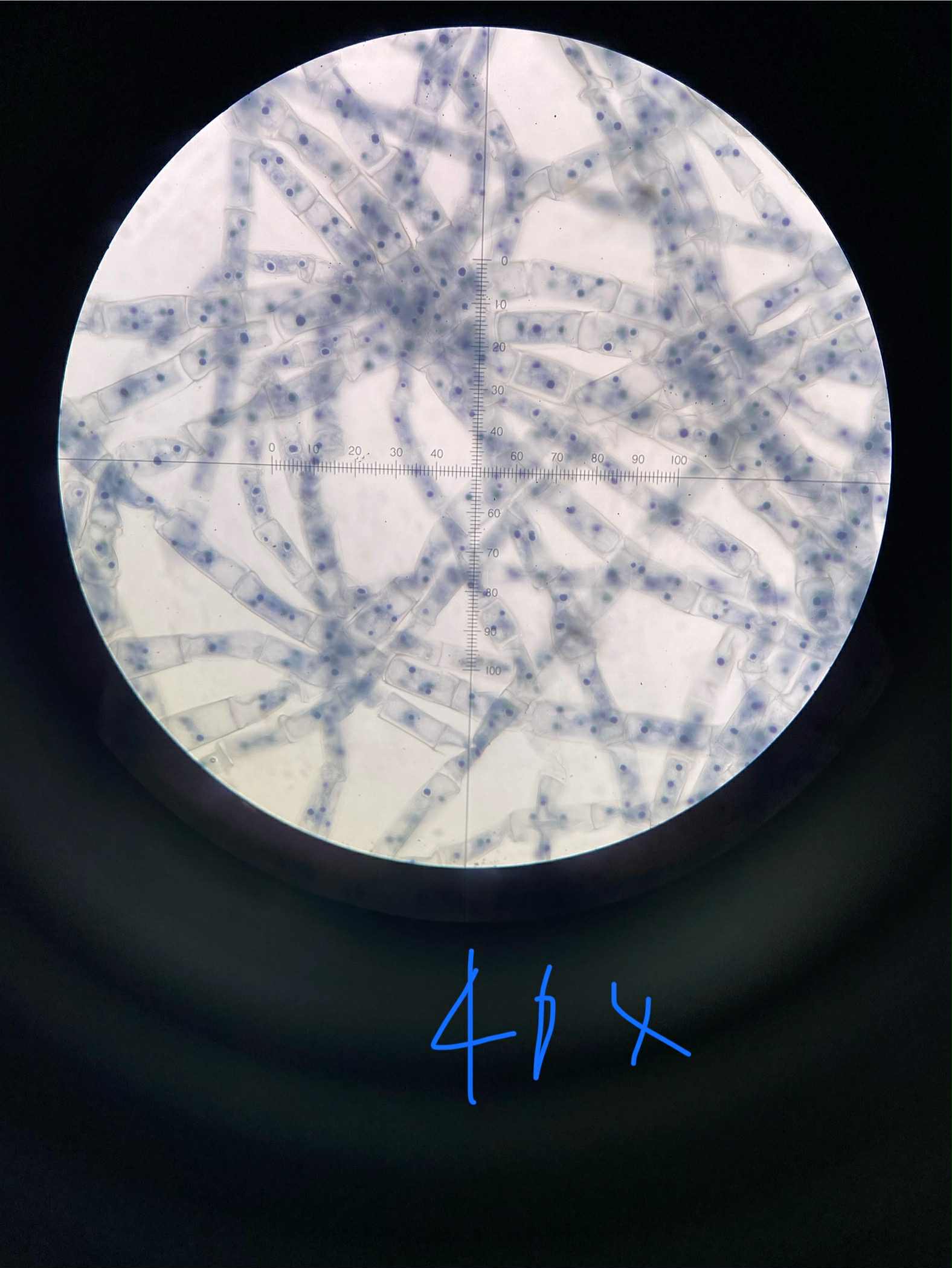
Oedogonium
chlorophyta
green
unbranched filaments
multicellular unbranched thallus
non-flagellated vegetative filaments; motile zoospore w ring of flagella
attach to substrates using a basal holdfast; apical caps (rings of old cell wall material left after cell division) indicate how many times a cell has divided
specimen
phylum
color
shape
cell arrangement
motility
characteristic
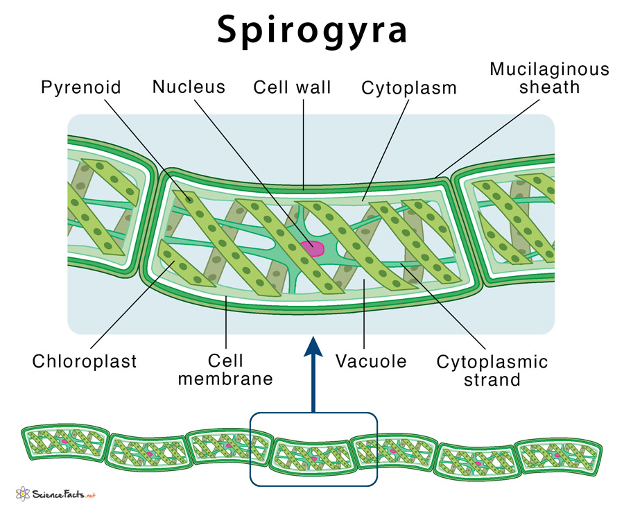
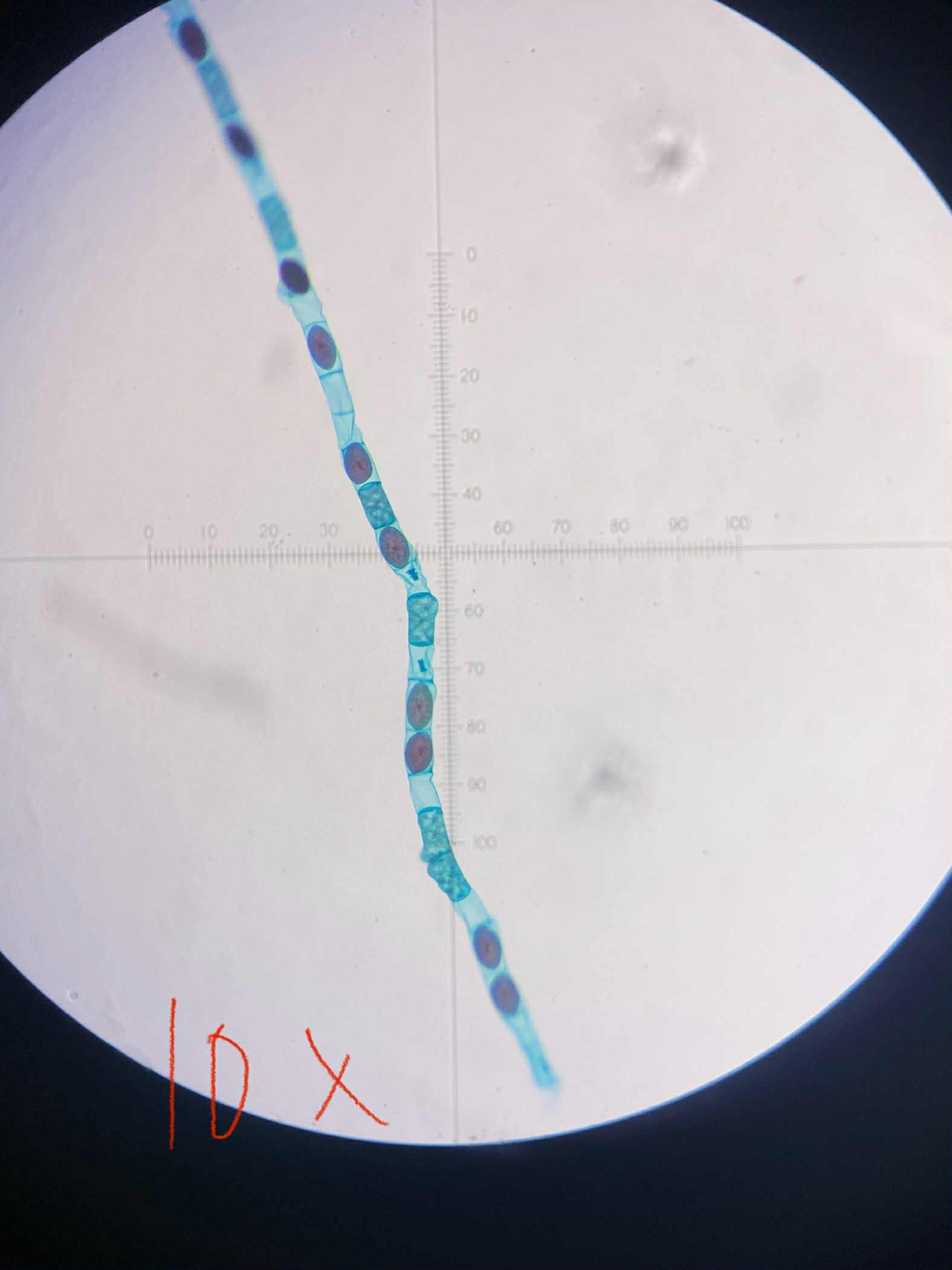
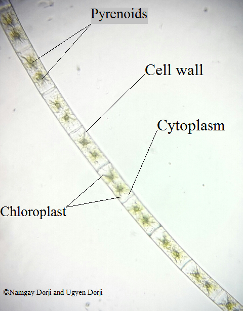
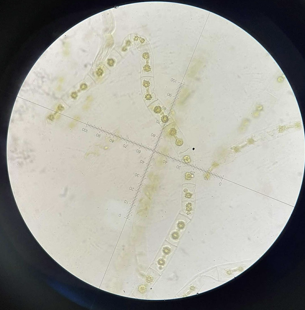
Spirogyra
chlorophyta
green
unbranched filaments
cells attached end to end to form filament
non-motile; capable of phototaxis
helical ribbon-shaped chloroplasts spiral around large central vacuole in each cell
specimen
phylum
color
shape
cell arrangement
motility
characteristic
Zygnema
charophyta
yellow-green; green
unbranched filaments
cell attached end-to-end to form unbranched filament
non-motile
non-flagellated, amoeboid gametes
specimen
phylum
color
shape
cell arrangement
motility
characteristic
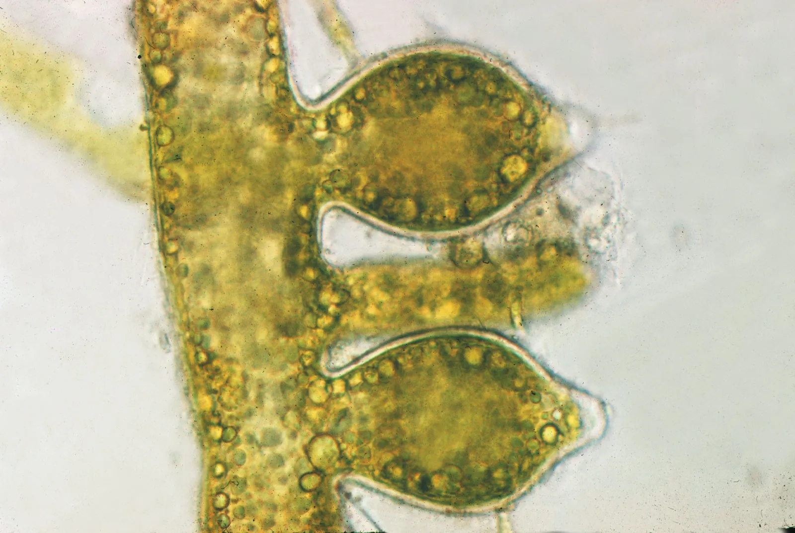
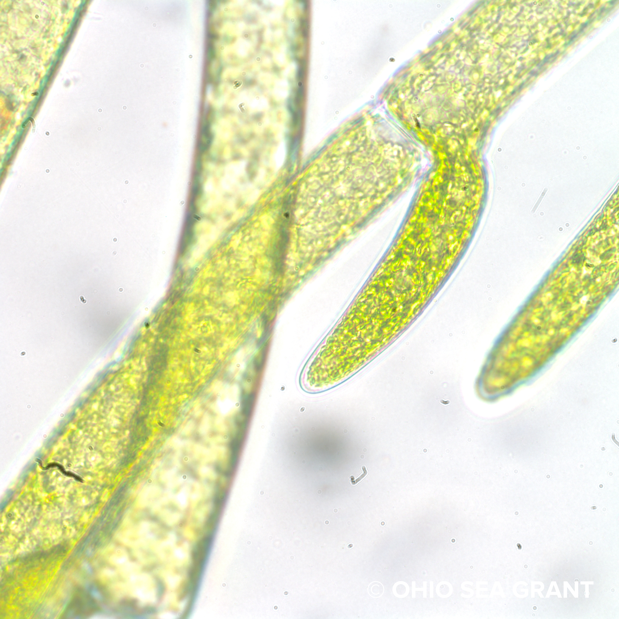
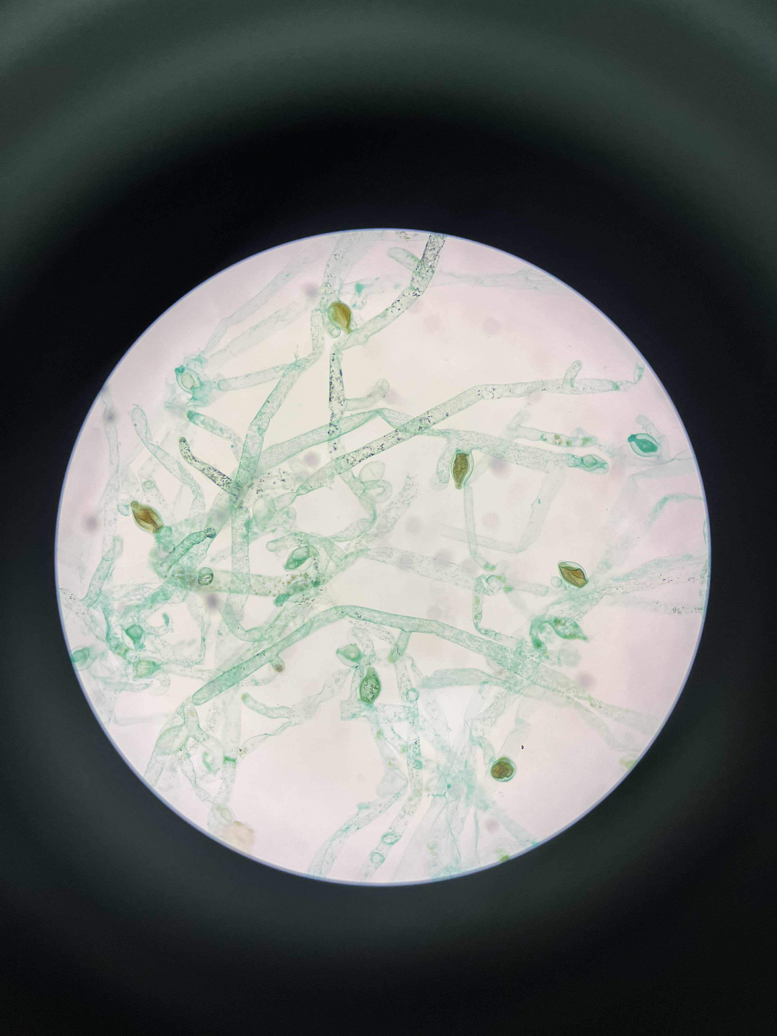
Vaucheria
xanthophyta
yellow-green (xanthophyll pigments)
branched, filamentous, tube-like thallus
coenocytic, aseptate thallus
sessile; motile zoospores and antherozoids
reserve food in the form of oil droplets, differing from the starch typical of other algae
specimen
phylum
color
shape
cell arrangement
motility
characteristic
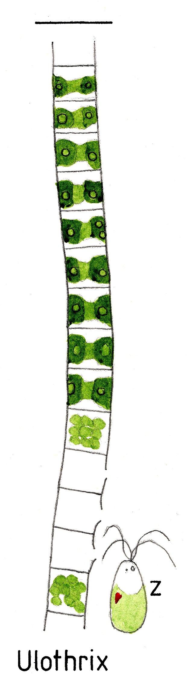
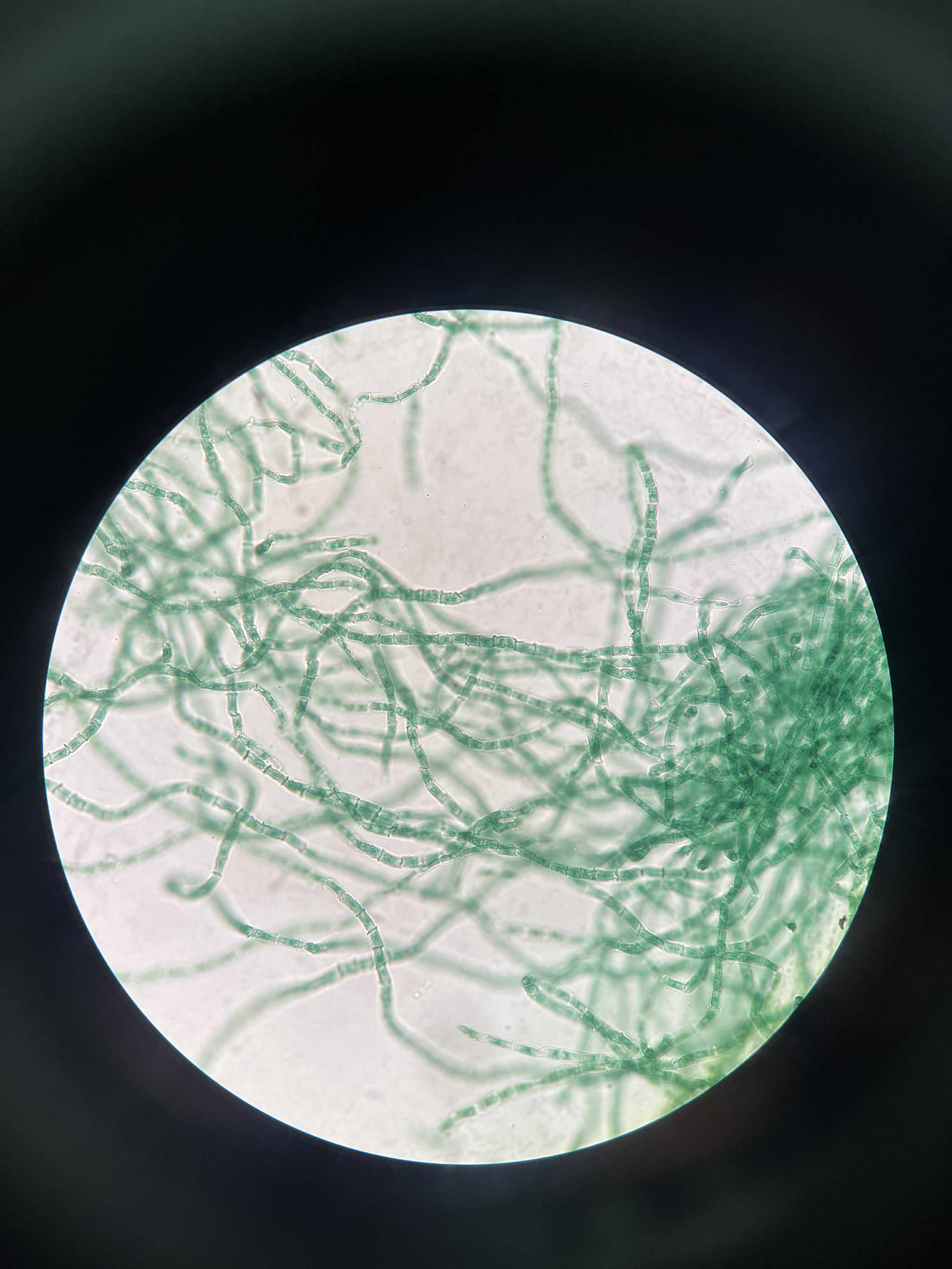
Ulorothrix
chlorophyta
green
unbranched filaments
uniseriate
non-motile; motile zoospores
girdle-shaped chloroplast; holdfast cell; cold temp
specimen
phylum
color
shape
cell arrangement
motility
characteristic
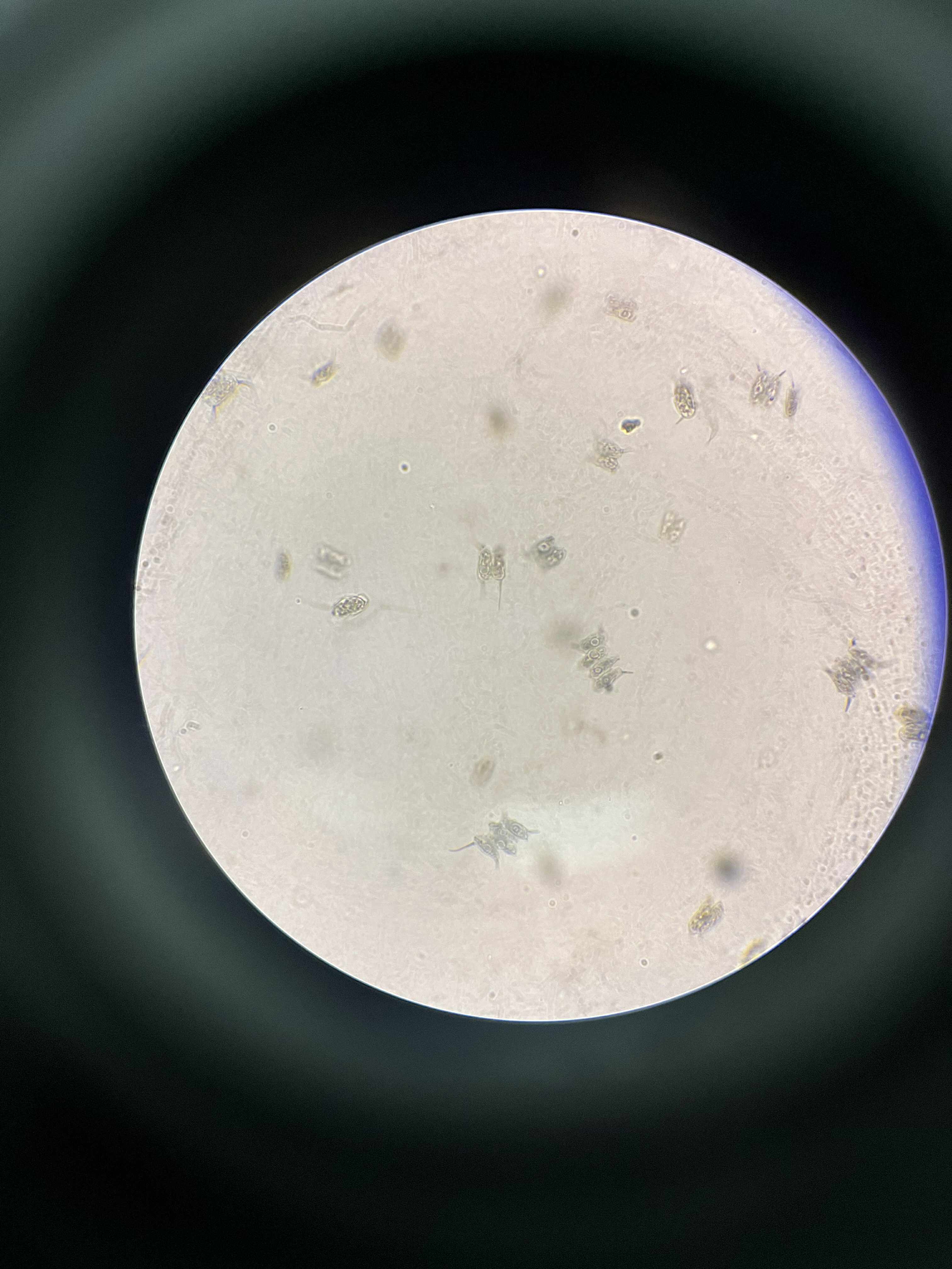
Desmids
charophyta
green
common shapes: globular, disc-like, spindle-like, and star-shaped
typically unicellular; each cell divided into 2 semi-cells
lack flagella
distinctive mirror-image semicells divided by a median constriction or isthmus
specimen
phylum
color
shape
cell arrangement
motility
characteristic
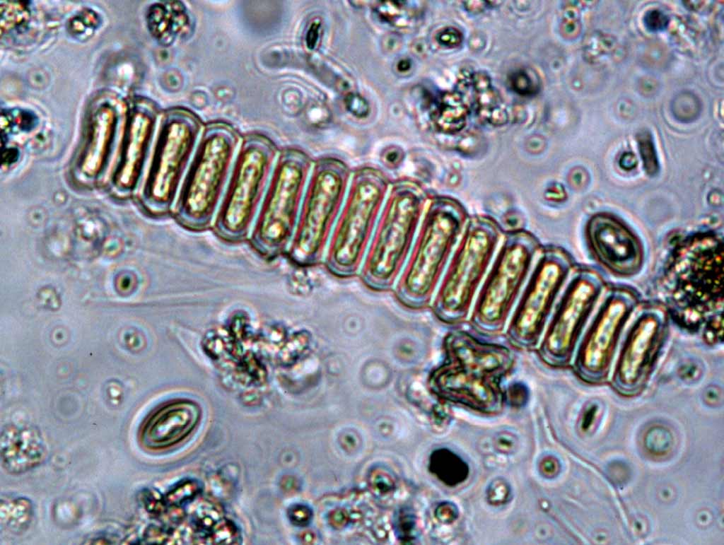
Scenedesmus
chlorophyta
green
cylindrical;ovoid;fusiform;lunate
aligned in a single row side by side
non-motile; no flagella
rigid, multi-layered cell wall with ornamentation (ridges, warts, spines); efficient at removing nitrogen and phosphorus from wastewater
specimen
phylum
color
shape
cell arrangement
motility
characteristic
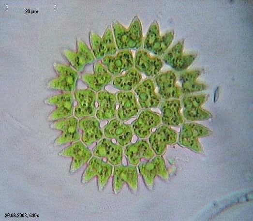
Pediastrum
chlorophyta
green
flat; disc-shaped circles/starlike
coloniall coenobium
nonmotile; motille spores and gametes
distinctive horn-like or bristle-like projections on its peripheral cells
specimen
phylum
color
shape
cell arrangement
motility
characteristic
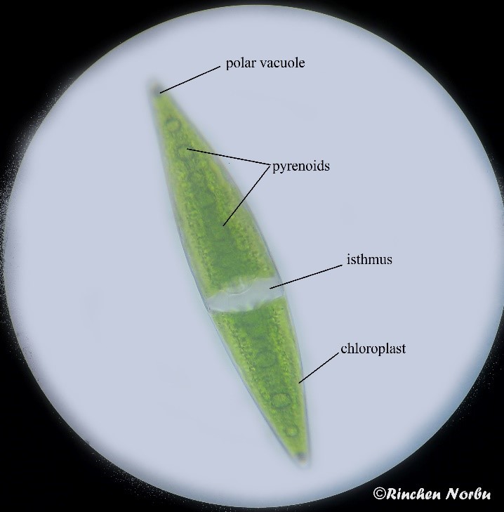
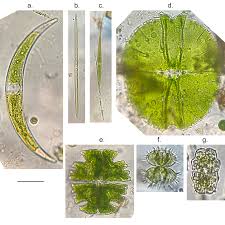
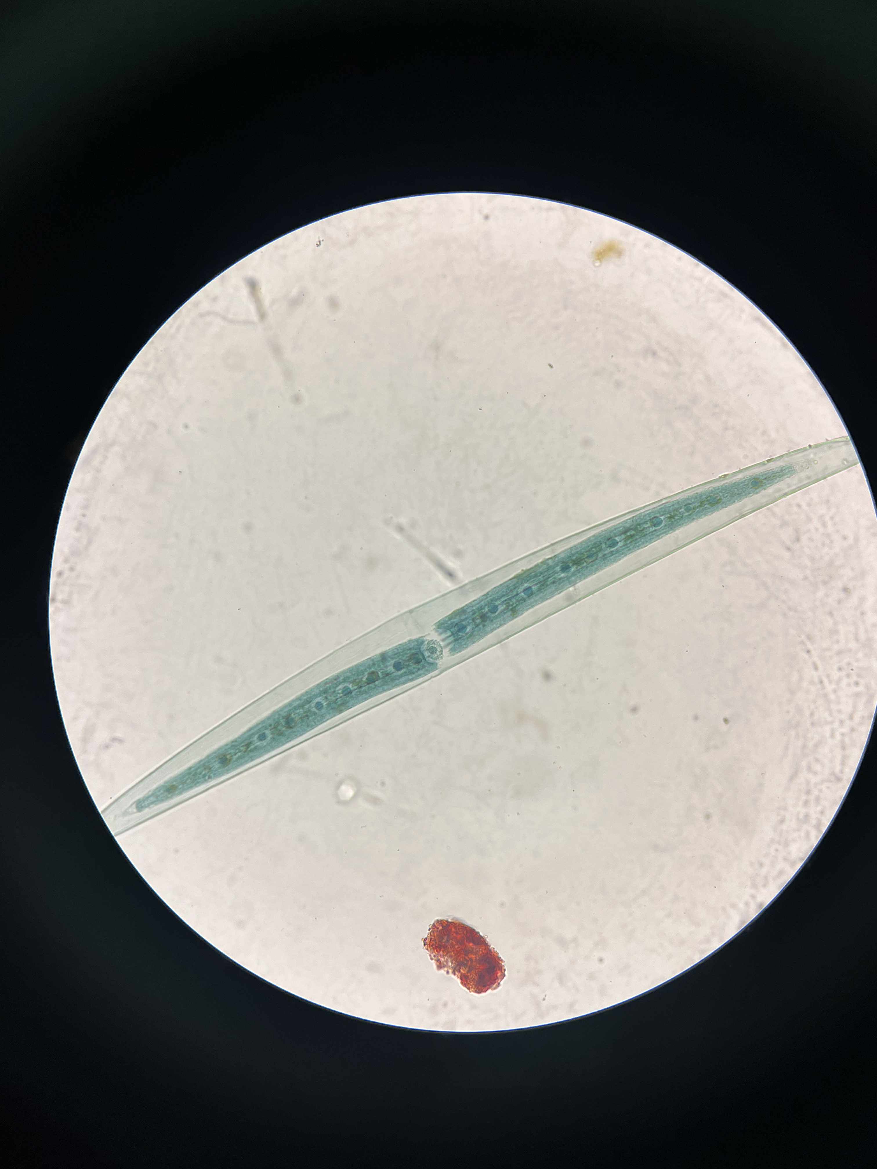
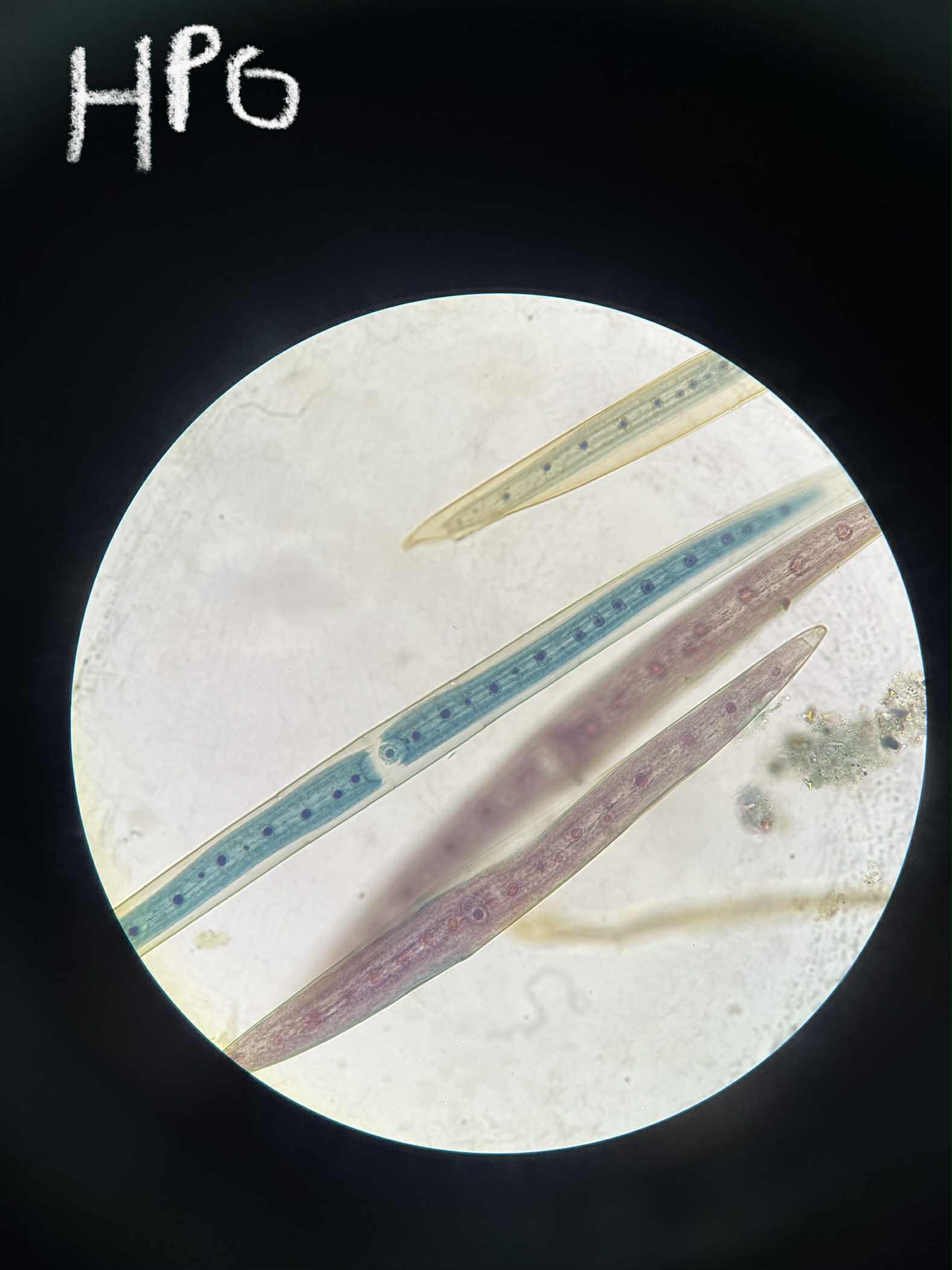
Closterium
charophyta
green
lunate/elongate; tips usually tapered
Singles
Non-motile
At the very tips of the cell, there are special vacuoles that contain vibrating crystals of barium or calcium sulfate, the function of which is currently unknown.
specimen
phylum
color
shape
cell arrangement
motility
characteristic
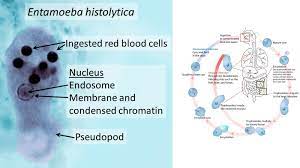
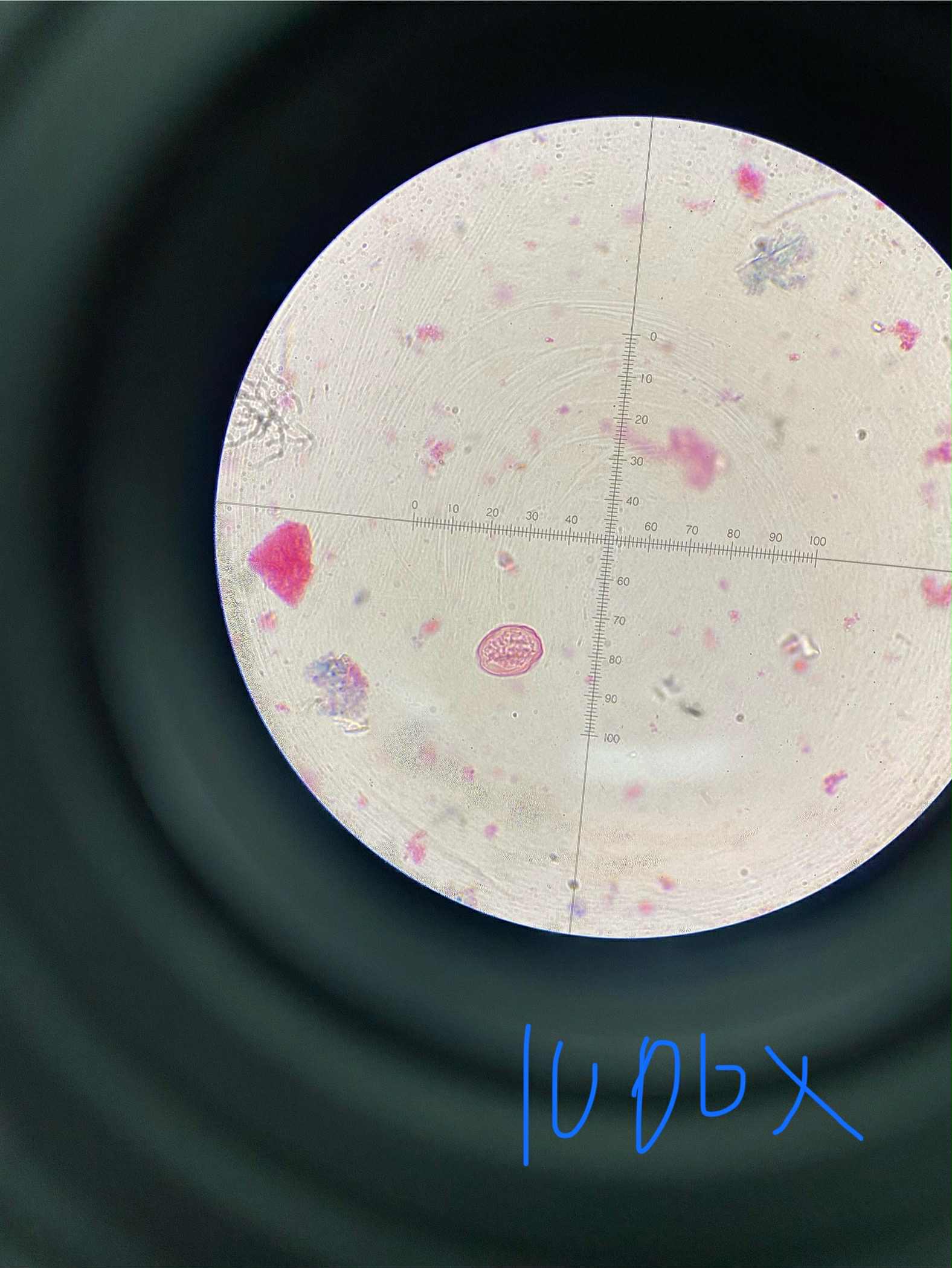
Entamoeba histolytica
Amoebozoa
Archamoebae
colorless
irregular/amoeboid (trophozoites); spherical (cysts)
single
active trophozoite stage is motile, pseudopodia (temporary projections)
Amebiasis
Prevention: Hygiene, food and water safety
Treatment: (initial) anti-parasitic drug e.g.metronidazolel → (luminal) for eliminating cysts
Life cycle:
Ingestion: Humans ingest mature cysts from contaminated food, water, or hands.
Excystation: In the small intestine, the cyst excysts to release the motile trophozoite.
Migration & Invasion: Trophozoites migrate to the large intestine, where they can invade the intestinal lining, causing amebic dysentery.
Multiplication: Trophozoites multiply by binary fission, and some form cysts.
Excretion: Cysts are passed in the feces, completing the cycle.
specimen
phylum
class
color
shape
cell arrangement
motility
disease
life cycle
prevention
treatment

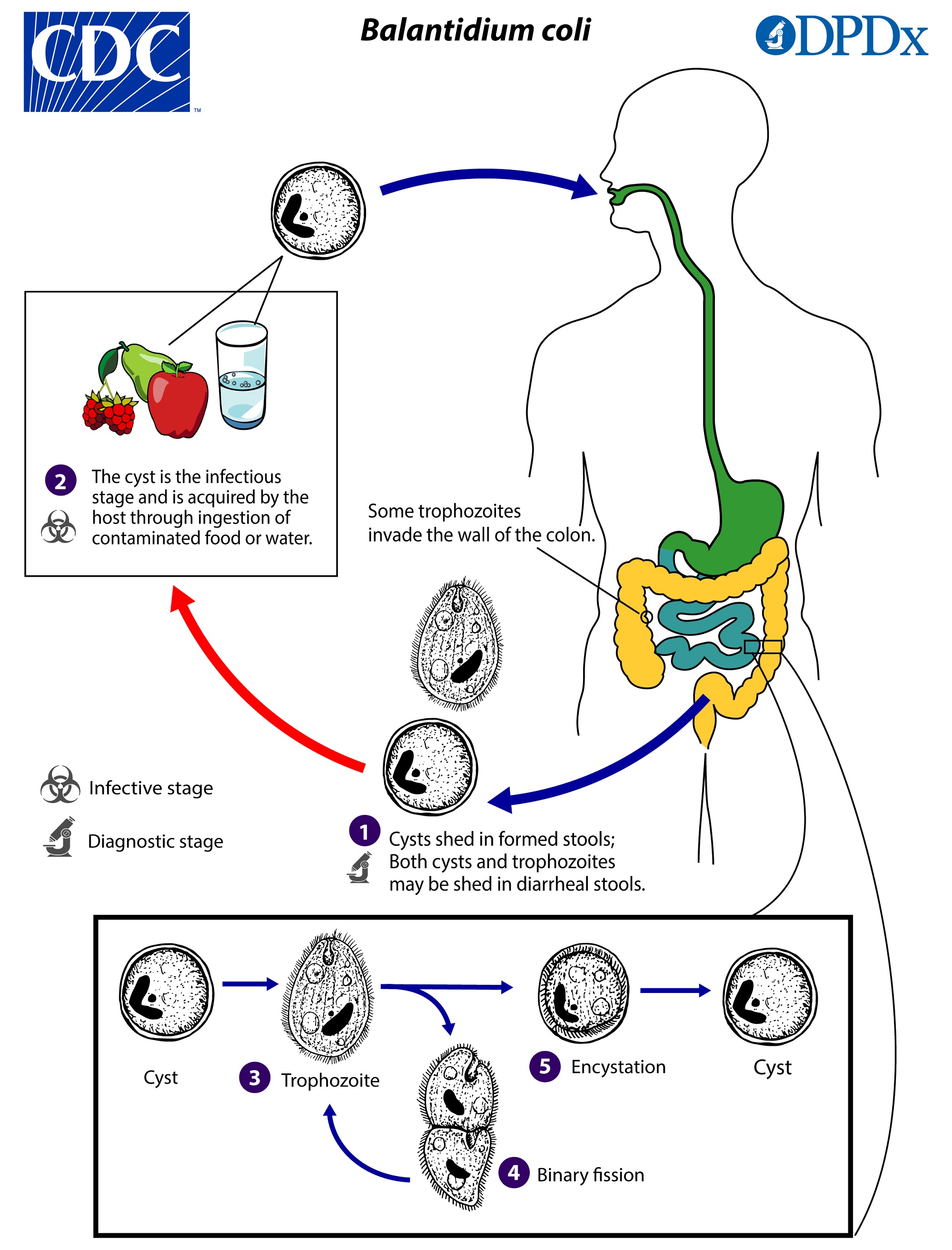
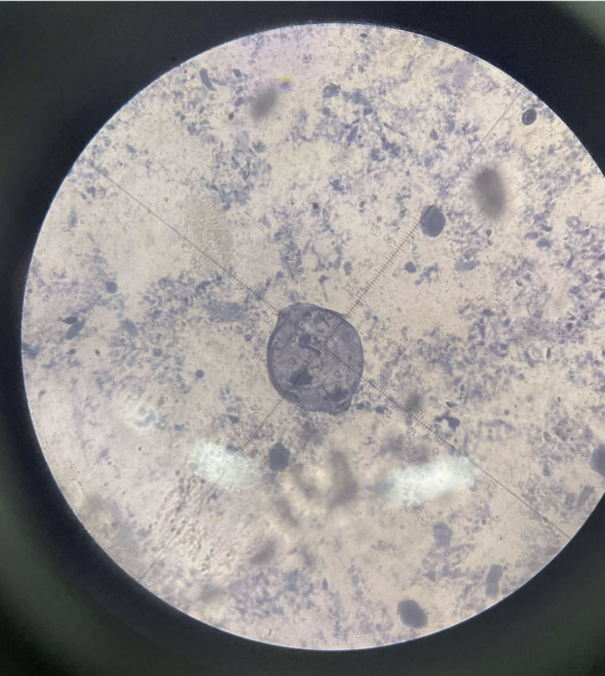
Balantidium coli
Ciliophora
Litostomatea
yellowish-green
ovoid/spherical
single cell
rapid movement due to trophozoite’s entire surface covered with cilia
Balantidiasis - an intestinal infection that is often asymptomatic but can manifest as diarrhea, dysentery, and abdominal pain
Strict adherence to good hygiene and proper sanitation especially in areas where pigs are raised, as they are a reservoir for the parasite.
Treatment: Antimicrobials
Life Cycle
Ingestion:
Humans become infected by ingesting the infective cysts through contaminated food or water.
Excystation:
Once in the small intestine, the cyst excysts, releasing the motile trophozoite.
Colonization:
The trophozoites then establish themselves in the mucosa of the large intestine.
Replication:
The trophozoites multiply in the large intestine.
Invasion and Pathology:
Some trophozoites can invade the colon wall, leading to ulceration and disease.
Encystation:
Trophozoites undergo encystation in the colon to produce infective cysts, which are then passed in the feces.
specimen
phylum
color
shape
cell arrangement
motility
found
disease
prevention
treatment
life cycle

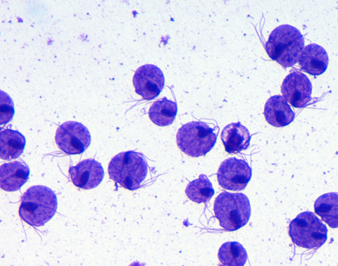
Giardia lamblia
Sarcomastigophora
Zoomastigophorea
colorless
(trophozoite) pear-shaped; (cyst) ovoid
Single (trophozoite 2 nuclei, cyst 4 nuclei)
trophozoite has flagella
Giardiasis - Diarrhea, abdominal cramps, bloating, gas, and fatty stools
Prevention: Hygiene, Water and Food Safety
Prescribed drugs
Ingestion: Humans ingest Giardia cysts, often from contaminated water or food.
Excystation: In the small intestine, exposure to digestive enzymes triggers the cysts to release motile trophozoites.
Trophozoite Activity: Trophozoites attach to the intestinal wall, reproduce by binary fission, and cause damage.
Encystation: As the parasite moves towards the colon, trophozoites transform into environmentally stable cysts.
Excretion: Cysts are passed in the feces, ready to infect another host
specimen
phylum
color
shape
cell arrangement
motility
found
disease
prevention
treatment
life cycle
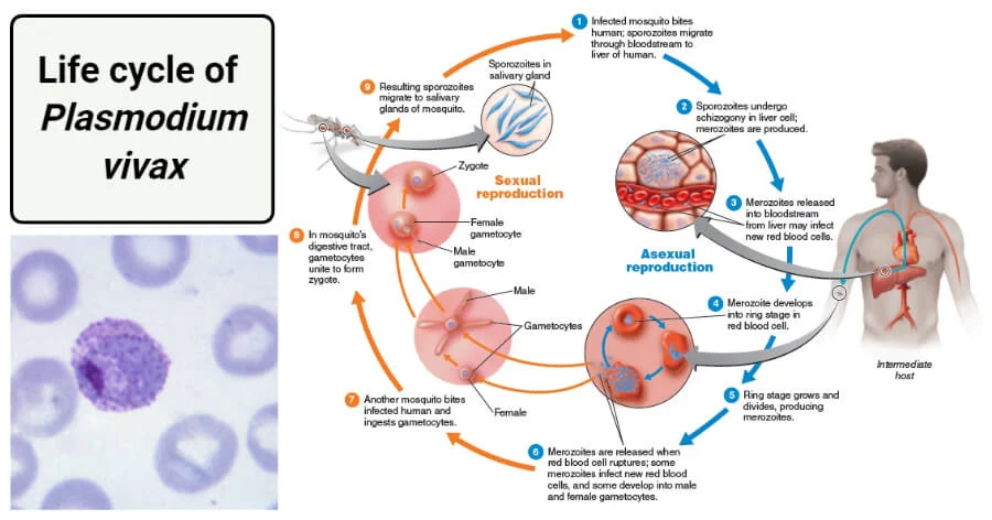
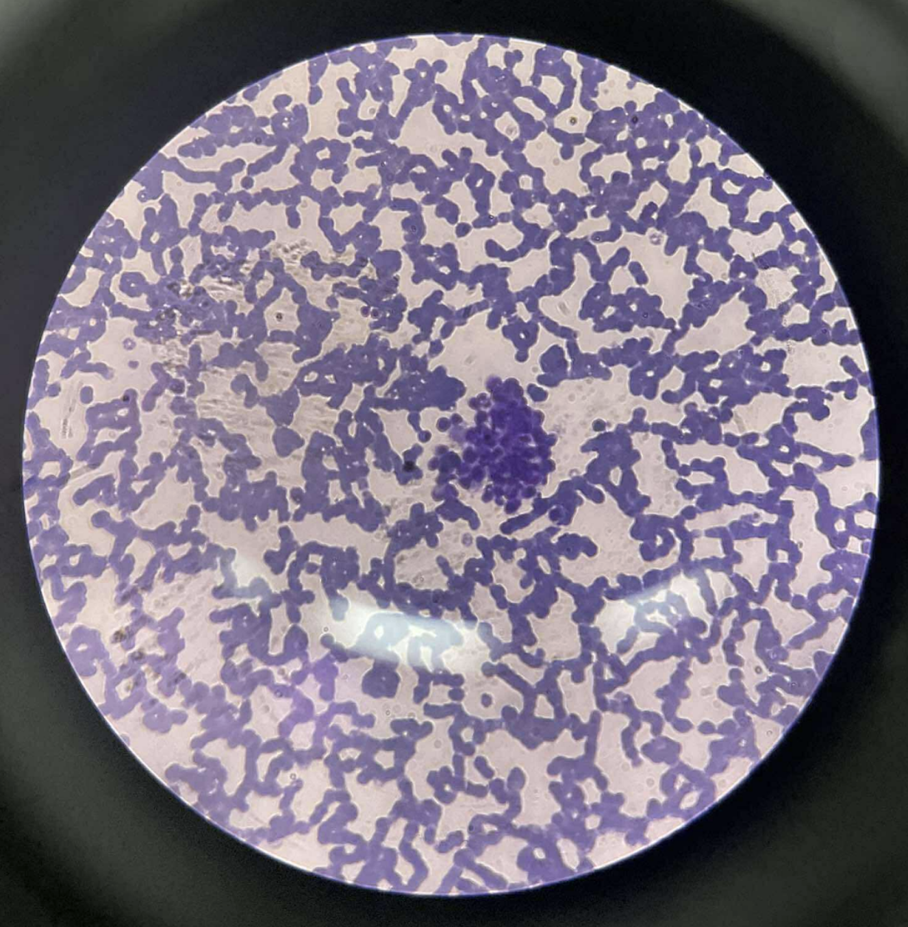
Plasmodium vivax
Apicomplexa
Aconoidasida
colorless
Shape:
Sporozoites: Lancet-shaped.
Trophozoites: Amoeboid, with an irregular, spreading cytoplasm.
Gametocytes: Round to oval shapes.
Cell Arrangement: The parasites grow and reproduce inside red blood cells and liver cells.
Motility: The parasites are non-motile.
Found: transmitted by female Anopheles mosquitoes.
Disease: P. vivax causes malaria, a febrile illness characterized by fever, chills, headaches, and digestive issues.
Prevention:
Mosquito control: Avoiding mosquito bites through the use of insect repellent and bed nets.
Antimalarial drugs: Chemoprophylaxis is used to prevent infection.
Treatment:
Blood stage treatment:
Chloroquine is the standard treatment for the symptomatic malaria parasite in the blood.
Liver stage treatment:
Primaquine is necessary to eradicate the dormant hypnozoite stage in the liver, which causes relapses.
Life Cycle:
Mosquito to human transmission:
An infected female Anopheles mosquito injects P. vivax sporozoites into a human host during a blood meal.
Liver stage (Exoerythrocytic phase):
Sporozoites migrate to the liver, where they multiply within liver cells (hepatocytes) and develop into liver-stage schizonts. Some may remain dormant as hypnozoites for long periods.
Merozoite release:
Rupturing schizonts release merozoites into the bloodstream.
Blood stage (Erythrocytic phase):
Merozoites infect red blood cells (specifically reticulocytes), where they develop into trophozoites, and then into schizonts.
Rupture and re-infection:
The schizonts rupture, releasing more merozoites to infect new red blood cells, repeating the cycle and causing symptoms.
Gametocyte formation:
Some parasites develop into gametocytes, which are then ingested by another mosquito during a blood meal.
Mosquito sexual reproduction:
Inside the mosquito, gametocytes undergo sexual reproduction to form sporozoites, which migrate to the salivary glands, completing the life cycle.
specimen
phylum
color
shape
cell arrangement
motility
found
disease
prevention
treatment
life cycle
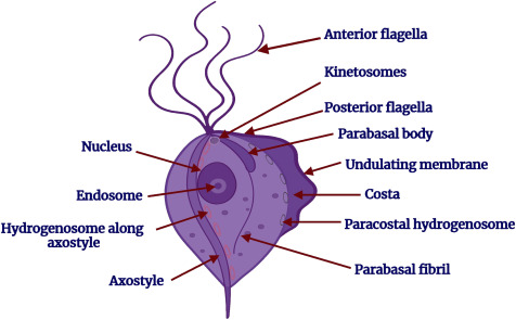
Trichomonas vaginalis
Trichomonada
Parabasalia
Colorless
pear-shaped (pyriform) or oval
single
Motility:
highly motile due to four anterior flagella and a posterior flagellum attached to an undulating membrane, giving it a jerky motion.
Found:
Resides in the lumen and mucosal surfaces of the urogenital tracts of humans, predominantly the vagina, urethra, and prostate.
Trichomoniasis, a sexually transmitted infection; foul-smelling yellow-green discharge, itching, and dysuria
Prevention: Safe sex practices, such as using condoms; Treatment of sexual partners;
specimen
phylum
color
shape
cell arrangement
motility
found
disease
prevention
treatment
life cycle